

Optical coherence tomography angiography (OCT-A) in retinitis pigmentosa and macular dystrophy patients: a retrospective study
- PMID: 34982219
- PMCID: PMC9061664
- DOI: 10.1007/s00417-021-05530-4
Optical coherence tomography angiography (OCT-A) in retinitis pigmentosa and macular dystrophy patients: a retrospective study
Abstract
Purpose: To evaluate macular vascular abnormalities in patients with macular dystrophies (MD) and retinitis pigmentosa (RP) through application of optical coherence tomography angiography (OCT-A).
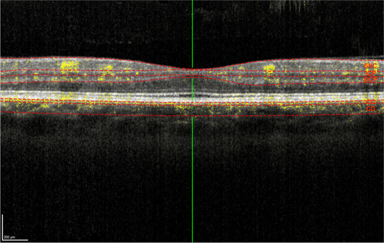
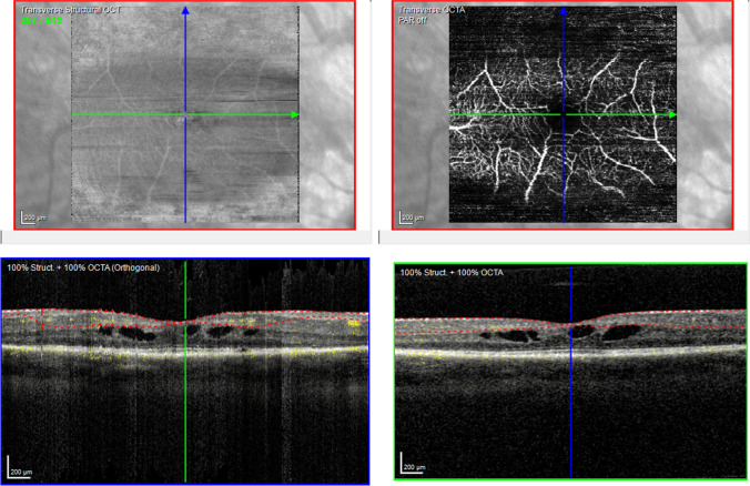
Methods: In this retrospective study, patients with MD and RP were examined by OCT-A and compared to healthy individuals. OCT-A images were analyzed regarding the diameter and surface area of the foveal avascular zone (FAZ) as well as flow (FL) in different retinal layers (superficial vascular complex (SVC), intermediate capillary complex (ICP), deep capillary complex (DCP), choriocapillaris (CC), and choroid (CD)).
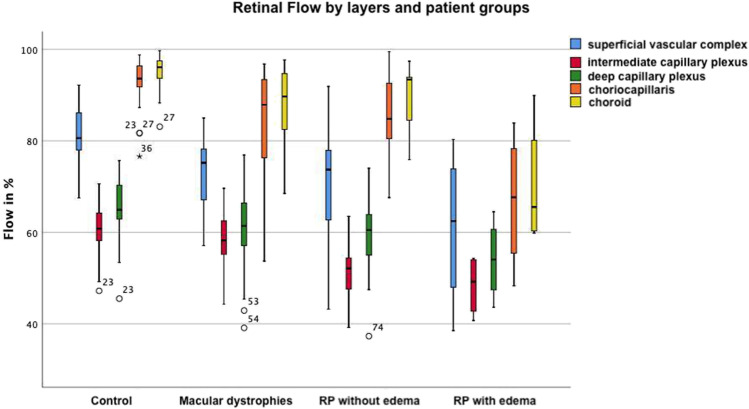
Results: Twenty-one patients with MD, 21 patients with RP without macular edema (RPnE), 8 patients with RP with edema (RPwE), and 41 healthy individuals were enrolled. The group of MD and RPnE patients showed none or only minor changes in FAZ. In RPwE patients, the FAZ was significantly smaller in vertical and horizontal measurements and surface area in SVC, whereas it was markedly enlarged in ICP. FL was significantly reduced compared to healthy individuals by an average of 13.2% in CD, 14.2% in CC, and 8.4% in DCP in all patient groups. In ICP, the reduction was 9.2% for RPnE and 12.7% for RPwE patients. The SVC showed reduced FL in the MD (8.1%) and RPnE (10.3%) group.
Conclusions: OCT-A is a valuable tool to examine retinal vascular abnormalities in patients with MD and RP. OCT-A revealed a reduced flow in various retinal layers in MD, RPnE, and RPwE. Alterations of the FAZ were less distinct in these groups which add to the variation reported previously.
Keywords: Inherited retinal dystrophy; Macula dystrophy; Optical coherence tomography angiography; Retinal imaging; Retinitis pigmentosa.
© 2022. The Author(s).
Conflict of interest statement
The authors declare no competing interests.
Figures


References
-
- De Silva SR, Arno G, Robson AG, Fakin A, Pontikos N, Mohamed MD, Bird AC, Moore AT, Michaelides M, Webster AR, Mahroo OA (2020) The X-linked retinopathies: physiological insights, pathogenic mechanisms, phenotypic features and novel therapies. Prog Retin Eye Res 100898. 10.1016/j.preteyeres.2020.100898 - PubMed
MeSH terms
LinkOut - more resources
Full Text Sources
Miscellaneous
