. 2022 Jan;11(1):e12179.
doi: 10.1002/jev2.12179.

Engineered small extracellular vesicles displaying ACE2 variants on the surface protect against SARS-CoV-2 infection

Affiliations

Engineered small extracellular vesicles displaying ACE2 variants on the surface protect against SARS-CoV-2 infection

Hark Kyun Kim et al. J Extracell Vesicles. 2022 Jan.

Abstract

Severe acute respiratory syndrome coronavirus 2 (SARS-CoV-2) entry is mediated by the interaction of the viral spike (S) protein with angiotensin-converting enzyme 2 (ACE2) on the host cell surface. Although a clinical trial testing soluble ACE2 (sACE2) for COVID-19 is currently ongoing, our understanding of the delivery of sACE2 via small extracellular vesicles (sEVs) is still rudimentary. With excellent biocompatibility allowing for the effective delivery of molecular cargos, sEVs are broadly studied as nanoscale protein carriers. In order to exploit the potential of sEVs, we design truncated CD9 scaffolds to display sACE2 on the sEV surface as a decoy receptor for the S protein of SARS-CoV-2. Moreover, to enhance the sACE2-S binding interaction, we employ sACE2 variants. sACE2-loaded sEVs exhibit typical sEVs characteristics and bind to the S protein. Furthermore, engineered sEVs inhibit the entry of wild-type (WT), the globally dominant D614G variant, Beta (K417N-E484K-N501Y) variant, and Delta (L452R-T478K-D614G) variant SARS-CoV-2 pseudovirus, and protect against authentic SARS-CoV-2 and Delta variant infection. Of note, sACE2 variants harbouring sEVs show superior antiviral efficacy than WT sACE2 loaded sEVs. Therapeutic efficacy of the engineered sEVs against SARS-CoV-2 challenge was confirmed using K18-hACE2 mice. The current findings provide opportunities for the development of new sEVs-based antiviral therapeutics.

Keywords: COVID-19; SARS-CoV-2; beta variant; delta variant; extracellular vesicles; soluble ACE2; spike.


Conflict of interest statement

Y.W.C. is the chief executive officer of ExoStemTech Inc. D.‐G.J. and J.H.P. are stockholders of ExoStemTech Inc. H.K.K, E.K., J.K., Y.W.C., D.‐G.J. have filed a patent application based on this work.

Figures

FIGURE 1
FIGURE 1
Schematic illustration of SARS‐CoV‐2 infection and the application of engineered sEVs for the neutralization of SARS‐CoV‐2. Mechanism of engineered sACE2 sEVs targeting the S protein of SARS‐CoV‐2 by exploiting the affinity of sACE2 for S. The resulting sEVs may represent an sEVs‐based antiviral therapy for blocking SARS‐CoV‐2 infection
FIGURE 2
FIGURE 2
Characterization of engineered sACE2 sEVs. (a) Cryo‐TEM images of HEK sEVs, sACE2(WT) sEVs, sACE2.v1 sEVs, and sACE2.v2 sEVs. (b) Size distribution of the engineered sEVs was measured using an nanoparticle tracking analysis. (c) Representative western blot analysis of cell lysates and sEVs for ACE2, Flag, TSG101, CANX, GM130, CD9, CD63 and ALIX. H: HEK sEVs, WT: sACE2(WT) sEVs, v1: sACE2.v1 sEVs, v2: sACE2.v2 sEVs. (d) CD81 staining of sEVs and fluorescence images of magnetic beads. (e) Flow cytometry of CD81 expression on sEVs. (f) sACE2 protein concentration in sEVs as determined via ELISA (n = 4). Scale bars, 50 nm. Data are presented as the mean ± s.d. *< 0.05; **< 0.01; ***< 0.001 (one‐way ANOVA with Tukey's multiple comparisons test)
FIGURE 3
FIGURE 3
Binding properties of engineered sEVs. (a) Scheme of ACE2 : SARS‐CoV‐2 S inhibitor assay. (b) The effect of HEK sEVs, sACE2(WT) sEVs, sACE2.v1 sEVs, and sACE2.v2 sEVs on the ACE2‐S interaction (n = 3). (c) Schematic illustration of the immunoprecipitation‐based binding assay of sEVs and the S protein. (d) S‐TagBFP protein immunoprecipitated from HEK293T cells transfected with the pCMV14 vector (Vec) and pCMV14‐SPIKE‐TagBFP (SPI) was detected with a TagBFP antibody. sEVs interacting with S‐TagBFP were detected using a Flag antibody. H: HEK sEVs, WT: sACE2(WT) sEVs, v1: sAJ. H. Sul, J. M. Jung, J. H. Park, Ji S Choi, Y. W. Cho, D. ?. G. JoCE2.v1 sEVs, v2: sACE2.v2 sEVs. Actin was used as a loading control for immunoblotting. Data are presented as the mean ± s.d. **< 0.01; ***< 0.001 (one‐way ANOVA with Tukey's multiple comparisons test d)
FIGURE 4
FIGURE 4
Protection against WT and mutant S‐containing pseudovirus. (a) Assessment of luciferase‐based WT S‐containing pseudovirus infectivity in HEK293T‐ACE2‐TagBFP cells in the presence of sEVs (n = 5). Values are normalized to virus alone (100%). (b) WT S‐containing pseudovirus infectivity related to the concentration of sACE2 in sEVs evaluated in Figure 2f. (c,d) Infectivity of pseudovirus bearing D614G S proteins (n = 3). (e,f) Infectivity of pseudovirus bearing Beta variant S (K417N, E484K, N501Y) proteins (n = 4). (g,h) Infectivity of pseudovirus bearing Delta variant S (L452R, E484K, D614G) proteins (n = 4). Data are presented as the mean ± s.d. *< 0.05; **< 0.01; ***< 0.001 (one‐way ANOVA with Tukey's multiple comparisons test)
FIGURE 5
FIGURE 5
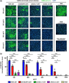
Protection against authentic Delta variant SARS‐CoV‐2 infection. (a) The immunostaining assays against SARS‐CoV‐2 N protein and DAPI for the Vero E6 cells infected by hCoV‐19/South Korea/KDCA5439/2021. Remdesivir (REM) was used as positive control. (b) The infectivity of Delta variant was assessed using the ratio of N protein‐positive cells to the total number of DAPI‐stained cell nuclei. Values are normalized to virus alone (100%) (n = 3). Scale bars, 200 μm. *< 0.05; **< 0.01; ***< 0.001 (one‐way ANOVA with Tukey's multiple comparisons test)
FIGURE 6
FIGURE 6
Anti‐SARS‐CoV‐2 activities of sACE2.v1 sEVs in K18‐hACE2 mice. (a) Schematic illustration of experimental design for K18‐hACE2 mice infected with SARS‐CoV‐2. (b) K18‐hACE2 mice were sacrificed at 3 days post infection (DPI) and viral titres in lungs were determined by plaque assay. Representative images of plaque assay and viral burden in the lungs (n = 6). (c) Viral RNA levels in the lungs at 3 DPI, as measure by qRT‐PCR. (d) qRT‐PCR analysis of expression in the lung homogenates of SARS‐CoV‐2‐infected K18‐hACE2 mice treated with PBS or sACE2.v1 sEVs. *< 0.05; **< 0.01; ***< 0.001 (unpaired Student's t‐test)

Similar articles

Cited by

References

    1. Barros, E. P. , Casalino, L. , Gaieb, Z. , Dommer, A. C. , Wang, Y. , Fallon, L. , Raguette, L. , Belfon, K. , Simmerling, C. , & Amaro, R. E. (2021). The flexibility of ACE2 in the context of SARS‐CoV‐2 infection. Biophysical Journal 120, 1072–1084. - PMC - PubMed
    1. Böker, K. O. , Lemus‐Diaz, N. , Rinaldi Ferreira, R. , Schiller, L. , Schneider, S. , & Gruber, J. (2018). The Impact of the CD9 Tetraspanin on Lentivirus Infectivity and Exosome Secretion. Molecular Therapy : The Journal of the American Society of Gene Therapy, 26, 634–647. - PMC - PubMed
    1. Boucheix, C. , Benoit, P. , Frachet, P. , Billard, M. , Worthington, R. E. , Gagnon, J. , & Uzan, G. (1991). Molecular cloning of the CD9 antigen. A new family of cell surface proteins. The Journal of Biological Chemistry, 266, 117–122. - PubMed
    1. Chan, K. K. , Dorosky, D. , Sharma, P. , Abbasi, S. A. , Dye, J. M. , Kranz, D. M. , Herbert, A. S. , & Procko, E. (2020). Engineering human ACE2 to optimize binding to the spike protein of SARS coronavirus 2. Science, 369, 1261–1265. - PMC - PubMed
    1. Cocozza, F. , Névo, N. , Piovesana, E. , Lahaye, X. , Buchrieser, J. , Schwartz, O. , Manel, N. , Tkach, M. , Théry, C. , & Martin‐Jaular, L. (2020). Extracellular vesicles containing ACE2 efficiently prevent infection by SARS‐CoV‐2 Spike protein‐containing virus. Journal of Extracellular Vesicles, 10, e12050. - PMC - PubMed

Publication types

Substances