. 2022 Jan 13;65(1):616-632.
doi: 10.1021/acs.jmedchem.1c01763. Epub 2022 Jan 4.

Heterobivalent Ligand for the Adenosine A2A-Dopamine D2 Receptor Heteromer

Affiliations

Heterobivalent Ligand for the Adenosine A2A-Dopamine D2 Receptor Heteromer

Daniel Pulido et al. J Med Chem. .

Abstract

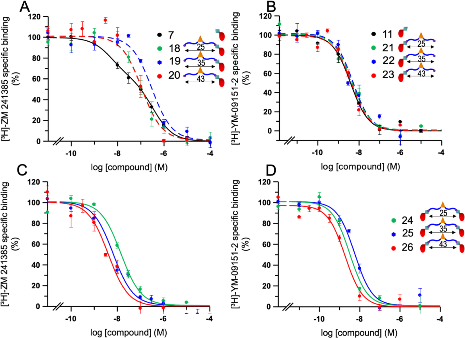
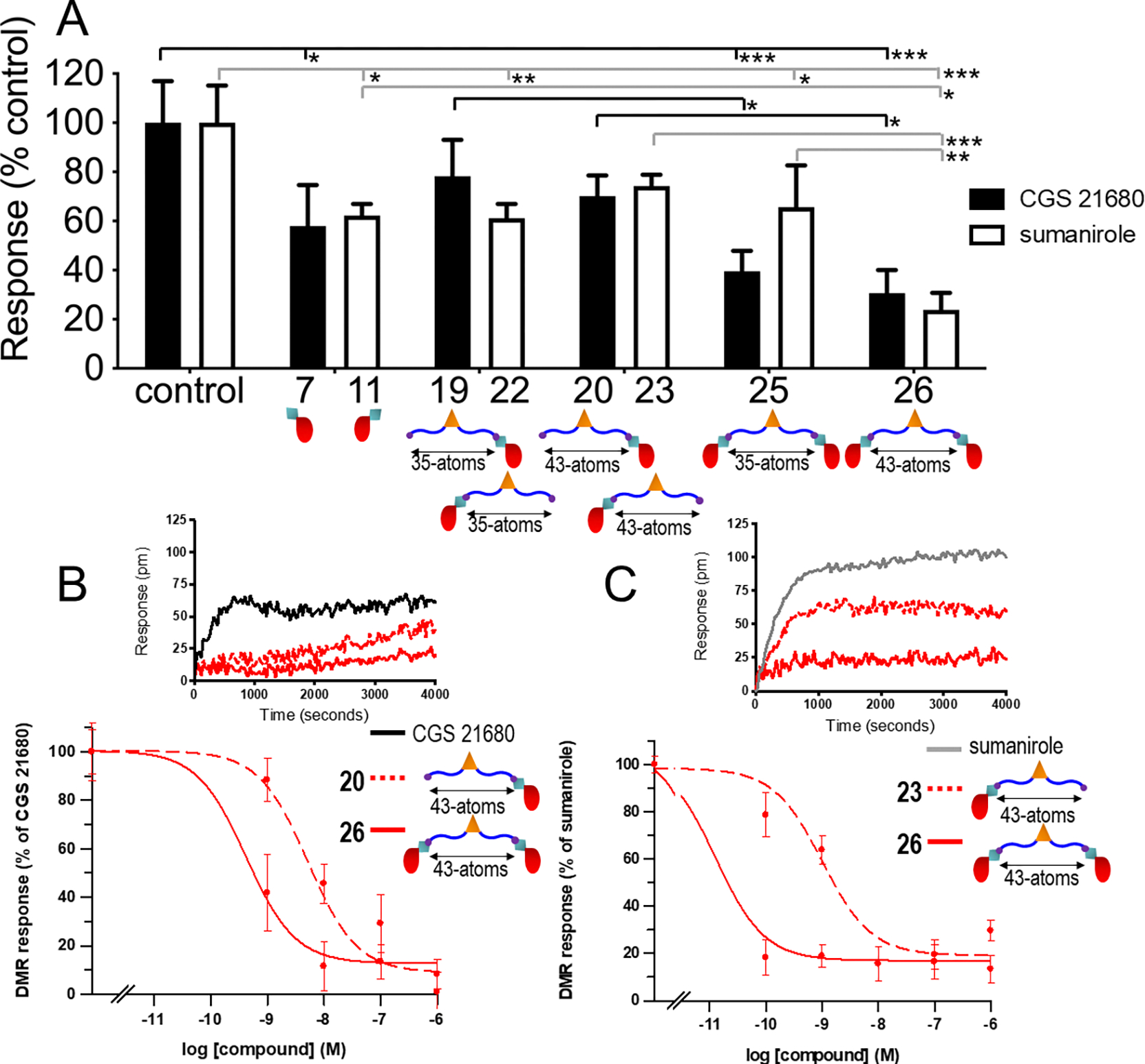
A G protein-coupled receptor heteromer that fulfills the established criteria for its existence in vivo is the complex between adenosine A2A (A2AR) and dopamine D2 (D2R) receptors. Here, we have designed and synthesized heterobivalent ligands for the A2AR-D2R heteromer with various spacer lengths. The indispensable simultaneous binding of these ligands to the two different orthosteric sites of the heteromer has been evaluated by radioligand competition-binding assays in the absence and presence of specific peptides that disrupt the formation of the heteromer, label-free dynamic mass redistribution assays in living cells, and molecular dynamic simulations. This combination of techniques has permitted us to identify compound 26 [KDB1 (A2AR) = 2.1 nM, KDB1 (D2R) = 0.13 nM], with a spacer length of 43-atoms, as a true bivalent ligand that simultaneously binds to the two different orthosteric sites. Moreover, bioluminescence resonance energy transfer experiments indicate that 26 favors the stabilization of the A2AR-D2R heteromer.


Conflict of interest statement

The authors declare no competing financial interest.

Figures

Figure 1.
Figure 1.
The heterobivalent ligand contains (i) a scaffold that can be properly derivatized (in orange), (ii) pharmacophore units (in red) that bind the orthosteric binding site of A2AR (7) and D2R (11) with high affinity, (iii) a spacer to cover the distance between both protomers of the heteromer (in blue), and (iv) a linker (in green) between the pharmacophore units and the spacer, adequate for the chemistry used for conjugation.
Figure 2.
Figure 2.
Evolution of bivalent ligands 25 and 26 in the A2AR-D2R heteromer, constructed via the TM 4/5 interface, as devised from MD simulations. (A) Molecular models of bivalent ligands 25 (spacer length of 35, n=m=2, see Figure 1) and 26 (spacer length of 43, n=4, m=1) bound to the A2AR-D2R heteromer. A MOE-based computational tool was used to model these ligands into the heteromer. (B) Representative structures (opaque sticks) and 100 structures collected every 10 ns (translucent lines) of 25 and 26 (the color code of the atoms is as in Figure 1), extracted from the simulations, whereas the structure of the A2AR-D2R heterodimer corresponds to the initial model. (C) The simultaneous binding of the pharmacophore units of 25 and 26 to both orthosteric sites was monitored by the salt bridge distance between the protonated amine of the NAPS pharmacophore and the Cγ atom of Asp3.32 of D2R and the hydrogen bond distance between the -NH2 group of the adenine moiety of the SCH-442,416 pharmacophore and the Oδ1 atom of Asn6.55 of A2AR (dashed gray line between gray spheres in the inset panel). The stability of the ligand-heteromer complex was analyzed via root mean-square deviations (rmsd) of the heavy atoms of 25 and 26 (red for the pharmacophore moieties and blue for linker/spacer/scaffold/spacer/linker moities) and the stability of the A2AR-D2R heteromer was also analyzed by rmsd of A2AR (green) and D2R (orange). Three replicas of 1 μs of each complex were run.
Figure 3.
Figure 3.. Competition experiments of the A2AR and D2R ligands 7, 11, 18–26.
Competition experiments of [3H]ZM 241385 for A2AR (A and C) or [3H]YM-09151–2 for D2R (B and D) vs. increasing concentrations of A2AR and D2R pharmacophores (7 and 11, respectively), A2AR and D2R monovalent compounds (18–20 and 21–23, respectively) (A and B), and heterobivalent ligands 24–26 (C and D), using membranes from sheep brain striatum that naturally express A2AR and D2R. Experimental data were fitted to the dimer receptor model equation (2) and (3) [see Experimental section]. Data are mean ± SEM from a representative competitive experiment (n=3–7) performed in triplicate. 100% corresponds to 0.14±0.02 (in A and C) or 0.15±0.01 (in B and D) pmol/mg of protein. For statistical significance analysis see Table 1.
Figure 4.
Figure 4.. Effect of TM3-TAT and TM5-TAT peptide of A2AR in the binding affinity of heterobivalent ligands 25 and 26 for A2AR.
Competition experiments of [3H]ZM 241385 vs increasing concentrations of heterobivalent ligands. 25 (A) or 26 (B) in the absence (solid line) or the presence of TM5-TAT (dash-dotted line) or of TM3-TAT (dashed line) peptides of A2AR, using membranes from sheep brain striatum. Experimental data were fitted to the dimer receptor model equation (3) [see Experimental section]. Data are mean ± SEM from a representative competitive experiment (n=3–6) performed in triplicate. 100% corresponds to 0.14±0.02 pmol/mg protein. For statistical significance analysis see Table 1.
Figure 5.
Figure 5.. Quantification of the antagonistic effect of the studied compounds on global cellular response induced by CGS 21680 or sumanirole.
Dynamic mass redistribution (DMR) assays were performed in CHO cells stably expressing D2R and A2AR. (A) Cells were treated with medium (control), with 10 nM of the A2AR ligands (pharmacophore derivative 7, monovalent compounds 19 and 20 or bivalent compounds 25 and 26, in black) or with 0.1 nM of the D2R ligands (pharmacophore derivative 11, monovalent compounds 22 and 23 and bivalent compounds 25 and 26, in white) for 30 minutes before the addition of 3 nM of CGS 21680 (black) or 100 nM of sumanirole (white). Values are mean ± SEM from 3–4 determinations carried out in triplicate. (B, C) Representative DMR curves in which cells were treated with medium (control, in black), with the monovalent compounds (dotted red) (10 nM of 20 in B or 0.1 nM of 23 in C), or with the heterobivalent compound (red) 26 (10 nM in B or 0.1 nM in in C) for 30 minutes before the addition of 3 nM of CGS 21680 (B) or 100 nM of sumanirole (C). Each curve is the mean of a representative optical trace experiment carried out in triplicates. The resulting shifts of reflected light wavelength (pm) were monitored over time. Dose-response curves of the antagonistic effect of heterobivalent compound 26 (red) and the corresponding monovalent ligands 20 and 23 (dotted red) on the DMR induced by 3 nM CGS 21680 (B) or 100 nM sumanirole (C). Data are mean ± SEM from 5–6 experiments and are presented as percentage of the maximal effect of CGS 21680 or sumanirole. Statistical significance was calculated by one-way ANOVA followed by Bonferroni’s multiple comparison post hoc test. *p<0.05, **p<0.01, ***p<0.001.
Figure 6.
Figure 6.. Influence of bivalent ligands on A2AR-D2R heterodimerization.
BRET experiments were performed using HEK-293T cells transiently transfected with A2AR-Rluc and D2R-YFP treated or not with 100 nM of the corresponding ligands. Data show mean ± SEM in % vs basal of 8–10 different experiments. Statistical significance was calculated by one-way ANOVA followed by Bonferroni’s multiple comparison post hoc test. *p < 0.05, **p < 0.01, ***p < 0.001.
Scheme 1.
Scheme 1.
Synthesis of monovalent ligands 18, 19, 20 and 23. a Reagents and conditions: (a) EDC·HCl, dry DMF, rt, 2 h, then 15, DIEA, dry DMF, rt, 90 min (80%); (b) 8, EDC·HCl, HOBt·H2O, DIEA, DMF, rt, 16 h (29%); (c) EDC·HCl, dry DMF, rt, 2 h, then 16, DIEA, dry DMF, rt, 90 min (79%); (d) 9, EDC·HCl, HOBt·H2O, DIEA, DMF, rt, 16 h (24%); (e) 1) Ac2O, Pyridine, 65 °C, 3 h, 2) 17, DIEA, dry DMF, rt, 1 h; (f) 10, EDC·HCl, HOBt·H2O, DIEA, DMF, rt, 24 h (37%); (g) 14, EDC·HCl, HOBt·H2O, DIEA, DMF, rt, 24 h (21%).
Scheme 2.
Scheme 2.
Synthesis of heterobivalent ligands 24, 25 and 26. a Reagents and conditions: (a) EDC·HCl, dry DMF, rt, 2 h, then 8, DIEA, dry DMF, rt, 2.5 h (75%); (b) 12, EDC·HCl, HOBt·H2O, DIEA, DMF, rt, 20 h (34%); (c) EDC·HCl, dry DMF, rt, 2 h, then 9, DIEA, dry DMF, rt, 2.5 h (63%); (d) 13, EDC·HCl, HOBt·H2O, DIEA, DMF, rt, 20 h (31%); (e) 1) Ac2O, Pyridine, rt, 16 h, 2) 10, DIEA, dry DMF, rt, 3 h (73%); (f) 14, EDC·HCl, HOBt·H2O, DIEA, DMF, rt, 24 h (23%).

References

    1. Newman AH; Battiti FO; Bonifazi A 2016 Philip S. Portoghese Medicinal Chemistry Lectureship: Designing Bivalent or Bitopic Molecules for G-Protein Coupled Receptors. The Whole Is Greater Than the Sum of Its Parts. J. Med. Chem. 2020, 63 (5), 1779–1797. 10.1021/acs.jmedchem.9b01105. - DOI - PMC - PubMed
    1. Huang B; St. Onge CM; Ma H; Zhang Y . Design of Bivalent Ligands Targeting Putative GPCR Dimers. Drug Discov. Today 2021, 26 (1), 189–199. 10.1016/j.drudis.2020.10.006. - DOI - PMC - PubMed
    1. Erez M; Takemori AE; Portoghese PS Narcotic Antagonistic Potency of Bivalent Ligands Which Contain β-Naltrexamine. Evidence for Bridging between Proximal Recognition Sites. J. Med. Chem. 1982, 25 (7), 847–849. 10.1021/jm00349a016. - DOI - PubMed
    1. Shonberg J; Scammells PJ; Capuano B Design Strategies for Bivalent Ligands Targeting GPCRs. ChemMedChem 2011, 6 (6), 963–974. 10.1002/cmdc.201100101. - DOI - PubMed
    1. Lane JR; Sexton PM; Christopoulos A Bridging the Gap: Bitopic Ligands of G-Protein-Coupled Receptors. Trends Pharmacol. Sci. 2013, 34 (1), 59–66. 10.1016/j.tips.2012.10.003. - DOI - PubMed

Publication types

LinkOut - more resources