Review
. 2022 May 9:51:135-156.
doi: 10.1146/annurev-biophys-090721-072718. Epub 2022 Jan 4.

Lipid-Protein Interactions in Plasma Membrane Organization and Function

Affiliations
Review

Lipid-Protein Interactions in Plasma Membrane Organization and Function

Taras Sych et al. Annu Rev Biophys. .

Abstract

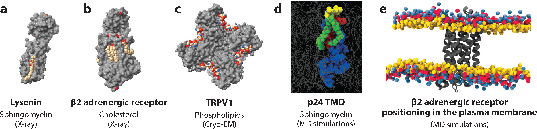
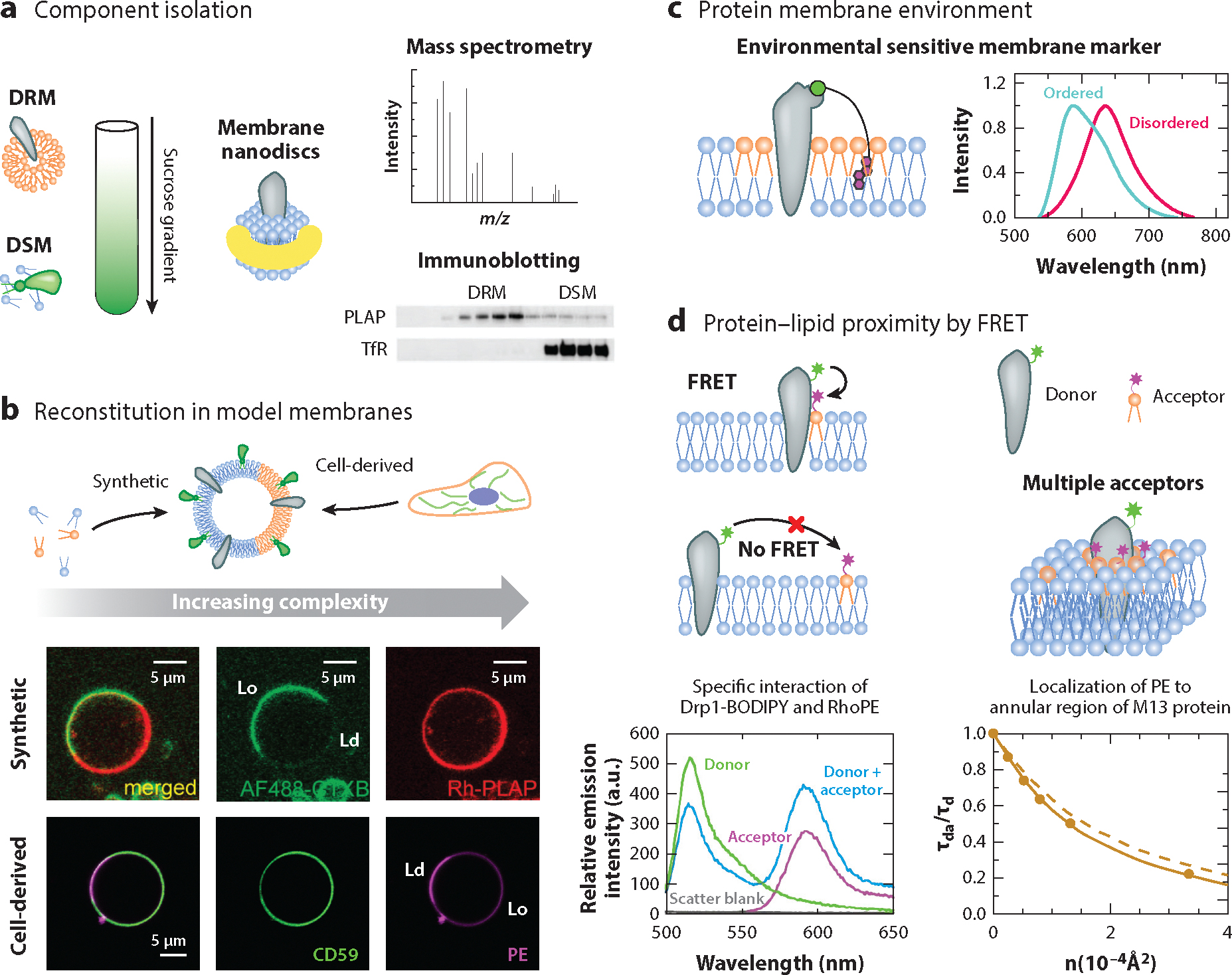
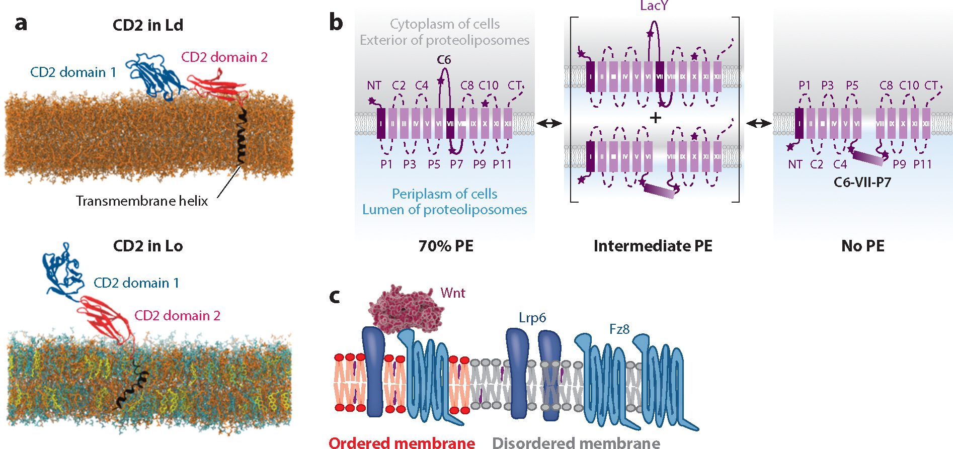
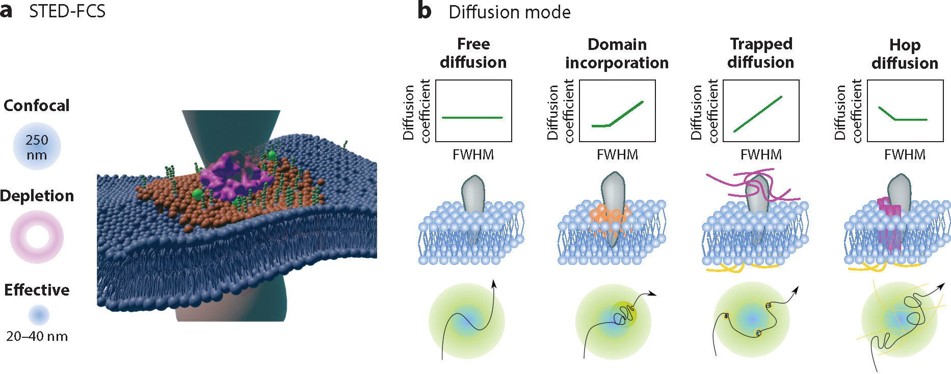
Lipid-protein interactions in cells are involved in various biological processes, including metabolism, trafficking, signaling, host-pathogen interactions, and transmembrane transport. At the plasma membrane, lipid-protein interactions play major roles in membrane organization and function. Several membrane proteins have motifs for specific lipid binding, which modulate protein conformation and consequent function. In addition to such specific lipid-protein interactions, protein function can be regulated by the dynamic, collective behavior of lipids in membranes. Emerging analytical, biochemical, and computational technologies allow us to study the influence of specific lipid-protein interactions, as well as the collective behavior of membranes on protein function. In this article, we review the recent literature on lipid-protein interactions with a specific focus on the current state-of-the-art technologies that enable novel insights into these interactions.

Keywords: collective membrane properties; cryo-EM; lipid-binding motifs; membrane biophysics; molecular dynamics; super-resolution imaging.


Figures

Figure 1
Figure 1
Structural studies of protein–lipid interactions. (a) The structure of the pore-forming protein lysenin (gray surface) with its sphingolipid ligand (colored spheres) determined by X-ray crystallography (30), available in the Protein Data Bank (PDB) as 3zxg. (b) The structure of β2 adrenergic receptor (gray surface) with its cholesterol ligands (colored spheres) determined by X-ray crystallography (49), available in the PDB as 3d4s. (c) The structure of the transient receptor potential protein TRPV1 (gray surface) with its phospholipid ligands (colored spheres) determined by cryogenic electron microscopy (cryo-EM) (41), available in the PDB as 5irz. (d) Sphingomyelin interacts with the transmembrane domain of COPI machinery protein p24, as determined by mass spectrometry and molecular dynamics (MD) simulations. Blue indicates the transmembrane domain (TMD) of p24; red indicates the sphingomyelin-binding pocket; yellow indicates the SM 18:0 head group; and green indicates the SM 18:0 backbone and N-acylated fatty acid. Panel d adapted with permission from Reference . (e) The position of β2 adrenergic receptor in the lipid bilayer, obtained by coarse-grained MD simulations. Lipids are shown with glycerol beads in yellow, phosphate beads in red, and choline beads in blue. Image available in the MemProtMD database (94) as 3d4s.
Figure 2
Figure 2
Methods to study collective lipid–protein interactions. (a) Lipids and proteins are isolated from plasma membranes by detergents or nanodiscs. The content of detergent-resistant membrane (DRM) and detergent-soluble membrane (DSM) fractions can be analyzed by biochemical methods or mass spectrometry (12). Representative Western blot showing DRM association of marker proteins [placental alkaline phosphatase (PLAP) and transferrin receptor (TfR)] from MDCK cells extracted with Triton X100. Panel a adapted with permission from Reference , copyright National Academy of Sciences (2003). (b) Reconstitution of proteins in synthetic [giant unilameller vesicles (GUVs)] or cell-derived [giant plasma membrane vesicles (GPMVs)] model membranes. Such vesicles are widely studied by fluorescence microscopy. The GPI-anchored proteins PLAP (62) and CD59 reconstituted in a GUV and a GPMV, respectively. In the synthetic GUV, the Lo domain is labeled by Cholera toxin B subunit (AF488-CTXB; green). PLAP is labeled with Rhodamine (Rh-PLAP; red), which incorporates preferentially into the Ld domain. In the cell-derived GPMV, Abberior Star Red-lipid (PE; magenta) localizes preferentially to the Ld domain. In contrast, GPI-anchored protein CD59-EGFP (CD59; green) incorporates preferentially to the Lo domain. Panel b adapted with permission from Reference , copyright American Chemical Society (2005). (c) Probing of local membrane order at the proximity of specific proteins with the environmentally sensitive membrane marker Nile Red. Nile Red is modified with a linker to specifically recognize a protein of interest and incorporate it into the lipid bilayer at the proximity of the protein. The fluorescence spectrum of Nile Red is sensitive to membrane order. (d) Elucidation of lipid–protein interaction by Förster resonance energy transfer (FRET). When the protein (donor) and the lipid (acceptor) are in close proximity (1–10 nm), energy transfer occurs. Lipids can localize to the annular region of the protein and act as multiple acceptors for a single protein donor. (Bottom left) Representative emission spectra of BODIPY-labeled wild-type (WT) Dynamin-related protein 1 (Drp1) (the donor) in the absence and presence of 1 mol% Rhodamine PE (RhoPE) lipid (the acceptor) in DOPC/DOPE/cardiolipin liposomes. FRET was detected by a decrease in donor emission intensity in the presence of the acceptor accompanied by a FRET-sensitized increase in acceptor emission upon donor excitation. The acceptor-only trace shows direct excitation of the acceptor at the donor excitation wavelength, which represents the background. Panel adapted with permission from Reference (84), copyright National Academy of Sciences (2021). (Bottom right) Donor (DCIA-labeled protein) fluorescence quenching by energy transfer to acceptor [(18:1)2-PE-NBD] in the DMoPC bilayer at different concentrations of the acceptor. The FRET efficiency is represented as a ratio between the fluorescence lifetime of the donor in the presence of the acceptor and the fluorescence lifetime of the donor in the absence of the acceptor. Circular points indicate experimental energy transfer efficiencies; the solid line indicates theoretical simulations obtained from the annular model for protein–lipid interaction; and the dashed line indicates simulations for random distribution of acceptors. Fitting indicates the localization of PE lipids in the annular region of the protein. Panel d adapted with permission from Reference , copyright Biophysical Society (2004).
Figure 3
Figure 3
Membrane biophysical properties determine protein geometry and interactions. (a) Simulation snapshots of the equilibrium configurations of CD2 in liquid-disordered and liquid-ordered bilayers, showing DOPC (orange), SM (cyan), Chol (yellow), CD2 domain 1 (dark blue), CD2 domain 2 (red), CD2 transmembrane helix (black), and lipid carbohydrate chains (light blue). Panel a adapted with permission from Reference . (b) Effect of changes in PE content on the orientation of lactose permease (LacY) in lipid bilayers. Transmembrane domain orientation is summarized for LacY in proteoliposomes containing 70% (left), intermediate (center), or 0% (right) PE. Panel b adapted with permission from Reference , copyright National Academy of Sciences (2015). (c) Wnt binding to its coreceptors (Fz8 and Lrp6) in the plasma membrane. Although the coreceptors are enriched in disordered domains, the ligand (Wnt3a) binds to the ordered domain pool of the receptors. Panel c adapted with permission from Reference .
Figure 4
Figure 4
Stimulated emission depletion fluorescence correlation spectroscopy (STED-FCS) to resolve lipid–protein interactions. (a) Principle of STED-FCS. Stimulated depletion reduces the effective focal spot down to 20–40 nm, increasing the resolution by an order of magnitude. (b) Lipid or protein diffusion modes can be determined by quantification of the protein diffusion coefficient (D) at different effective focal point diameters [apparent full width at half max (FWHM)]. Protein diffusion modes are characteristic for the types of interactions of the protein in the lipid bilayer. At free diffusion, D remains constant. At transient domain incorporation, D decreases together with FWHM, but when FWHM approaches domain size, D saturates or, in some cases, slightly increases due to incorporation in domains with a specific D. At trapped diffusion, D decreases with decreasing FWHM due to transient binding to lipids, the actin cytoskeleton, or the extracellular matrix. Hop diffusion manifests when the protein diffuses fast within plasma membrane compartments (introduced by actin meshwork, for example), but hopping between compartments is significantly slower. Thus, D increases toward small FWHM.

Similar articles

Cited by

References

    1. Abankwa D, Gorfe AA, Inder K, Hancock JF. 2010. Ras membrane orientation and nanodomain localization generate isoform diversity. PNAS 107(3):1130–35 - PMC - PubMed
    1. Anderluh A, Hofmaier T, Klotzsch E, Kudlacek O, Stockner T, et al. 2017. Direct PIP 2 binding mediates stable oligomer formation of the serotonin transporter. Nat. Commun. 8:14089. - PMC - PubMed
    1. Andrade DM, Clausen MP, Keller J, Mueller V, Wu C, et al. 2015. Cortical actin networks induce spatiotemporal confinement of phospholipids in the plasma membrane—a minimally invasive investigation by STED-FCS. Sci. Rep. 5:11454. - PMC - PubMed
    1. Azbazdar Y, Ozalp O, Sezgin E, Veerapathiran S, Duncan AL, et al. 2019. More favorable palmitic acid over palmitoleic acid modification of Wnt3 ensures its localization and activity in plasma membrane domains. Front. Cell Dev. Biol. 7:281. - PMC - PubMed
    1. Bacia K, Schuette CG, Kahya N, Jahn R, Schwille P. 2004. SNAREs prefer liquid-disordered over “raft” (liquid-ordered) domains when reconstituted into giant unilamellar vesicles. J. Biol. Chem. 279(36):37951–55 - PubMed

Publication types

LinkOut - more resources