. 2022 Apr;66(4):391-401.
doi: 10.1165/rcmb.2021-0364OC.

The Type 2 Asthma Mediator IL-13 Inhibits Severe Acute Respiratory Syndrome Coronavirus 2 Infection of Bronchial Epithelium

Affiliations

The Type 2 Asthma Mediator IL-13 Inhibits Severe Acute Respiratory Syndrome Coronavirus 2 Infection of Bronchial Epithelium

Luke R Bonser et al. Am J Respir Cell Mol Biol. 2022 Apr.

Abstract

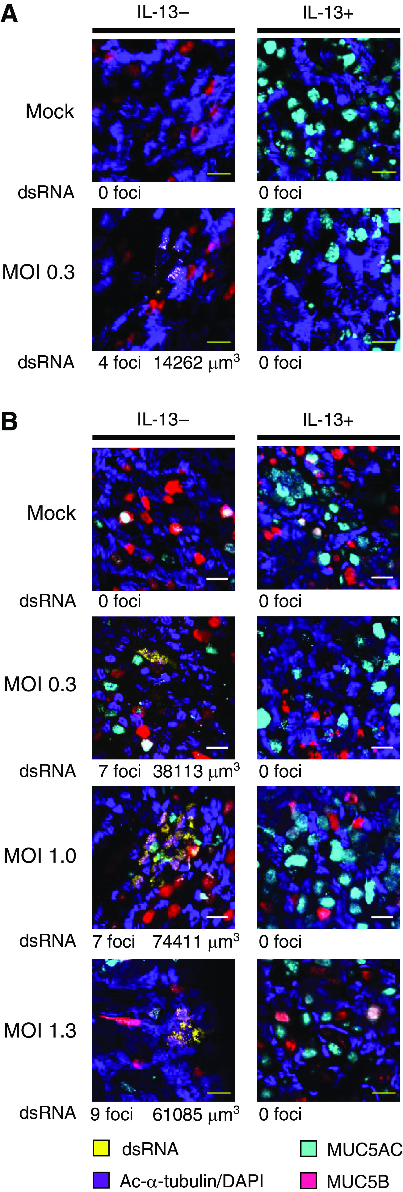
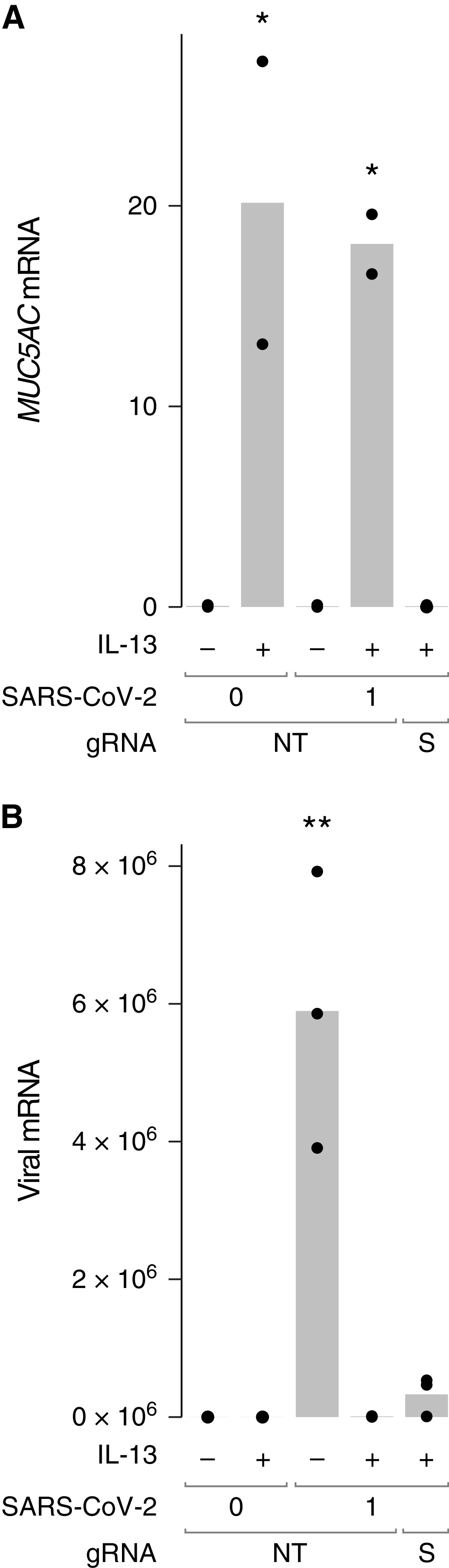
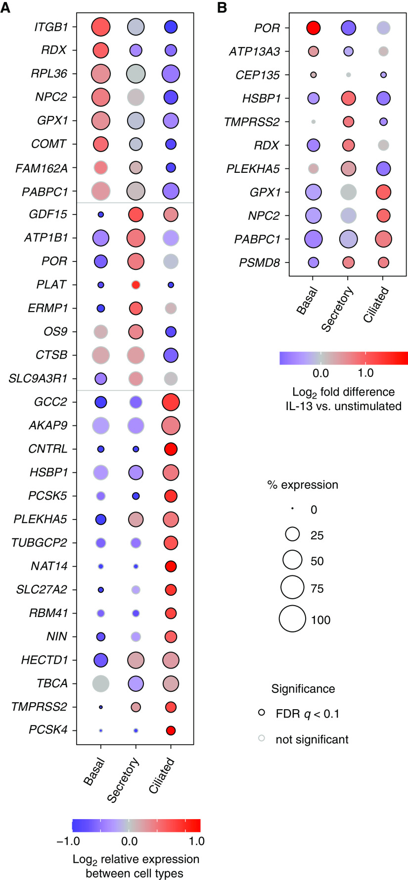
Asthma is associated with chronic changes in the airway epithelium, a key target of severe acute respiratory syndrome coronavirus 2 (SARS-CoV-2). Many epithelial changes, including goblet cell metaplasia, are driven by the type 2 cytokine IL-13, but the effects of IL-13 on SARS-CoV-2 infection are unknown. We found that IL-13 stimulation of differentiated human bronchial epithelial cells (HBECs) cultured at air-liquid interface reduced viral RNA recovered from SARS-CoV-2-infected cells and decreased double-stranded RNA, a marker of viral replication, to below the limit of detection in our assay. An intact mucus gel reduced SARS-CoV-2 infection of unstimulated cells, but neither a mucus gel nor SPDEF, which is required for goblet cell metaplasia, were required for the antiviral effects of IL-13. Bulk RNA sequencing revealed that IL-13 regulated 41 of 332 (12%) mRNAs encoding SARS-CoV-2-associated proteins that were detected in HBECs (>1.5-fold change; false discovery rate < 0.05). Although both IL-13 and IFN-α each inhibit SARS-CoV-2 infection, their transcriptional effects differed markedly. Single-cell RNA sequencing revealed cell type-specific differences in SARS-CoV-2-associated gene expression and IL-13 responses. Many IL-13-induced gene expression changes were seen in airway epithelium from individuals with type 2 asthma and chronic obstructive pulmonary disease. IL-13 effects on airway epithelial cells may protect individuals with type 2 asthma from COVID-19 and could lead to identification of novel strategies for reducing SARS-CoV-2 infection.

Keywords: COVID-19; IL-13; SARS-CoV-2; airway epithelium; asthma.


Figures

Figure 1.
Figure 1.
IL-13 stimulation and mucus reduce severe acute respiratory syndrome coronavirus 2 (SARS-CoV-2) virus RNA levels in infected human bronchial epithelial cells (HBECs). (A) HBECs from donor 10-75 were left unstimulated (−) or stimulated with IL-13 (+), washed with a DTT-containing solution (Mucus Removed) or left unwashed (Mucus Intact), and inoculated with SARS-CoV-2 (0.3 plaque-forming units [pfu] based on titration in Vero E6 cells). SARS-CoV-2 mRNA was measured 48 hours after infection. (B) In a second experiment, cells from donor 14-30 were studied using the same protocol, except that three different inocula (0.3, 1.0, and 1.3 pfu) from another virus preparation were used. Each point represents a separate Transwell culture (n = 3 per condition except as shown). **P < 0.01 and ***P < 0.0001 for the effects of IL-13 by ANOVA with Tukey-Kramer post hoc tests. For cells not stimulated with IL-13, viral RNA load was lower in infections performed with mucus intact compared with infections performed with mucus removed (P < 0.0001 for all viral inocula in both experiments, except for P = 0.01 for the 0.3 pfu inoculum in the second experiment, by ANOVA with Tukey-Kramer post hoc tests). For viral RNA, 1 unit represents the amount of viral RNA present in 1 pfu from the viral stock, based on titration in Vero E6 cells.
Figure 2.
Figure 2.
IL-13 stimulation reduces SARS-CoV-2 replication in HBECs. (A and B) Additional HBEC cultures derived from cells from donor 10-75 (A) and donor 14-30 (B) were inoculated with virus after removal of mucus as part of the same experiments shown in Figure 4. After 48 hours, cells were stained with antibodies against double-stranded RNA (dsRNA) (yellow), the ciliated cell marker acetylated α tubulin (Ac-α-tubulin) and DAPI (both imaged in the same channel, purple), MUC5B (red), and MUC5AC (cyan). We surveyed the entire sample (16.6 μm) for dsRNA staining and acquired stacks encompassing each dsRNA-stained focus. Numbers of dsRNA-stained foci and total volumes of dsRNA staining are shown below representative images for each condition. Scale bars, 20 μM. MOI = multiplicity of infection.
Figure 3.
Figure 3.
SPDEF targeting has little effect on the amount of viral RNA observed in HBECs pretreated with IL-13. HBEC cultures from donor 19-07 were subjected to two successive electroporations with guide RNAs (gRNAs) targeting a nonhuman, nontargeting control sequence (NT) or SPDEF (S) and fully differentiated. HBECs were left unstimulated (−) or stimulated with IL-13 (+) and washed with a DTT-containing solution before inoculation with 1 pfu SARS-CoV-2. RNA was harvested at 72 hours after infection. (A) MUC5AC mRNA was measured to assess SPDEF targeting. *P < 0.05 compared with unstimulated or IL-13–stimulated SPDEF-targeted cells by ANOVA with Tukey-Kramer post hoc test. (B) SARS-CoV-2 RNA. **P < 0.01 compared with all other conditions. Each point represents a separate Transwell culture (n = 2–3 per condition as shown). For viral RNA, 1 unit represents the amount of viral RNA present in 1 pfu from the viral stock, based on titration in Vero E6 cells.
Figure 4.
Figure 4.
SARS-CoV-2–associated genes are highly expressed in HBECs, and many are regulated by cytokines. HBECs from six donors were cultured without cytokine (−), or with IL-13, IFN-α, a combination of IL-13 and IFN-α, IFN-γ, or IL-17 and analyzed by RNA sequencing (RNA-seq). (A) Comparison of read counts between SARS-CoV-2–associated genes, including ACE2 and TMPRSS2, and all detected genes (⩾1 read per million mapped reads in ⩾50% of samples) in unstimulated HBECs. (B and C) Heatmap illustrating canonical cytokine-regulated genes (B), and cytokine regulated SARS-CoV-2–associated genes (C) (false discovery rate [FDR] q ⩽ 0.05; absolute fold change ⩾ 1.5 for any cytokine).
Figure 5.
Figure 5.
Single-cell RNA sequencing (scRNA-seq) reveals cell type–specific expression of many SARS-CoV-2–associated genes and cell type–specific effects of IL-13. (A) Cell type–specific expression in unstimulated HBECs. Genes were selected from a set of 113 differentially expressed SARS-CoV-2–associated genes listed in Table E3. (B) Cell type–specific differences in IL-13 responses. For 11 SARS-CoV-2– associated genes, IL-13 increased expression in at least one cell type and decreased expression in at least one other cell type (FDR q < 0.1 for both). Gene expression was determined by aggregating data from all cells from experiments with four donors. (A and B) Coloring of each dot indicates expression level relative to other cell types (A) or in IL-13–stimulated cells compared with unstimulated cells (B). The size of each dot is proportional to the percentage of cells with at least one read mapped to the gene, and black circles at the perimeter of each dot indicate that expression levels are significantly different (q < 0.1) compared with other cell types (A) or in IL-13–stimulated compared with unstimulated cells of the same type (B).
Figure 6.
Figure 6.
Expression of many IL-13–regulated SARS-CoV-2–associated genes correlates with an IL-13 signature in asthma. Correlation of IL-13–induced, SARS-CoV-2–associated genes with a type 2/IL-13 signature (the three-gene mean [TGM]) in endobronchial brushing samples from participants with asthma (red) and healthy control subjects (cyan). Values for gene expression represent log2 of normalized read counts from bulk RNA-seq. The eight SARS-CoV-2 genes with the highest Pearson’s correlations (R) are shown, and associated P values are adjusted for multiple comparisons. Correlations for the full set of IL-13–induced SARS-CoV-2–associated genes are shown in Table E4.

Update of

Comment in

  • IL-13 Protects against SARS-CoV-2?
    Peebles RS Jr. Peebles RS Jr. Am J Respir Cell Mol Biol. 2022 Apr;66(4):351-352. doi: 10.1165/rcmb.2021-0562ED. Am J Respir Cell Mol Biol. 2022. PMID: 35085479 Free PMC article. No abstract available.

References

    1. Williamson EJ, Walker AJ, Bhaskaran K, Bacon S, Bates C, Morton CE, et al. Factors associated with COVID-19-related death using OpenSAFELY. Nature . 2020;584:430–436. - PMC - PubMed
    1. Hadjadj J, Yatim N, Barnabei L, Corneau A, Boussier J, Smith N, et al. Impaired type I interferon activity and inflammatory responses in severe COVID-19 patients. Science . 2020;369:718–724. - PMC - PubMed
    1. Bastard P, Rosen LB, Zhang Q, Michailidis E, Hoffmann H-H, Zhang Y, et al. HGID Lab; NIAID-USUHS Immune Response to COVID Group; COVID Clinicians; COVID-STORM Clinicians; Imagine COVID Group; French COVID Cohort Study Group; Milieu Intérieur Consortium; CoV-Contact Cohort; Amsterdam UMC Covid-19 Biobank; COVID Human Genetic Effort Autoantibodies against type I IFNs in patients with life-threatening COVID-19. Science . 2020;370:eabd4585. - PMC - PubMed
    1. Combes AJ, Courau T, Kuhn NF, Hu KH, Ray A, Chen WS, et al. UCSF COMET Consortium Global absence and targeting of protective immune states in severe COVID-19. Nature . 2021;591:124–130. - PMC - PubMed
    1. Centers for Disease Control and Prevention 2020https://www.cdc.gov/coronavirus/2019-ncov/need-extra-precautions/evidenc....

Publication types