Case Reports
. 2022 Apr 12;6(7):2275-2289.
doi: 10.1182/bloodadvances.2021005703.

A somatic UBA2 variant preceded ETV6-RUNX1 in the concordant BCP-ALL of monozygotic twins

Affiliations
Case Reports

A somatic UBA2 variant preceded ETV6-RUNX1 in the concordant BCP-ALL of monozygotic twins

Benedicte Bang et al. Blood Adv. .

Abstract

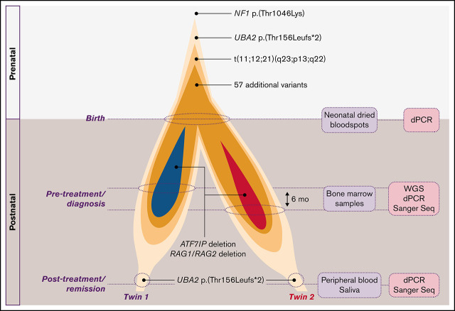
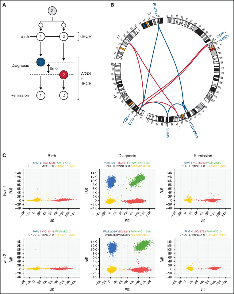
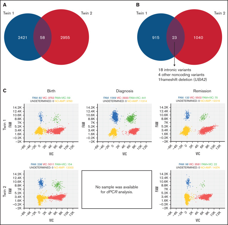
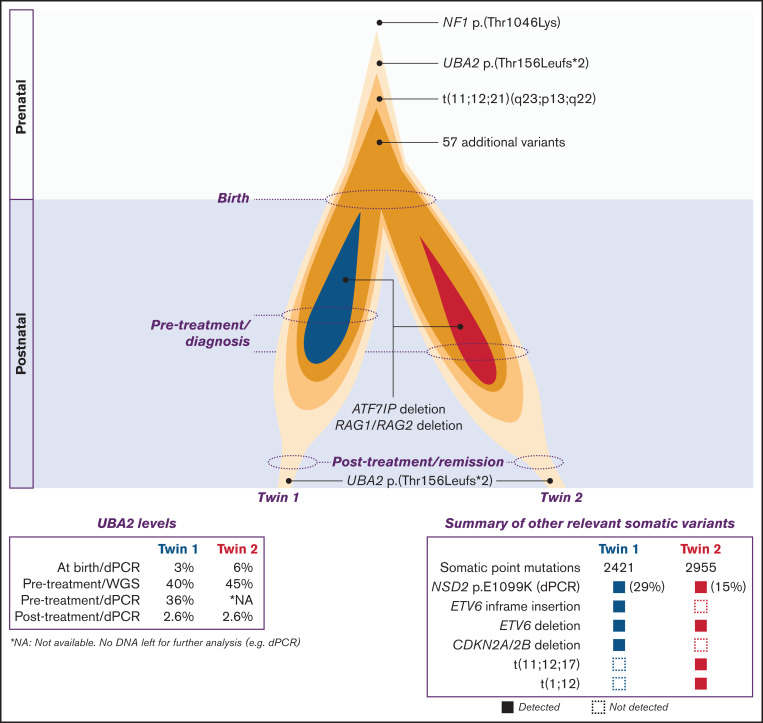
Genetic analysis of leukemic clones in monozygotic twins with concordant acute lymphoblastic leukemia (ALL) has proved a unique opportunity to gain insight into the molecular phylogenetics of leukemogenesis. Using whole-genome sequencing, we characterized constitutional and somatic single nucleotide variants/insertion-deletions (indels) and structural variants in a monozygotic twin pair with concordant ETV6-RUNX1+ B-cell precursor ALL (BCP-ALL). In addition, digital PCR (dPCR) was applied to evaluate the presence of and quantify selected somatic variants at birth, diagnosis, and remission. A shared somatic complex rearrangement involving chromosomes 11, 12, and 21 with identical fusion sequences in leukemias of both twins offered direct proof of a common clonal origin. The ETV6-RUNX1 fusion detected at diagnosis was found to originate from this complex rearrangement. A shared somatic frameshift deletion in UBA2 was also identified in diagnostic samples. In addition, each leukemia independently acquired analogous deletions of 3 genes recurrently targeted in BCP-ALLs (ETV6, ATF7IP, and RAG1/RAG2), providing evidence of a convergent clonal evolution only explained by a strong concurrent selective pressure. Quantification of the UBA2 deletion by dPCR surprisingly indicated it persisted in remission. This, for the first time to our knowledge, provided evidence of a UBA2 variant preceding the well-established initiating event ETV6-RUNX1. Further, we suggest the UBA2 deletion exerted a leukemia predisposing effect and that its essential role in Small Ubiquitin-like Modifier (SUMO) attachment (SUMOylation), regulating nearly all physiological and pathological cellular processes such as DNA-repair by nonhomologous end joining, may hold a mechanistic explanation for the predisposition.


Figures

None
Graphical abstract
Figure 1.
Figure 1.
Schematic overview and SV analysis. (A) Monozygotic twins developed BCP-ALL with a 6-month difference in latency. Sampling took place at birth (dried neonatal blood spots), diagnosis (bone marrow), and in remission (peripheral blood, 2 years 11 months [Tw1] and 2 years 5 months [Tw2] after diagnosis). WGS and dPCR analysis were applied to samples as illustrated to uncovered constitutional and somatic variants and quantify key mutations, respectively. Blue and red circles represent Tw1’s and Tw2’s leukemia, respectively. (B) Circos plot showing somatic SVs in leukemias. Only chromosomes involved in rearrangements are displayed (1, 11, 12, 17, and 21). Colored lines illustrate how breakpoints have fused. Genes disrupted by or in close proximity of the breakpoints are indicated. Blue lines represent the shared complex rearrangement t(11;12;21)(q23;p13;q22), involving 2 inversions on chromosome 11 and generating a ETV6-RUNX1 fusion. Redlines represent SVs unique to Tw2: 2 subclonal translocations t(1;12) and 1 t(11;12;17). (C) dPCR detection and quantification of shared complex rearrangement t(11;12;21)(q23;p13;q22) at birth, diagnosis, and in remission of both twins. TaqMan assay targeted chromosome 11;12 junction sequence. Clusters of dPCR chip wells positive for internal reference control RPPH1 (red), target region (blue), reference and target (green), and with no amplification (yellow). Complex rearrangement readily detected at diagnosis but beyond detection at birth and in remission. Detection limit: 1 in 1000 copies. Images acquired from QuantStudio 3D Analysis Suite Cloud Software, version 3.1.6-PRC-build2 with default parameters. Z, zygote.
Figure 2.
Figure 2.
Shared and unique somatic SNVs/indels in twins’ leukemias. (A) All somatic SNVs/indels across the genome. Two thousand, four hundred twenty-one and 2955 unique to Tw1 and Tw2, respectively, and 58 shared. (B) SNVs/indels in protein coding genes. Nine hundred fifteen and 1040 unique to Tw1 and 2, respectively: 23 shared. (C) dPCR detection and quantification of UBA2 deletion (NM_005499.2: c.463_470del; NP_005490.1: p.(Thr156Leufs*2) at birth, diagnosis, and in remission. Diagnostic sample of Tw2 not available for analysis. Clusters of dPCR chip wells positive for wildtype allele (red), mutant allele (blue), wildtype and mutant allele (green), and with no amplification (yellow). UBA2 deletion was detected at birth (both twins), diagnosis (Tw1; Tw2 lacked sample for analysis), and, unexpectedly, also in remission (both twins). Detection limit: 1 in 1000 copies. Images acquired from QuantStudio 3D Analysis Suite Cloud Software, version 3.1.6-PRC-build2 with default parameters.
Figure 3.
Figure 3.
Illustration of clonal evolution from in utero leukemia initiation (prenatal) to remission (postnatal). Strikingly, UBA2 deletion proceeded ETV6-RUNX1 fusion generated by the shared complex rearrangement in utero. Fifty-seven additional shared SNVs/indels were acquired during the prenatal period. Clonal evolution of preleukemic clones established prenatally in both twins continued separately, mainly postnatally, acquiring SVs and SNVs/indels unique to each twins’ leukemia. Genes known to be recurrent targets of secondary events in BCP-ALL, ATF7IP, RAG1/RAG2, and ETV6, were targeted by unique analogous deletions in both twins. The UBA2 deletion persisted subclonally in remission of both twins. dPCR results stated in percent refers to the fraction of mutant target DNA in the analyzed sample.

Similar articles

Cited by

References

    1. Mullighan CG. Molecular genetics of B-precursor acute lymphoblastic leukemia. J Clin Invest. 2012;122(10):3407-3415. - PMC - PubMed
    1. Lim JY, Bhatia S, Robison LL, Yang JJ. Genomics of racial and ethnic disparities in childhood acute lymphoblastic leukemia. Cancer. 2014;120(7):955-962. - PMC - PubMed
    1. Hunger SP, Mullighan CG. Acute lymphoblastic leukemia in children. N Engl J Med. 2015;373(16):1541-1552. - PubMed
    1. Chiaretti S, Zini G, Bassan R. Diagnosis and subclassification of acute lymphoblastic leukemia. Mediterr J Hematol Infect Dis. 2014;6(1):e2014073. - PMC - PubMed
    1. Bhojwani D, Yang JJ, Pui CH. Biology of childhood acute lymphoblastic leukemia. Pediatr Clin North Am. 2015;62(1):47-60. - PMC - PubMed

Publication types

MeSH terms