. 2022 Apr 18;145(2):670-683.
doi: 10.1093/brain/awab317.

Inactivating Celsr2 promotes motor axon fasciculation and regeneration in mouse and human

Affiliations

Inactivating Celsr2 promotes motor axon fasciculation and regeneration in mouse and human

Quan Wen et al. Brain. .

Abstract

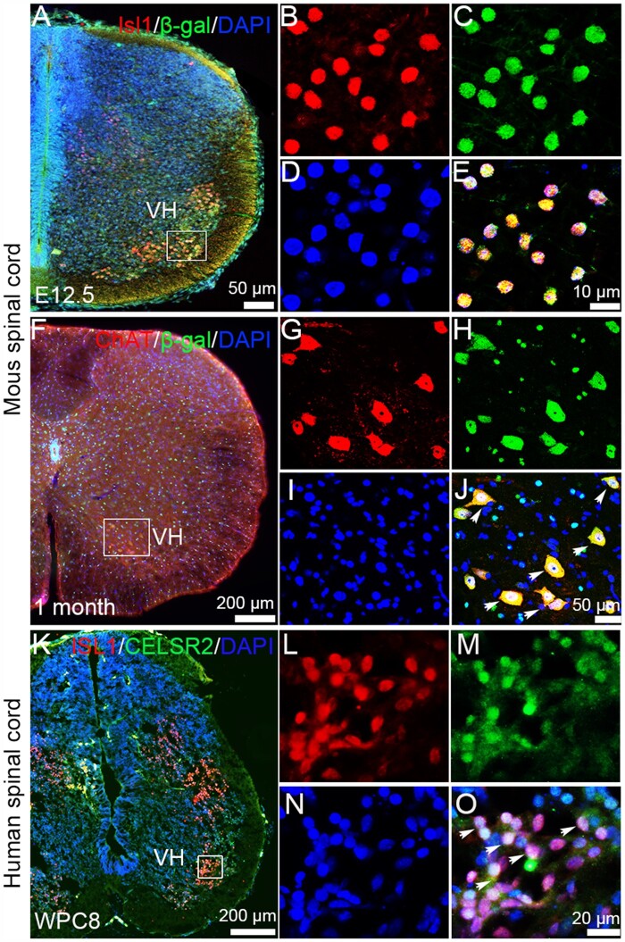
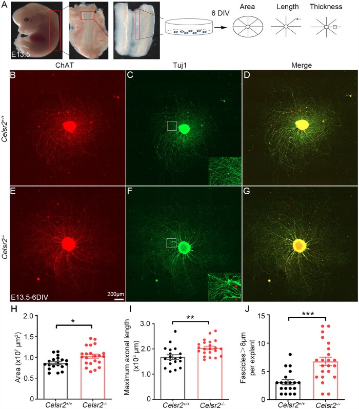
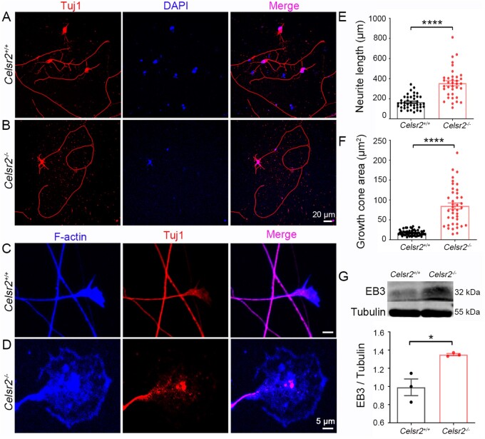
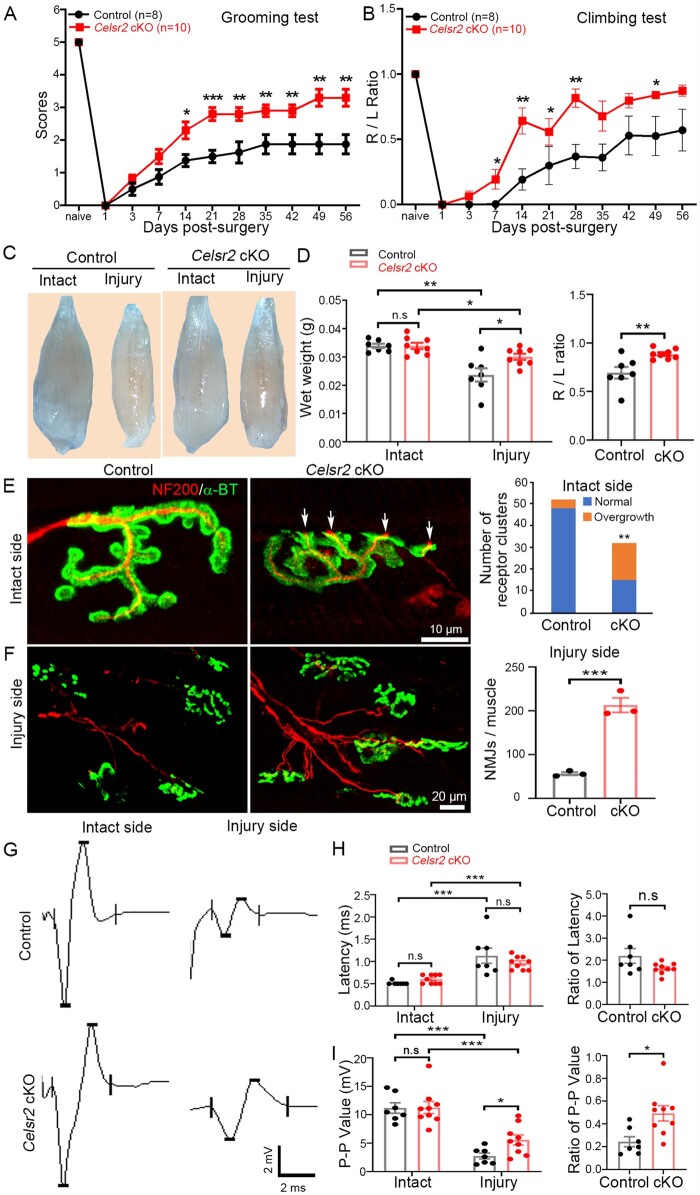
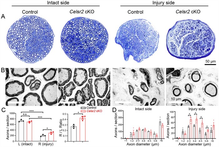
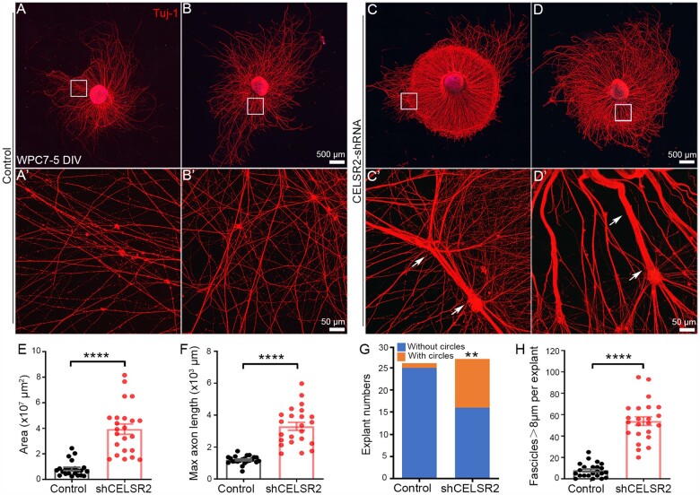
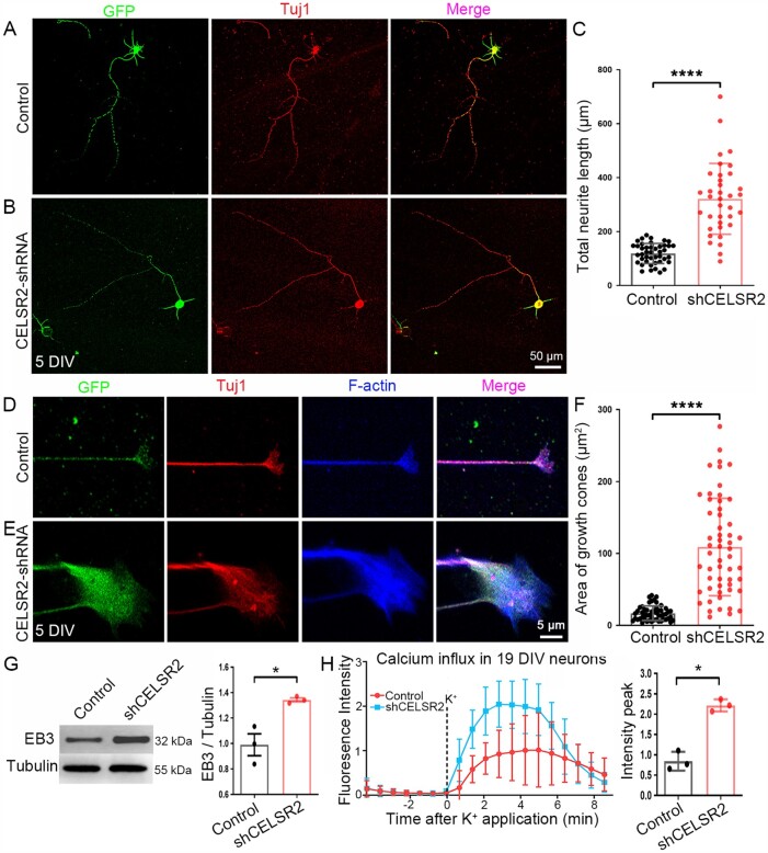
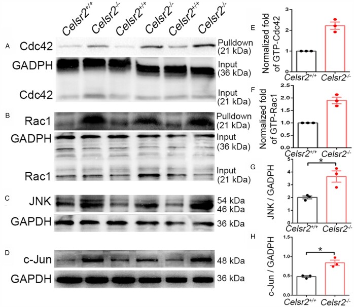
Understanding new modulators of axon regeneration is central to neural repair. Our previous work demonstrated critical roles of atypical cadherin Celsr2 during neural development, including cilia organization, neuron migration and axon navigation. Here, we address its role in axon regeneration. We show that Celsr2 is highly expressed in both mouse and human spinal motor neurons. Celsr2 knockout promotes axon regeneration and fasciculation in mouse cultured spinal explants. Similarly, cultured Celsr2 mutant motor neurons extend longer neurites and larger growth cones, with increased expression of end-binding protein 3 and higher potassium-induced calcium influx. Mice with Celsr2 conditional knockout in spinal motor neurons do not exhibit any behavioural deficits; however, after branchial plexus injury, axon regeneration and functional forelimb locomotor recovery are significantly improved. Similarly, knockdown of CELSR2 using shRNA interference in cultured human spinal motor explants and motor neurons increases axonal fasciculation and growth. In mouse adult spinal cord after root avulsion, in mouse embryonic spinal cords, and in cultured human motor neurons, Celsr2 downregulation is accompanied by increased levels of GTP-bound Rac1 and Cdc42, and of JNK and c-Jun. In conclusion, Celsr2 negatively regulates motor axon regeneration and is a potential target to improve neural repair.

Keywords: axon regeneration; brachial plexus injury; human embryos; root avulsion; spinal motor neurons.


Figures

Figure 1
Figure 1
Celsr2 expression is enriched in mouse and human spinal motor neurons. Using Celsr2LacZ transgenic mice, Celsr2 expression is detected by anti-β-gal immunostaining. (AE) E12.5 spinal sections were immunostained for Isl1 (red), β-gal (green) and counterstained for DAPI (blue). The merged image showed that all Isl1-positive cells in the ventral horn (VH) co-labelled by β-gal immunoreactivity (E). (FJ) In adult spinal sections, all ChAT-positive (red) spinal motor neurons co-expressed β-gal (green) indicated by arrows (J). (KO) In WPC8 human spinal sections, ISL1 and CELSR2 double immunofluorescent staining showed that CELSR2 was expressed in ISL1-positive neurons in the ventral horn (VH) indicated by arrows (O). BE, GJ and LO correspond to boxed areas in A, F and K, respectively. Nuclei were counterstained by DAPI (blue).
Figure 2
Figure 2
Celsr2 knockout improves axon growth in mouse spinal motor explant culture. (A) Schema of the experimental procedure for explant culture and analysis. (BG) Spinal motor neuron explants from E13.5 Celsr2+/+ (BD) and Celsr2−/− mouse embryos (E–G) were cultured for 6 DIV and then immunostained for ChAT (B and E; red) and Tuj1 (C and F; green). Both signals co-localize (D and G; yellow). Representative axonal bundles are indicated in the insets of C and F. (HJ) Quantification of the maximal area covered by growing axons in 107 µm2 (H; control: 0.86 ± 0.03, mutant: 1.02 ± 0.05; P < 0.05), maximal axon length in 103 µm (I; control: 1.68 ± 0.09, mutant: 2.04 ± 0.07; P < 0.001) and number of large axon bundles (>8 µm in diameter; J; control: 3.11 ± 0.45, mutant: 6.77 ± 0.74; P < 0.001). These parameters are increased significantly in the mutant compared to the control. ***P < 0.001; **P < 0.01; *P < 0.05; Student’s t-test; n = 19 in the control and n = 22 in the mutant.
Figure 3
Figure 3
Celsr2 knockout contributes to neurite growth in primary spinal motor neuron culture. (A and B) E13.5 spinal motor neurons from Celsr2+/+ (A) and Celsr2–/– (B) mouse embryos were cultured for 6 DIV and immunostained for Tuj1 (red). DAPI counterstained nuclei (blue). (C and D) Double immunostaining of cultured neurons for F-actin (blue) and Tuj1 (red) disclosed the axon shafts and growth cones. (E and F) Statistical analysis of total neurite length (E; control: 165.25 ± 10.52 μm, mutant: 354.19 ± 24.89 μm; P < 0.0001, n = 41 in the control and n = 36 in the mutant) and growth cone areas (F; control: 17.48 ± 0.91 µm2, mutant: 84.89 ± 7.75 µm2; P < 0.0001, n = 58 in the control and n = 40 in the mutant). (G) Protein extracts from E13.5 ventral horns of cervical spinal segments was subjected to western blots using anti-EB3 and β-III tubulin (tubulin). There was a dramatic increase of EB3 in the mutant compared to the control (control: 0.99 ± 0.09, mutant: 1.35 ± 0.01; P < 0.05, n = 3 animals in each group). *P < 0.05; ****P < 0.0001; Student’s t-test.
Figure 4
Figure 4
Celsr2 cKO in spinal motor neurons improves functional recovery and NMJ formation after root avulsion/reimplantation. (A and B) After root avulsion/reimplantation, the function of the affected forelimb was assessed using the grooming (A) and climbing test (B). Scores were significantly higher in Celsr2 cKO (Isl1-Cre; Celsr2f/–) compared to littermate controls (Celsr2f/–) at Days 14, 21, 28, 35, 42, 49 and 56 post-injury. During the climbing test, usage of injured (R) and intact (L) forelimbs was compared; the R/L ratio was increased in the Celsr2 cKO. *P < 0.05; **P < 0.01; Student’s t-test. (C and D) Biceps collected 56 days after injury were more atrophic on the injured than the intact side, in both mutant and control mice (C). Muscle wet weight on the intact side was comparable in both groups, whereas it was higher on the injured side in mutants versus controls, as reflected by the increased the R/L ratio (injured side to intact side) (D). Wet weight, control: 0.0339 ± 0.0008 g, mutant: 0.0339 ± 0.0011 g on the intact side, P < 0.05; control: 0.0237 ± 0.0024 g, mutant: 0.0300 ± 0.0010 g on the injured side, P < 0.05; the R/L ratio: 0.69 ± 0.06 and 0.89 ± 0.02 in the control and the mutant, respectively, P < 0.01. *P < 0.05; **P < 0.01; Student’s t-test; n = 7 in the control and n = 8 in the mutant. (E and F) NMJs were examined using anti-NF200 and anti-ɑ-BT double staining 56 days post-surgery. On intact sides (E), several cholinergic receptor clusters showed axonal terminals overgrowth in Celsr2 cKO (17/32; indicated by arrows), but rarely in the control (4/52). On injury sides (F), there were more numerous growing axons and NMJs in the mutant than in the control (control: 56.33 ± 3.53, mutant: 213.00 ± 16.56 NMJs/muscle, P < 0.001, n = 3 animals in each group). **P < 0.01; ***P < 0.001; Student’s t-test for NMJ number comparison and chi-square for receptor cluster comparison. (GI) EMG of biceps was recorded 56 days post-surgery (G). The latencies were increased after injury, with no difference between both groups (H; control: 0.514 ± 0.014, mutant: 0.600 ± 0.033 ms on the intact side; 1.129 ± 0.170 ms for control and 1.632 ± 0.083 ms for mutant on the injured side; ratio of latency: 2.195 ± 0.335 and 1.632 ± 0.083 in the control and the mutant, respectively; P > 0.05 in all comparisons). Denervation resulted in a significant decrease of the peak–peak amplitude in both groups, but the amplitudes and the ratio of the injured to the intact muscle was significantly higher in the mutant compared to the control (I; control: 11.182 ± 0.923 mV, mutant: 11.269 ± 1.055 mV on the intact side, P > 0.05; control: 2.740 ± 0.523 mV, mutant: 5.572 ± 0.873 mV on the injured side, P < 0.05; ratio: 0.244 ± 0.044 and 0.492 ± 0.067 in the control and the mutant, respectively, P < 0.05). *P < 0.05; **P < 0.01; ***P < 0.001; n.s, not significant; Student’s t-test; n = 7 in the control and n = 9 in the mutant.
Figure 5
Figure 5
Improved axon regeneration in Celsr2 cKO mice after root avulsion/reimplantation. (A) Toluidine Blue staining of musculocutaneous nerves from intact and injured sides 56 days after root avulsion/reimplantation. (B) Electron microscope images of musculocutaneous nerves from intact and injured sides. On the intact side, axons were more closely packed in the mutant than in the control. (C and D) Statistical analysis showed that axon number was comparable on the intact side in the two genotypes but higher in the mutant on the injured side (C; control: 734.0 ± 24.8, mutant: 696.3 ± 20.2 axons/section on the intact side, P > 0.05; control: 201.0 ± 21.5, mutant: 300.3 ± 25.8 axons/section on the injured side, P < 0.05; the ratio: 0.244 ± 0.044 and 0.492 ± 0.067, P < 0.05; n = 7 in the control and n = 9 in the mutant). The ratio of the injured to the intact side (R/L) was higher in the mutant (C; control: 0.27 ± 0.02, mutant: 0.43 ± 0.03, P < 0.05; n = 3 in each group). The distribution of axons according to their diameter showed no differences in the two genotypes on the intact side and an increased number of axons with 2–6 µm diameter on the operated side in the mutant relative to the control (D). *P < 0.05; **P < 0.01; ***P < 0.001; n.s, not significant; Student’s t-test.
Figure 6
Figure 6
CELSR2 knockdown increases axonal regeneration in human embryonic spinal motor explant culture. (AD) Cultured spinal motor neuron explants from WPC7 human embryos were transfected with a CELSR2 scrambled shRNA as control (A and B) and with CELSR2-shRNA (C and D). After 5 DIV, cultured explants were immunostained for Tuj1. A′, B′, C′ and D′ are enlarged areas from A, B, C and D, respectively. Upon CELSR2-shRNA knockdown, growing axons grew in circles (one example indicated in C) and formed large axonal bundles (arrows in C′ and D′). (EH) The maximal area in 107 µm2 (E; control: 0.82 ± 0.12, CELSR2-shRNA: 3.93 ± 0.40; P < 0.0001, n = 23 in each group), maximal axon length in 103 µm (F; control: 1.20 ± 0.05, and CELSR2-shRNA: 3.30 ± 0.25; P < 0.0001, n = 23 in each group), explants with axons growing into circles (G; 1/26 in the control and 11/27 in the CELSR2-shRNA) and the number of large axon bundles (H; control : 7.83 ± 1.37, mutant: 53.96 ± 3.98, P < 0.0001, n = 23 in each group) were significantly increased in the CELSR2-shRNA knockdown explants compared to control. ****P < 0.0001; Student’s t-test (E, F and H). **P < 0.01; chi-square test (G).
Figure 7
Figure 7
CELSR2 knockdown promotes axonal growth in primary human spinal motor neuron culture. (AC) CELSR2 scrambled shRNA (A, control) and CELSR2-shRNA (B) were used to transfect cultured primary spinal motor neurons from WPC7 and WPC8 human embryos. Transfected neurons were visualized by virus-encoded GFP (green). After 5 DIV, neurons were immunostained for Tuj1 (red). GFP- and Tuj1-immunoreactivity overlapped in the somas and neurites as shown in the merged images. A significant increase of total neurite length was observed in CELSR2-shRNA transfected neurons (C; in µm, control: 118.70 ± 5.66, CELSR2-shRNA: 321.28 ± 21.90, P < 0.0001, n = 43 in the control and n = 36 in the CELSR2-shRNA). (DF) Double immunostaining for F-actin (blue) and Tuj1 (red) reveal axon shafts and growth cones in control (D) and CELSR2-shRNA (E) transfected neurons (GFP labelling, green). The growth cone area was significantly increased in CELSR2-shRNA versus control transfected neurons (F; control: 16.92 ± 1.40 µm2, and CELSR2-shRNA: 109.00 ± 9.12, P < 0.0001, n = 52 in the control and 55 in the CELSR2-shRNA). (G) Western blot analysis of 5-DIV cultured neurons with antibodies to EB3 and β-III tubulin (tubulin, reference) showed an increase of EB3 levels in CELSR2-shRNA transfected neurons (control: 0.99 ± 0.09, CELSR2-shRNA: 1.34 ± 0.02, P < 0.05, n = 3 independent experiments). (H) Intracellular calcium influx was evaluated in 19 DIV-cultured neurons by measuring Fluoro-4 AM fluorescence intensity. The curves were drawn from captured images before and after potassium application. There was an increase of the fluorescent peaks in CELSR2-shRNA transfected neurons (control: 0.84 ± 0.13, and CELSR2-shRNA: 2.22 ± 0.09, P < 0.05, n = 3 independent experiments). *P < 0.05; ****P < 0.0001; Student’s t-test.
Figure 8
Figure 8
Celsr2 knockout increases Cdc42/Rac1 and JNK/c-Jun signalling in injured ventral horns. (AD) Western blot analysis of GST-pulldown proteins and samples from spinal ventral columns 3 days after root avulsion, using anti-Cdc42 (A), anti-Rac1 (B), anti-JNK (C), anti-c-Jun (D) antibodies. Anti-GADPH antibody was used as reference. (EH) GTP-bound Cdc42 (E; control: 1 ± 0, mutant: 2.22 ± 0.17, P < 0.05) and Rac1 (F; control: 1 ± 0, mutant: 1.90 ± 0.13, P < 0.05) proteins, as well as concentrations of JNK (G; control: 2.01 ± 0.12, mutant: 3.65 ± 0.44, P < 0.05) and c-Jun (H; control: 0.48 ± 0.02, mutant: 0.84 ± 0.07, P < 0.05) were increased in mutant compared to control samples. *P < 0.05; Student’s t-test; n = 3 animals in each group.

Comment in

References

    1. Liu K, Tedeschi A, Park KK, He Z.. Neuronal intrinsic mechanisms of axon regeneration. Annu Rev Neurosci. 2011;34:131–152. - PubMed
    1. Mahar M, Cavalli V.. Intrinsic mechanisms of neuronal axon regeneration. Nat Rev Neurosci. 2018;19(6):323–337. - PMC - PubMed
    1. Ruven C, Chan TK, Wu W.. Spinal root avulsion: An excellent model for studying motoneuron degeneration and regeneration after severe axonal injury. Neural Regen Res. 2014;9(2):117–118.8. - PMC - PubMed
    1. Cullheim S, Carlstedt T, Linda H, Risling M, Ulfhake B.. Motoneurons reinnervate skeletal muscle after ventral root implantation into the spinal cord of the cat. Neuroscience. 1989;29(3):725–733. - PubMed
    1. Carlstedt T, Grane P, Hallin RG, Noren G.. Return of function after spinal cord implantation of avulsed spinal nerve roots. Lancet. 1995;346(8986):1323–1325. - PubMed

Publication types