

Intramedullary parasite eggs, latent for three decades, mimicking acute transverse myelitis
- PMID: 34983433
- PMCID: PMC8725547
- DOI: 10.1186/s12879-021-07013-7
Intramedullary parasite eggs, latent for three decades, mimicking acute transverse myelitis
Abstract
Background: Intramedullary parasitic infection is extremely uncommon, and clinical presentation of Brown-Sequard syndrome is even rarer.
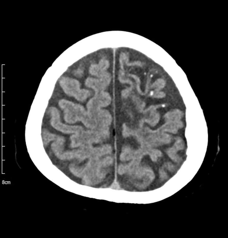
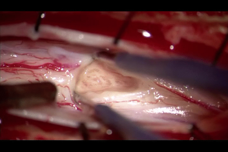
Case presentation: The authors report a case involving a 57-year-old woman with Brown-Sequard syndrome, in whom magnetic resonance imaging and clinical and epidemiological features were similar to those of acute transverse myelitis. Myelotomy suggested inflammation caused by latent parasite eggs in the spinal cord. Antiparasitic and steroid therapies were administered postoperatively. To the author's knowledge, this is the first report to describe a surgical experience for Taenia solium eggs in the spinal cord.
Conclusion: Intramedullary parasitic infection is a diagnostic challenge that requires careful discrimination from other diseases. If parasite infection is suspected in a progressively deteriorating patient, myelotomy should be considered for rapid and accurate treatment.
Keywords: Acute transverse myelitis; Brown-Sequard syndrome; Central nervous system parasitic infections; Neurocysticercosis; Parasite eggs; Spinal cord neoplasm.
© 2021. The Author(s).
Conflict of interest statement
All authors declare that there are no competing interests.
Figures


Similar articles
-
Intramedullary spinal cord neurocysticercosis presenting as Brown-Séquard syndrome.BMC Neurol. 2015 Jan 16;15:1. doi: 10.1186/s12883-014-0245-5. BMC Neurol. 2015. PMID: 25595849 Free PMC article.
-
Brown-Séquard syndrome as a presentation of idiopathictransverse myelitis.Arch Argent Pediatr. 2024 Feb 1;122(1):e202202978. doi: 10.5546/aap.2022-02978.eng. Epub 2023 Aug 24. Arch Argent Pediatr. 2024. PMID: 37594647 English, Spanish.
-
Idiopathic transverse myelitis presenting as the Brown-Sequard syndrome.Spinal Cord. 2009 Feb;47(2):176-8. doi: 10.1038/sc.2008.23. Epub 2008 Mar 11. Spinal Cord. 2009. PMID: 18332885
-
Intramedullary spinal cord metastasis of lung adenocarcinoma presenting as Brown-Sequard syndrome.Surg Neurol. 2004 Jan;61(1):72-6. doi: 10.1016/s0090-3019(03)00298-2. Surg Neurol. 2004. PMID: 14706385 Review.
-
[Non-traumatic acute transverse spinal cord syndromes].Praxis (Bern 1994). 2005 Jul 27;94(30-31):1151-9. doi: 10.1024/0369-8394.94.30.1151. Praxis (Bern 1994). 2005. PMID: 16117470 Review. German.
Cited by
-
Treatment outcome in patients with spinal neurocysticercosis: a systematic review of published cases and case series.Future Microbiol. 2025 Jan;20(1):45-56. doi: 10.1080/17460913.2024.2428526. Epub 2024 Nov 15. Future Microbiol. 2025. PMID: 39545648
References
-
- Dzikowiec M, Góralska K, Błaszkowska J. Neuroinvasions caused by parasites. Ann Parasitol. 2017;63:243–253. - PubMed
-
- Siddharth Shah SD. Cysticercosis of the spine: a review. Arch Parasitol. 2017;1:110.
-
- Del Brutto OH, García HH. Taenia solium: biological characteristics and life cycle. In: Cysticercosis of the human nervous system. Berlin, Heidelberg: Springer; 2014. 10.1007/978-3-642-39022-7_2.
Publication types
MeSH terms
Grants and funding
LinkOut - more resources
Full Text Sources
Medical
