

Protective effects of Quercus acuta Thunb. fruit extract against UVB-induced photoaging through ERK/AP-1 signaling modulation in human keratinocytes
- PMID: 34983480
- PMCID: PMC8728912
- DOI: 10.1186/s12906-021-03473-1
Protective effects of Quercus acuta Thunb. fruit extract against UVB-induced photoaging through ERK/AP-1 signaling modulation in human keratinocytes
Abstract
Background: Quercus acuta Thunb. (Fagaceae) or Japanese evergreen oak is cultivated as an ornamental plant in South Korea, China, Japan, and Taiwan and used in traditional medicine. The acorn or fruit of Quercus acuta Thunb. (QAF) is the main ingredient of acorn jelly, a traditional food in Korea. Its leaf was recently shown to have potent xanthine oxidase inhibitory and anti-hyperuricemic activities; however, there have been no studies on the biological activity of QAF extracts. Solar ultraviolet light triggers photoaging of the skin, which increases the production of reactive oxygen species (ROS) and expression of matrix metalloproteinase (MMPs), and destroys collagen fibers, consequently inducing wrinkle formation. The aim of this study was to investigate the effect of water extracts of QAF against UVB-induced skin photoaging and to elucidate the underlying molecular mechanisms in human keratinocytes (HaCaT).
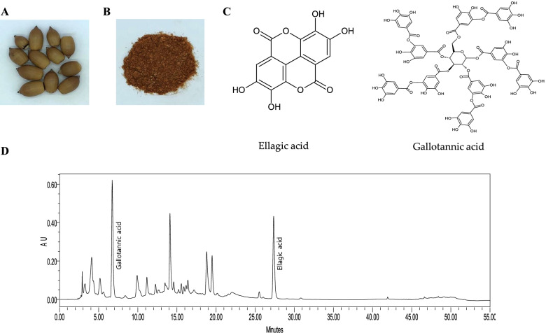
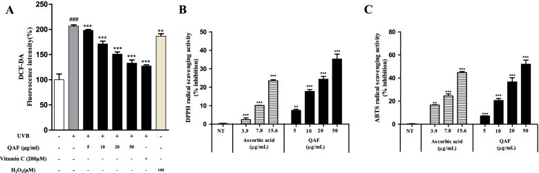
Methods: In this study, we used HPLC to identify the major active components of QAF water extracts. Anti-photoaging effects of QAF extracts were evaluated by analyzing ROS procollagen type I in UVB-irradiated HaCaT keratinocytes. Antiradical activity was determined using 2,2-diphenyl-1-picrylhydrazyl and 2,20-azino-bis (3-ethylbenzothiazoline-6-sulphonic acid) assays. The expression of MMP-1 was tested by western blotting and ELISA kits. QAF effects on phosphorylation of the MAPK (p38, JNK, and ERK) pathway and transcription factor AP-1, which enhances the expression of MMPs, were analyzed by western blots.
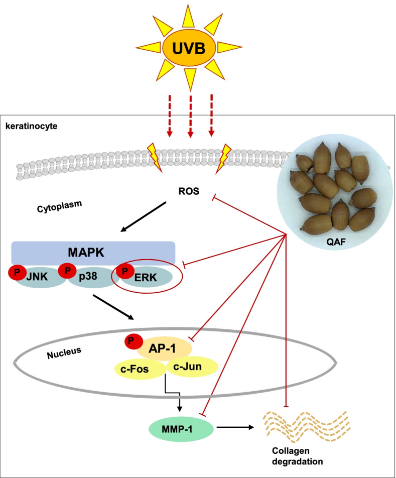
Results: We identified two major active components in QAF water extracts, gallotannic acid and ellagic acid. The QAF aqueous extracts recovered UVB-induced cell toxicity and reduced oxidative stress by inhibiting intracellular ROS generation in HaCaT cells. QAF rescued UVB-induced collagen degradation by suppressing MMP-1 expression. The anti-photoaging activities of QAF were associated with the inhibition of UVB-induced phosphorylation of extracellular signal-regulated kinase (ERK) and activator protein 1 (AP-1). Our findings indicated that QAF prevents UVB-induced skin damage due to collagen degradation and MMP-1 activation via inactivation of the ERK/AP-1 signaling pathway. Overall, this study strongly suggests that QAF exerts anti-skin-aging effects and is a potential natural biomaterial that inhibits UVB-induced photoaging.
Conclusion: These results show that QAF water extract effectively prevents skin photoaging by enhancing collagen deposition and inhibiting MMP-1 via the ERK/AP-1 signaling pathway.
Keywords: Activator protein 1; Extracellular signal-regulated kinases; Matrix metalloproteinase-1; Mitogen-activated protein kinase; Photoaging; Quercus acuta Thunb. fruit; Ultraviolet B.
© 2022. The Author(s).
Conflict of interest statement
The authors declare that they have no competing interests.
Figures


Similar articles
-
Hydrangea serrata (Thunb.) Ser. Extract Attenuate UVB-Induced Photoaging through MAPK/AP-1 Inactivation in Human Skin Fibroblasts and Hairless Mice.Nutrients. 2019 Mar 1;11(3):533. doi: 10.3390/nu11030533. Nutrients. 2019. PMID: 30823635 Free PMC article.
-
Anti-Photoaging Effect of Phaseolus angularis L. Extract on UVB-Exposed HaCaT Keratinocytes and Possibilities as Cosmetic Materials.Molecules. 2023 Feb 1;28(3):1407. doi: 10.3390/molecules28031407. Molecules. 2023. PMID: 36771069 Free PMC article.
-
Oleracone C from Portulaca oleracea attenuates UVB-induced changes in matrix metalloproteinase and type I procollagen production via MAPK and TGF-β/Smad pathways in human keratinocytes.Int J Cosmet Sci. 2023 Apr;45(2):166-176. doi: 10.1111/ics.12828. Epub 2023 Jan 12. Int J Cosmet Sci. 2023. PMID: 36415152
-
[Anti-photoaging effects and mechanisms of active ingredients of Chinese medicine: a review].Zhongguo Zhong Yao Za Zhi. 2022 Jul;47(14):3709-3717. doi: 10.19540/j.cnki.cjcmm.20220415.601. Zhongguo Zhong Yao Za Zhi. 2022. PMID: 35850827 Review. Chinese.
-
Epithelial-mesenchymal interaction mechanisms leading to the over-expression of neprilysin are involved in the UVB-induced formation of wrinkles in the skin.Exp Dermatol. 2016 Aug;25 Suppl 3:2-13. doi: 10.1111/exd.13083. Exp Dermatol. 2016. PMID: 27539896 Review.
Cited by
-
Role of autophagy in skin photoaging: A narrative review.Medicine (Baltimore). 2024 Feb 23;103(8):e37178. doi: 10.1097/MD.0000000000037178. Medicine (Baltimore). 2024. PMID: 38394552 Free PMC article. Review.
-
A Comparative Study of Skin Changes in Different Species of Mice in Chronic Photoaging Models.Int J Mol Sci. 2023 Jun 28;24(13):10812. doi: 10.3390/ijms241310812. Int J Mol Sci. 2023. PMID: 37445996 Free PMC article.
-
Current Knowledge on Interactions of Plant Materials Traditionally Used in Skin Diseases in Poland and Ukraine with Human Skin Microbiota.Int J Mol Sci. 2022 Aug 25;23(17):9644. doi: 10.3390/ijms23179644. Int J Mol Sci. 2022. PMID: 36077043 Free PMC article. Review.
-
Antiphotoaging Effect of AGEs Blocker™ in UVB-Irradiated Cells and Skh:HR-1 Hairless Mice.Curr Issues Mol Biol. 2023 May 9;45(5):4181-4199. doi: 10.3390/cimb45050266. Curr Issues Mol Biol. 2023. PMID: 37232735 Free PMC article.
-
In Vitro Evaluation of the Healing Potential and Proteomic Study of Quercus robur L. Leaf Extracts in Human Keratinocytes.Molecules. 2025 May 14;30(10):2152. doi: 10.3390/molecules30102152. Molecules. 2025. PMID: 40430324 Free PMC article.
References
-
- Hashem MA, Jun KY, Lee E, Lim S, Choo HY, Kwon Y. A rapid and sensitive screening system for human type I collagen with the aim of discovering potent anti-aging or anti-fibrotic compounds. Mol Cells. 2008;26(6):625–630. - PubMed
-
- Bergfeld WF. The aging skin. Int J Fertil Womens Med. 1997;42(2):57–66. - PubMed
MeSH terms
Substances
LinkOut - more resources
Full Text Sources
Medical
Research Materials
Miscellaneous
