. 2022 Jan 5;20(1):1.
doi: 10.1186/s12915-021-01182-2.

Co-option of the limb patterning program in cephalopod eye development

Affiliations

Co-option of the limb patterning program in cephalopod eye development

Stephanie Neal et al. BMC Biol. .

Abstract

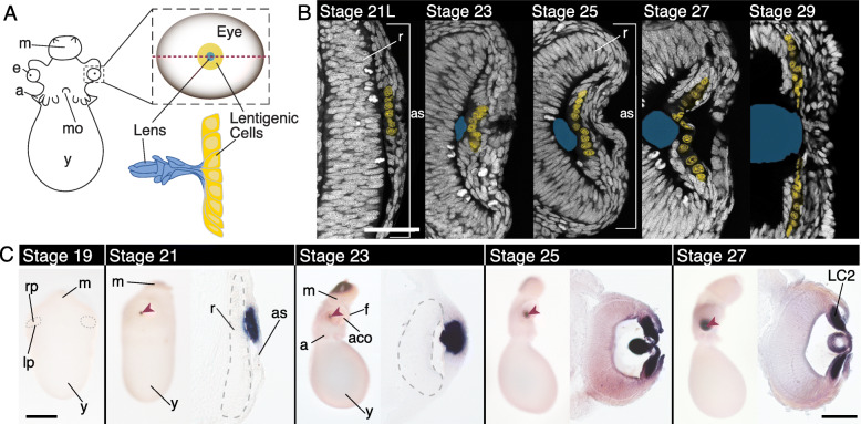
Background: Across the Metazoa, similar genetic programs are found in the development of analogous, independently evolved, morphological features. The functional significance of this reuse and the underlying mechanisms of co-option remain unclear. Cephalopods have evolved a highly acute visual system with a cup-shaped retina and a novel refractive lens in the anterior, important for a number of sophisticated behaviors including predation, mating, and camouflage. Almost nothing is known about the molecular-genetics of lens development in the cephalopod.

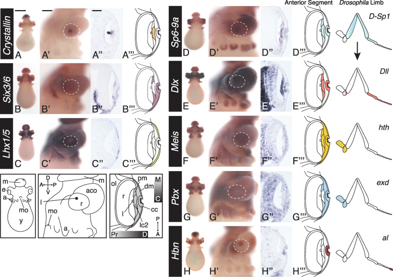
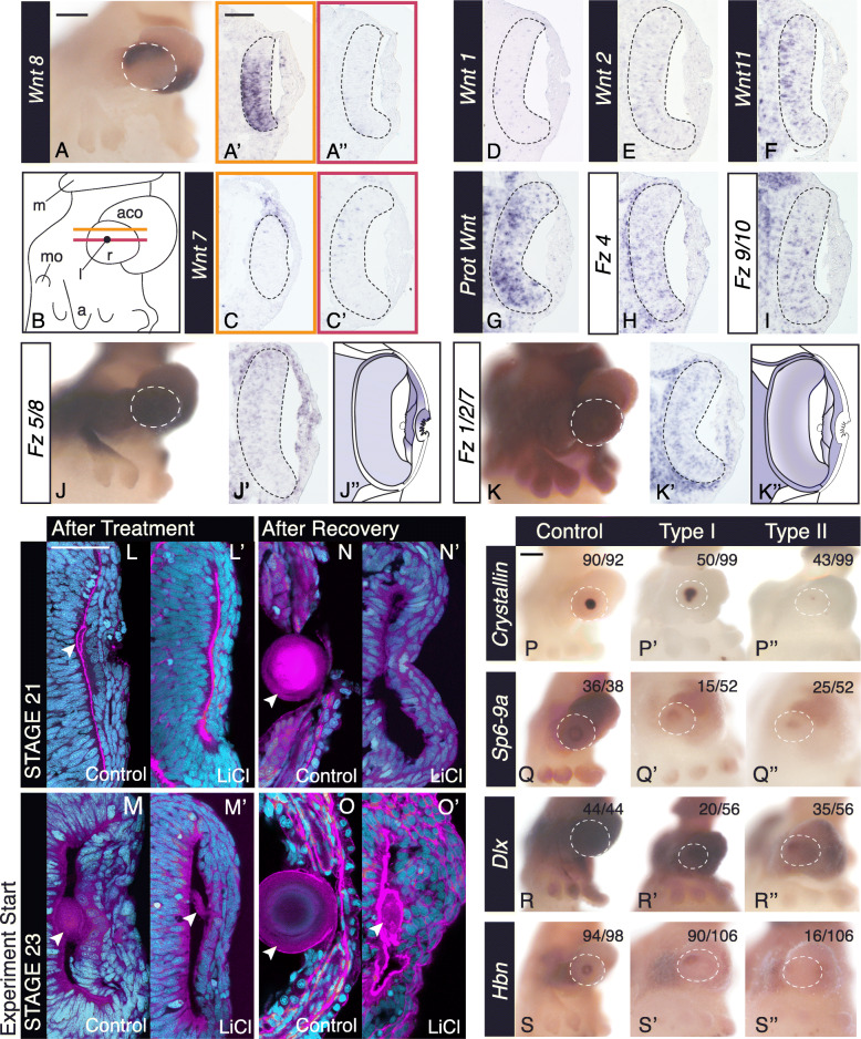
Results: Here we identify the co-option of the canonical bilaterian limb patterning program during cephalopod lens development, a functionally unrelated structure. We show radial expression of transcription factors SP6-9/sp1, Dlx/dll, Pbx/exd, Meis/hth, and a Prdl homolog in the squid Doryteuthis pealeii, similar to expression required in Drosophila limb development. We assess the role of Wnt signaling in the cephalopod lens, a positive regulator in the developing Drosophila limb, and find the regulatory relationship reversed, with ectopic Wnt signaling leading to lens loss.

Conclusion: This regulatory divergence suggests that duplication of SP6-9 in cephalopods may mediate the co-option of the limb patterning program. Thus, our study suggests that this program could perform a more universal developmental function in radial patterning and highlights how canonical genetic programs are repurposed in novel structures.

Keywords: Cephalopod; Dlx; Eye; Eye evolution, Lens; Limb patterning; Spiralia; Vision; Wnt.


Conflict of interest statement

The authors declare that they have no competing interests.

Figures

Fig. 1
Fig. 1
Lentigenic cell differentiation and DpS-Crystallin expression in the squid. A Cartoon diagram of a squid embryo (anterior), en face cartoon of the developing eye (red dotted line shows cross-section plane throughout the paper), and developing lentigenic cells and lens (cartoon of lens and lentigenic cells based on [43]). B Cross-section of the developing anterior segment at Arnold stages 21 late, 23, 25, 27, and 29 identifying differentiation of lentigenic cells [49]. White: Sytox-Green labeling nuclei, Yellow: False-colored lentigenic cell nuclei corresponding to the LC2 population identified by nuclear morphology [43, 44, 46]. Blue is the outline of the lens, as identified using phalloidin staining (not shown). First evidence of LC2 cells is late stage 21. Lentigenic cell number multiplies and distribution grows across the anterior segment (as) throughout development. Scale is 50 μm. C In situ hybridization of DpS-Crystallin in whole-mount and cryo-section. Stage 19 is an anterior view, the boundary between the retina placode and the lip cells is highlighted with a dotted line. No DpS-Crystallin expression is apparent at this stage. Stages 21–27 are shown in a lateral view of the embryo on the left and a cross-section of the eye on the right. Anterior of the embryo is down in the sections. The retina is outlined with a dashed grey line in stage 21 and 23. DpS-Crystallin expression corresponds with LC2 population. Scale is 500 μm in whole mount images. Scale is 100 μm in sectioned images. as, anterior segment; a, arm; aco, anterior chamber organ; e, eye; f, funnel; LC2, lentigenic cell population; lp, lip; m, mantle; mo, mouth; rp, retina placode; r, retina; y, yolk. Red arrow highlights the lens
Fig. 2
Fig. 2
Limb patterning program expressed in the developing anterior segment. For each gene: left to right, anterior whole-mount view, lateral whole-mount view (anterior left), cross-section (anterior is down), cartoon summary is specific to only the anterior segment expression, excluding brain and retina expression for clarity. Dotted white outline in lateral view outlines the perimeter of the eye. AC Defining cell populations in the developing anterior segment at stage 23. A, A’, A” DpS-Crystallin expression in the anterior segment at stage 23, expressed in the proximal, central cells corresponding with the LC2 cells (lc2). Expression is also apparent in the lens. B, B’, B” Expression of DpSix3/6. B” Expression is apparent in the distal, central cup cells (cc) and the proximal-marginal (pm) anterior segment cells. The proximal-central cells (lc2) lack expression of DpSix3/6. C, C’, C” DpLhx1/5 expression. C” Expression of DpLhx1/5 is found in the distal-marginal cell (dm) population. Expression is excluded from the central cup (cc). DG Expression of the limb patterning program genes. Summary of the proximodistal expression of each Drosophila homolog during proximodistal patterning of the limb is shown on the right. H Prd-like homolog Homeobrain (Hbn) expression in the distal, central cup cells. a, arms; aco, anterior chamber organ; cc, cup cells; dm, distal-marginal cells; e, eye; l, lens; lc2, LC2 cells; m, mantle; mo, mouth; pm, proximal-marginal cells; r, retina; y, yolk. Anterior segment highlighted in grey in the cartoon. Orientation abbreviations: M, marginal; C, central; Pr, proximal; D, Distal; A, anterior; P, posterior. Scale for whole-mount anterior view is 500 μm. Scale for lateral whole-mount view 200 μm. Scale for sectioned images 50 μm
Fig. 3
Fig. 3
Wnt signaling pathway expression in the developing squid eye. AG Wnt gene expression at stage 23. Based on expression, Wnt7, Wnt8, Wnt2, Wnt11, and Prot Wnt are possible candidates to signal the anterior segment. A Lateral, whole-mount expression of Wnt8. A’ Dorsal retina expression of Wnt8. Location of the section indicated by the orange line in B. A” Central section lacking retina expression. Location of the section indicated by the red line in B. B Cartoon of the lateral whole-mount embryo at stage 23. Orange and red lines correspond to the location of the two sections shown in A, A’, and C, C’. DG Expression of other Wnt homologs in central sections. HK Expression of Frizzled receptors at stage 23. Fz1/2/7 shows asymmetric expression and Fz5/8 shows specific exclusion from the central cup cells. J and K are lateral view of the whole mount expression. J” and K” are cartoons of expression in J’ and K’ respectively. Gradients of expression show intracellular asymmetries in the anterior segment. Black dotted line in sectioned images shows the perimeter of the retina. LO Anterior segment and lens morphology after Wnt agonist treatment (LiCl). Embryos were cryosectioned and stained with Sytox-green (nuclei, cyan) and phalloidin (F-actin, magenta). L and L’ Control and LiCl agonist treatments started at stage 21, treated for 24 h and fixed immediately. M and M’ Control and Wnt agonist (LiCl) treatments started at stage 23 for 24 h and fixed immediately. N and N’ Control and Wnt agonist (LiCl) treatments started at stage 21, treated for 24 h and allowed to recover for 48 h and fixed. O and O’ Control and Wnt agonist (LiCl) treatments started at stage 23, treated for 24 h and allowed to recover for 48 h and fixed. Arrowhead highlights the lens. PS In situ hybridization of anterior segment markers after 24-h control and LiCl treatments starting at stage 23. Phenotypes are characterized as Type I (mild) and Type II (severe). The white dotted line outlines the eye in the lateral image. The number of eyes scored in control and the two phenotypes is found in LiCl-treated animals in the top right corner. Scale for all lateral whole-mount view images is 200 μm. Scale for all sectioned images is 50 μm. Anterior is down in all sectioned images. White dotted line in whole mount images identifies the perimeter of the eye. m, mantle; a, arms; aco, anterior chamber organ; mo, mouth; r, retina; l, lens
Fig. 4
Fig. 4
Ectopic Wnt signaling activation leads to loss of the lens. A Model for lentigenic cell differentiation at stage 21. LC2 lentigenic cells differentiate on the dorsal side of the eye first, with a wave moving ventrally. Type I DpS-Crystallin embryos have been interrupted in progress. B En face summary of sample radial expression of the limb patterning program across developmental contexts [14, 16, 24]

Similar articles

Cited by

References

    1. Shubin N, Tabin C, Carroll S. Fossils, genes and the evolution of animal limbs. Nature. 1997;388(6643):639–648. doi: 10.1038/41710. - DOI - PubMed
    1. Erwin DH, Davidson EH. The last common bilaterian ancestor. Development. 2002;129:3021–3032. doi: 10.1242/dev.129.13.3021. - DOI - PubMed
    1. Pueyo JI, Couso JP. Parallels between the proximal-distal development of vertebrate and arthropod appendages: homology without an ancestor? Curr Opin Genet Dev. 2005;15(4):439–446. doi: 10.1016/j.gde.2005.06.007. - DOI - PubMed
    1. Panganiban G, Nagy L, Carroll SB. The role of the Distal-less gene in the development and evolution of insect limbs. Curr Biol. 1994;4(8):671–675. doi: 10.1016/s0960-9822(00)00151-2. - DOI - PubMed
    1. Panganiban G, Irvine SM, Lowe C, Roehl H, Corley LS, Sherbon B, Grenier JK, Fallon JF, Kimble J, Walker M, Wray GA, Swalla BJ, Martindale MQ, Carroll SB. The origin and evolution of animal appendages. Proceedings of the National Academy of Sciences. 1997;94(10):5162–5166. doi: 10.1073/pnas.94.10.5162. - DOI - PMC - PubMed

Publication types

LinkOut - more resources