Review
. 2022 Jan 4;29(1):1.
doi: 10.1186/s12929-021-00784-w.

Monoclonal antibodies for COVID-19 therapy and SARS-CoV-2 detection

Affiliations
Review

Monoclonal antibodies for COVID-19 therapy and SARS-CoV-2 detection

Yu-Chyi Hwang et al. J Biomed Sci. .

Abstract

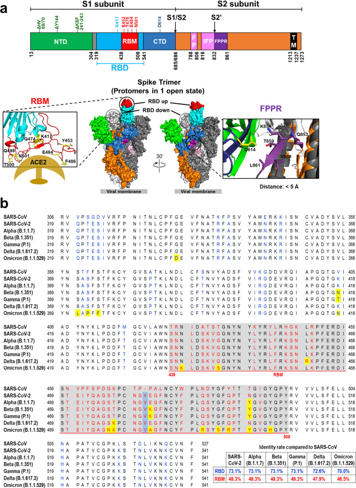
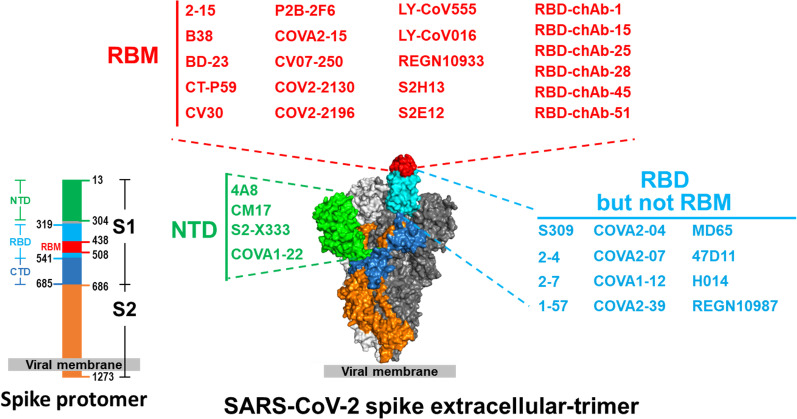
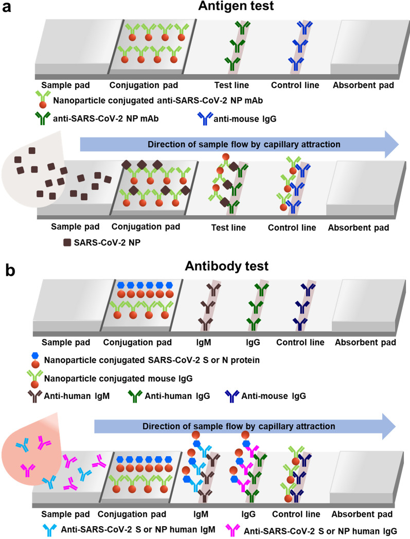
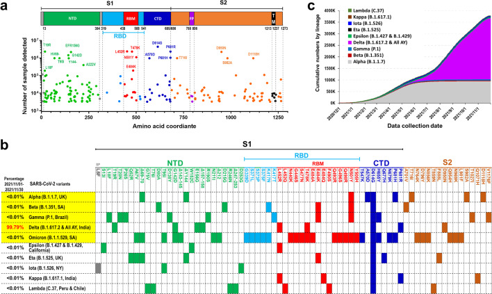
The coronavirus disease 2019 (COVID-19) pandemic is an exceptional public health crisis that demands the timely creation of new therapeutics and viral detection. Owing to their high specificity and reliability, monoclonal antibodies (mAbs) have emerged as powerful tools to treat and detect numerous diseases. Hence, many researchers have begun to urgently develop Ab-based kits for the detection of severe acute respiratory syndrome coronavirus 2 (SARS-CoV-2) and Ab drugs for use as COVID-19 therapeutic agents. The detailed structure of the SARS-CoV-2 spike protein is known, and since this protein is key for viral infection, its receptor-binding domain (RBD) has become a major target for therapeutic Ab development. Because SARS-CoV-2 is an RNA virus with a high mutation rate, especially under the selective pressure of aggressively deployed prophylactic vaccines and neutralizing Abs, the use of Ab cocktails is expected to be an important strategy for effective COVID-19 treatment. Moreover, SARS-CoV-2 infection may stimulate an overactive immune response, resulting in a cytokine storm that drives severe disease progression. Abs to combat cytokine storms have also been under intense development as treatments for COVID-19. In addition to their use as drugs, Abs are currently being utilized in SARS-CoV-2 detection tests, including antigen and immunoglobulin tests. Such Ab-based detection tests are crucial surveillance tools that can be used to prevent the spread of COVID-19. Herein, we highlight some key points regarding mAb-based detection tests and treatments for the COVID-19 pandemic.

Keywords: Angiotensin converting enzyme II (ACE2); Coronavirus disease 2019 (COVID-19); Cytokine storm; Receptor binding motif (RBM); Receptor-binding domain (RBD); Severe acute respiratory syndrome coronavirus 2 (SARS-CoV-2); Spike; Therapeutic antibody; Viral detection.


Conflict of interest statement

No potential conflicts of interest are disclosed.

Figures

Fig. 1
Fig. 1
SARS-CoV-2 Spike protein. a Structure SARS-CoV-2 spike protein. Different domains of the SARS-CoV-2 spike protein: N-terminal domain (NTD), receptor-binding domain (RBD), receptor-binding motif (RBM), subdomain 1 and 2, protease cleavage sites (S1/S2/S2′), fusion peptide (FP), internal fusion peptide (IFP), fusion peptide proximal region (FPPR), and transmembrane region (TM). HV69/70, Y144, and KSF241-243 are frequently deleted residues in the NTD of SARS-CoV-2 variants of concern. K417, E452, E484, T478, N501 and D614 are the most frequently mutated residues in the RBD of SARS-CoV-2 variants of concern. Key residues of the receptor-binding motif in the S protein of SARS-CoV-2 that interact with ACE2 are shown (lower left). The SARS-CoV-2 S protein trimeric complex is shown in a “one-up” RBD conformation. The two RBD-down protomers are depicted in light and dark gray. The RBD-up protomer is colored according to its domains; RBM in red, non-RBM RBD in light blue, NTD in green, S2 in orange, FP and IFP in pink, and FPPR in purple. The dashed circle indicates the RBD site of an RBD-down conformation protomer. Inter-atomic contacts between aspartate 614 (yellow) in a reference S monomer (dark blue) and five residues (purple) in its adjacent S protein monomer chain (dark gray) within 5 Å. These five contacts might be destabilizing and create a hydrophilic-hydrophobic repulsion that is lost upon replacement of aspartate by glycine in the D614G mutation (lower right). b RBD sequences of SARS-CoV (GeneBank: AAP30030.1), SARS-CoV-2 (GeneBank: QVW76257.1), and SARS-CoV-2 variants of concern, including B.1.1.7 (Alpha), B.1.351 (Beta), P.1 (Gamma), B.1.617.2 (Delta), and B.1.1.529 (Omicron). The amino acids encoded by SARS-CoV-2 that are altered in comparison to SARS-CoV are colored blue (RBD) or red (RBM). The amino acid inserted in SARS-CoV-2 is denoted by a light blue background. The amino acids substituted in variants of concern are denoted by a yellow background. The residues 438–508 comprise the RBM of SARS-CoV-2 and are shown with grey background
Fig. 2
Fig. 2
Epitopes of anti-spike and anti-RBD nAbs mapped to a surface model of SARS-CoV-2 spike trimer. The identified epitope regions are depicted as surface regions (PDB: 6VSB). Some of the shown anti-spike nAbs have known exact epitopes; for others the exact epitopes are unknown. Ab names are color-coded by the domains they recognized: N-terminal domain (NTD), light green; receptor binding motif (RBM), red; and receptor binding domain (RBD) but not RBM, cyan
Fig. 3
Fig. 3
Example of COVID-19 lateral flow immunoassays (LFIA). a For antigen detection, a sample containing viral antigens is dropped on the sample pad and flows by capillary action up to the absorbent pad. The sample with viral N protein (NP) directly binds to the anti-NP Ab conjugated with nanoparticles, such as colloidal gold particles or latex nanocomposites. Then, the nanoparticle-conjugated immunocomplexes are released from the conjugation pad. The free epitope of NP is captured to the second anti-NP Ab in the test line. Unbound conjugated Abs will be recognized by immobilized anti-mouse IgG in the control line. b For Ab detection, patient serum or plasma specimens are dropped on the sample pad. The sample fluid flows through the conjugation pad which contains nanoparticle-conjugated SARS-CoV-2 spike (S) or N proteins to form antigen-Ab immunocomplexes. The immunocomplexes flow to the test line and are then captured by specific isotype immunoglobulins such as IgM and IgG. Unbound control nanoparticle-conjugated mouse IgG is captured by anti-mouse IgG at the control line
Fig. 4
Fig. 4
SARS-CoV-2 spike mutations. a Top 200 identified SARS-CoV-2 spike mutations. Each dot indicates an amino acid mutation in the S protein. The colors indicate different domains of the SARS-CoV-2 spike protein; NTD N-terminal domain, RBD receptor-binding domain, RBM receptor-binding motif, CTD C-terminal domain, S2 subdomain 2, FP fusion peptide, TM transmembrane region. The altered amino acids of the top 20 SARS-CoV-2 spike mutations are shown as indicated. The data from 4,450,473 sequences were collected from GISAID and COVID CG (updated to 2021-11-22). b Nonsynonymous mutation positions in spike protein of newly emerged SARS-CoV-2 variants. The B.1.1.7 (Alpha), B.1.351 (Beta), P.1 (Gamma), B.1.617.2 (Delta) and B.1.1.529 (Omicron) variants are classified as variants of concern by WHO. The percentage of India B.1.617.2 (Delta) variant includes B.1.617.2 and its all AY sub-lineages. c Confirmed COVID-19 cases comprising SARS-CoV-2 variants. The data from 4,337,516 sequences were collected from GISAID and COVID CG (from 2020-12-01 to 2021-11-30) and grouped by lineage
Fig. 5
Fig. 5
Neutralization of SARS-CoV-2 variants by therapeutic mAbs. The neutralization abilities of therapeutic mAbs against wild-type, D614G and newly emerged SARS-CoV-2 variants, including B.1.1.7 (Alpha), B.1.351 (Beta), P.1 (Gamma), B.1.427 and B.1.429 (Epsilon), B.1.526 (Iota), B.1.617.1 (Kappa), and B.1.617.2 (Delta). Symbols and colors indicate the range of half maximal inhibitory concentration (IC50) values toward authentic SARS-CoV-2 virus. +++ with blue, IC50 < 10 ng/ml; ++ with light blue, IC50 = 10–100 ng/ml; + with white, IC50 = 100–1000 ng/ml; —, IC50 with red > 1000 ng/ml; ND with grey, no determined; #, preliminary results reported on the website of Celltrion Healthcare Co., Ltd.; *, the range of IC50 values toward pseudotype SARS-CoV-2 virus. RBD, receptor-binding domain; RBM, receptor-binding motif. The mutant amino acids in RBD of each SARS-CoV-2 spike protein are shown as indicated

References

    1. WHO Coronavirus disease (COVID-2019) situation reports. World Health Organization. https://www.who.int/emergencies/diseases/novel-coronavirus-2019/situatio.... Accessed 23 Mar 2021.
    1. Fan E, Beitler JR, Brochard L, Calfee CS, Ferguson ND, Slutsky AS, Brodie D. COVID-19-associated acute respiratory distress syndrome: is a different approach to management warranted? Lancet Respir Med. 2020;8(8):816–821. - PMC - PubMed
    1. Bradley BT, Maioli H, Johnston R, Chaudhry I, Fink SL, Xu H, Najafian B, Deutsch G, Lacy JM, Williams T, Yarid N, Marshall DA. Histopathology and ultrastructural findings of fatal COVID-19 infections in Washington State: a case series. Lancet. 2020;396(10247):320–332. - PMC - PubMed
    1. Struyf T, Deeks JJ, Dinnes J, Takwoingi Y, Davenport C, Leeflang MM, Spijker R, Hooft L, Emperador D, Dittrich S, Domen J, Horn SRA, Van den Bruel A, Cochrane C-DTAG. Signs and symptoms to determine if a patient presenting in primary care or hospital outpatient settings has COVID-19 disease. Cochrane Database Syst Rev. 2020;7:13665. - PMC - PubMed
    1. Chen N, Zhou M, Dong X, Qu J, Gong F, Han Y, Qiu Y, Wang J, Liu Y, Wei Y, Xia J, Yu T, Zhang X, Zhang L. Epidemiological and clinical characteristics of 99 cases of 2019 novel coronavirus pneumonia in Wuhan, China: a descriptive study. Lancet. 2020;395(10223):507–513. - PMC - PubMed