Review
. 2022 Jan 4;20(1):4.
doi: 10.1186/s12951-021-01181-z.

Nanomedicine-enabled chemotherapy-based synergetic cancer treatments

Affiliations
Review

Nanomedicine-enabled chemotherapy-based synergetic cancer treatments

Wencheng Wu et al. J Nanobiotechnology. .

Abstract

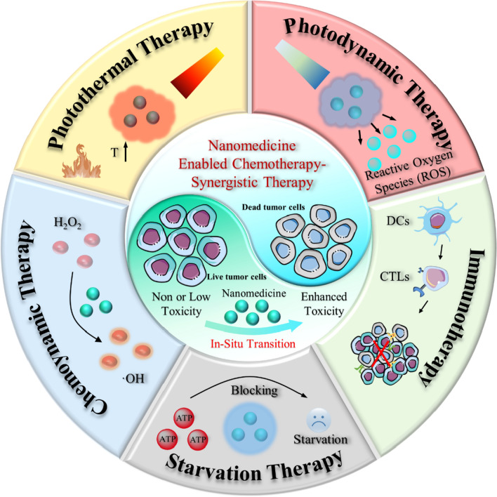
Chemotherapy remains one of the most prevailing regimens hitherto in the fight against cancer, but its development has been being suffering from various fatal side effects associated with the non-specific toxicity of common chemical drugs. Advances in biomedical application of nanomedicine have been providing alternative but promising approaches for cancer therapy, by leveraging its excellent intrinsic physicochemical properties to address these critical concerns. In particular, nanomedicine-enabled chemotherapy has been established as a safer and promising therapeutic modality, especially the recently proposed nanocatalytic medicine featuring the capabilities to generate toxic substances by initiating diverse catalytic reactions within the tumor without directly relying on highly toxic but non-selective chemotherapeutic agents. Of special note, under exogenous/endogenous stimulations, nanomedicine can serve as a versatile platform that allows additional therapeutic modalities (photothermal therapy (PTT), photodynamic therapy (PDT), chemodynamic therapy (CDT), etc.) to be seamlessly integrated with chemotherapy for efficacious synergistic treatments of tumors. Here, we comprehensively review and summarize the representative studies of multimodal synergistic cancer treatments derived from nanomedicine and nanocatalytic medicine-enabled chemotherapy in recent years, and their underlying mechanisms are also presented in detail. A number of existing challenges and further perspectives for nanomedicine-synergized chemotherapy for malignant solid tumor treatments are also highlighted for understanding this booming research area as comprehensively as possible.

Keywords: Chemotherapy; Nanomedicine; Synergisitic cancer treatments.


Conflict of interest statement

All authors declare no financial/commercial conflicts of interest.

Figures

Scheme 1
Scheme 1
Summary of the nanomedicine-enabled chemotherapy and derived synergistic cancer therapy
Fig. 1
Fig. 1
Nanomedicine enhanced chemotherapy through different mechanisms. a, b Schematic diagram of the fabrication of PVCL-GMA NGs of different diameters and dendrimers decorated PVCL-GMA NGs (NGs-G2) used for efficient drug delivery in vivo. c, d Schematic representation of pH-responsive drug release and cancer cells targeting behavior of nanomedicine
Fig. 2
Fig. 2
a Schematic representation of active tumor penetration of nanomedicine mediated by cationization-initiated transcytosis. b The structures with or without GGT-responsive cationized drug-conjugated nanomedicine. d The biodistribution of CPT within 24 h after intravenous administration of different nanomedicines
Fig. 3
Fig. 3
a Schematic illustration of DOX-CaO2/MnO2-MS NPs for largely promoted drug transport. b Schematic diagram of the synthesis process of M-R@D-PDA-PEG-FA-D and the combination of photothermal chemotherapy and gene targeting for the treatment of tumors
Fig. 4
Fig. 4
a Schematic diagram of the in vivo working mechanism of DSF@Cu-HMSNs enhanced chemotherapy. b TEM image and SAED pattern of PEG-Cu-HMSNs. c Accumulated release of copper ions from PEG-Cu-HMSNs at varied pHs. d The electron spin resonance (ESR) spectroscopy under different conditions (only H2O2; DSF@PEG/Cu-HMSNs + H2O2, pH = 7.4; DSF@PEG/Cu-HMSNs + H2O2, pH = 6.5). e Chemical mechanism of Cu-mediated reactions. f Cell viabilities of 4T1 cells after different treatments. g Schematic illustration of the synthesis of DSF/DOX@ZIF-8@Cu-TA. h TEM images of different nanoparticles. i Schematic diagram of the in vivo DSF/DOX@ZIF-8@Cu-TA triggered synergistic chemotherapy and CDT
Fig. 5
Fig. 5
Cu-engineered HMPB for PTT enhanced intratumoral chelating for tumor-specific synergistic therapy based on the nontoxicity-to-toxicity transition of DSF. a Schematic diagram of the fabrication of DSF@PVP/Cu-HMPB and b the in vivo PVP/Cu-HMPB triggered PTT enhanced chemotherapy. c TEM images and corresponding elemental mapping of DSF@PVP/Cu-HMPB. Scale bars 50 nm. d Temperature change curves of PVP/Cu-HMPB under light irradiation and subsequently after turning off the light source. e Absorbance of DSF@PVP/Cu-HMPB solution in the range of 200–600 nm at different time intervals. f Cell viabilities of 4T1 cells after being treated by different conditions for 24 h. g Weight of tumors in different groups of BALB/c nude mice at the end of treatment. h The blood-circulation lifetime of PVP/Cu-HMPB. i Biodistribution of PVP/Cu-HMPB-derived Cu2+ at different time durations after injecting PVP/Cu-HMPB. j Bioluminescence images of mice's main organs. k, l Body weight of mice in different groups
Fig. 6
Fig. 6
Design principle and therapeutic mechanism of Silicene@Silica-AQ4N. a Synthetic route of Silicene@Silica-AQ4N. b Illustration of the Silicene@Silica-AQ4N triggered synergistic photonic hyperthermia amplified chemotherapy in vivo. c The TEM, Dark-field TEM images, and corresponding elemental mapping of Silicene@Silica nanosheets. d Temperature change curves of Silicene@Silica-RGD under laser irradiation with different power densities. e The drug release of Silicene@Silica-AQ4N in PBS (pH = 7.4) under irradiated with laser irradiation with different power densities. f, g Cell viabilities of 4T1 cells treated by different conditions. h Temperature change curves at the tumor site of mice during different treatments. i Tumor volume of mice in different groups
Fig. 7
Fig. 7
a Schematic representation of molecular structures of PUFAylated cabazitaxel prodrug (LTK-CTX) and chlorin e6 prodrug (L-Ce6) and the working mechanism of PSPC nanoassemblies in vivo. b Morphology presentation of the LTK-CTX nanoassemblies (NAs), L-Ce6 NAs, and PSPC NAs. Scale bars, 100 nm. c Confocal imaging of A375 cells stained with DCFH-DA for 30 min after different treatments. d Confocal imaging of A375 cells stained with γ-H2AX (red) and DAPI(blue) in different groups. e Flow cytometry analysis of A375 cell apoptosis treated by varied conditions. f Tumor volume change curves of mice in different groups. g Body weight change of mice in different groups post-administration. h Scheme showing different stages of Dox-PEG-PS@MIL-100 NPs in H2O2 and corresponding size change and PDT assisted tumor penetration. i The tumor volumes change curves of mice
Fig. 8
Fig. 8
Nanomedicine-enabled tumor synergistic starvation/chemotherapy based on Gox-mediated glucose glycolysis. a Schematic diagram of the synthesis and use of TG-GV for combination chemo/starvation therapy. b TEM image of TG-GVs. c The release curves of TPZ from TG-GVs solutions with or no glucose addition. d The cell viabilities of 4T1 cells after undergoing various treatments for 24 h. e The recorded tumor volume change in different groups during the treatments. f Schematic of in vivo working mechanism of A/Q4N/GOx@ZIF-8@CM. g, h TEM images of AQ4N/GOx@ZIF-8 and AQ4N/GOx@ZIF-8@CM. i CLSM images of HepG2 cells stained with oxidative stress/hypoxia detection probe after treated by different conditions. Scale bar, 20 μm. j, k Viabilities of LO2 and HepG2 cells after various treatments. l The photographs of tumors tissues. m HIF-1α, VEGF immunohistochemical, and H&E staining images of tumor tissues
Fig. 9
Fig. 9
Nanomedicine induced starvation to generate differential stress sensitization for enhanced tumor-specific chemotherapy. a Schematic representation of the fabrication of liposomal nanomedicine and its interactions with tumor or normal cells. b TEM -based study of Lip-(2DG + Dox). Scale bar, 50 nm. c DLS measurements of different liposomes. d, e Cell viabilities of HeLa tumor cells and normal HUVEC cells after different treatments
Fig. 10
Fig. 10
A novel nanomedicine based on the self-assembling among Cu2+, DOX, and NLG919 for chemotherapy sensitized immunotherapy. a Schematic diagram of the synthetic route and in vivo working mechanism of Cu-DON. b Immunofluorescence images of CRT and HMGB1 in 4T1 cells after different treatments. Scale bar, 25 μm. c Z-stack CLSM images of 4T1 tumor spheroids after being treated by DOX and Cu-DON. Scale bar, 200 μm. d, e Tumor volume change curves of the primary tumor and distant tumor volume
Fig. 11
Fig. 11
Engineered sequential pH/redox responsive nanoparticles (PC7A-ss-DOX) for precisely delivering chemodrug (DOX) and immunoadjuvant (imidazoquinolines, IMDQs) for synergistic chemo-immunotherapy. a Schematic diagram of the chemical structure of PC7A-ss-DOX and the sequential immune activation mechanism by PC7A-ss-DOX. b, c TEM images of PC7A-ss-DOX at different pH values. Scale bar, 100 nm. d In vitro DOX release from PC7A-ss-DOX under different conditions. e Immunofluorescence images of CRT after various treatments. Scale bar, 20 μm. f ATP levels in tumor cells after different treatments. g The content of HMGB1 in the tumor cells treated by different conditions and schematic diagram of DC maturation under TLR priming. h RAW-Blue reporter cells number after being stimulated by different materials. i Percentage of mature DCs in different groups: (1) B16-OVA; (2) PC7A-ss-DOX treated B16-OVA; (3) B16-OVA + iPDPA-IMDQ; (4) PC7A-ss-DOX treated B16-OVA + iPDPA-IMDQ. The tumor growth curves of j primary tumors upon different treatments and corresponding k distant tumors of mice

References

    1. Chabner BA, Roberts TG. Chemotherapy and the war on cancer. Nat Rev Cancer. 2005;5:65–72. - PubMed
    1. Lin H, Chen Y, Shi J. Nanoparticle-triggered in situ catalytic chemical reactions for tumour-specific therapy. Chem Soc Rev. 2018;47:1938–1958. - PubMed
    1. Matera C, Gomila AMJ, Camarero N, Libergoli M, Soler C, Gorostiza P. Photoswitchable antimetabolite for targeted photoactivated chemotherapy. J Am Chem Soc. 2018;140:15764–15773. - PubMed
    1. Nair RK, Christie C, Ju D, Shin D, Pomeroy A, Berg K, Peng Q, Hirschberg H. Enhancing the effects of chemotherapy by combined macrophage-mediated photothermal therapy (PTT) and photochemical internalization (PCI) Lasers Med Sci. 2018;33:1747–1755. - PMC - PubMed
    1. Zou Y, Wei J, Xia Y, Meng F, Yuan J, Zhong Z. Targeted chemotherapy for subcutaneous and orthotopic non-small cell lung tumors with cyclic RGD-functionalized and disulfide-crosslinked polymersomal doxorubicin. Signal Transduct Target Ther. 2018;3:32. - PMC - PubMed

Substances