. 2021 Dec 31;30(6):401-414.
doi: 10.5607/en21006.

Oleanolic Acid Inhibits Neuronal Pyroptosis in Ischaemic Stroke by Inhibiting miR-186-5p Expression

Affiliations

Oleanolic Acid Inhibits Neuronal Pyroptosis in Ischaemic Stroke by Inhibiting miR-186-5p Expression

Shi-Chang Cai et al. Exp Neurobiol. .

Abstract

Ischaemic stroke is a common condition leading to human disability and death. Previous studies have shown that oleanolic acid (OA) ameliorates oxidative injury and cerebral ischaemic damage, and miR-186-5p is verified to be elevated in serum from ischaemic stroke patients. Herein, we investigated whether OA regulates miR-186-5p expression to control neuroglobin (Ngb) levels, thereby inhibiting neuronal pyroptosis in ischaemic stroke. Three concentrations of OA (0.5, 2, or 8 μM) were added to primary hippocampal neurons subjected to oxygen-glucose deprivation/reperfusion (OGD/R), a cell model of ischaemic stroke. We found that OA treatment markedly inhibited pyroptosis. qRT-PCR and western blot revealed that OA suppressed the expression of pyroptosis-associated genes. Furthermore, OA inhibited LDH and proinflammatory cytokine release. In addition, miR-186-5p was downregulated while Ngb was upregulated in OA-treated OGD/R neurons. MiR-186-5p knockdown repressed OGD/R-induced pyroptosis and suppressed LDH and inflammatory cytokine release. In addition, a dual luciferase reporter assay confirmed that miR-186-5p directly targeted Ngb. OA reduced miR-186-5p to regulate Ngb levels, thereby inhibiting pyroptosis in both OGD/R-treated neurons and MCAO mice. In conclusion, OA alleviates pyroptosis in vivo and in vitro by downregulating miR-186-5p and upregulating Ngb expression, which provides a novel theoretical basis illustrating that OA can be considered a drug for ischaemic stroke.

Keywords: Ischaemic stroke; Neuroglobin; Oleanolic acid; Pyroptosis; miR-186-5p.


Conflict of interest statement

CONFLICT OF INTEREST

The authors declare that there is no conflict of interest.

Figures

Fig. 1
Fig. 1
OGD/R treatment leads to hippocampal neuron pyroptosis. An OGD/R cell model was established in primary hippocampal neurons. (A) Pyroptosis was detected by flow cytometry using caspase-1 and PI staining. (B) ELISA was applied to measure IL-1β and IL-18 expression. (C) LDH release in the cell supernatant. (D) Pyroptosis-related markers, including NLRP3, pro-caspase-1, cleaved caspase-1, GSDMD-FL, and GSDMD-N, were assessed using western blot assay. The results are shown as the mean±SD. n=3 per group. *p<0.05, **p<0.01, ***p<0.001.
Fig. 2
Fig. 2
OGD/R-induced pyroptosis is ameliorated by OA. (A) Flow cytometry was utilized to analyse the impact of OA at different doses on OGD/R-induced pyroptosis. (B) Levels of IL-1β and IL-18 were analysed by ELISA after OA treatment. (C) LDH release after OA treatment. (D) Expression of pyroptosis-related markers was evaluated using western blot assay. The results are shown as the mean±SD. n=3 per group. *p<0.05, **p<0.01, ***p<0.001.
Fig. 3
Fig. 3
OA application reverses the levels of miR-186-5p and Ngb caused by OGD/R. (A) The levels of miR-186-5p were evaluated as shown by qRT-PCR. (B, C) The mRNA (B) and protein (C) expression of Ngb was measured using qRT-PCR and western blotting, respectively. The results are shown as the mean±SD. n=3 per group. **p<0.01, ***p<0.001.
Fig. 4
Fig. 4
miR-186-5p knockdown alleviates OGD/R-induced pyroptosis. The miR-186-5p inhibitor or inhibitor NC was transfected into neurons. (A) Expression of miR-186-5p was measured using qRT-PCR. (B) Pyroptosis was assessed using flow cytometry. (C) Expression of IL-1β and IL-18 was detected by ELISA. (D) LDH release was restored by inhibition of miR-186-5p. (E) Pyroptosis-related markers were detected by western blot assay. The results are shown as the mean±SD. n=3 per group. *p<0.05, **p<0.01, ***p<0.001.
Fig. 5
Fig. 5
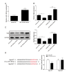
miR-186-5p overexpression inhibits Ngb expression. (A) qRT-PCR was used to evaluate the level of miR-186-5p after neurons were incubated with miR-186-5p mimics. (B, C) mRNA (B) and protein (C) levels of Ngb were detected by qRT–PCR and western blotting in neurons. (D) The binding site was predicted using TargetScan version 7.2, and luciferase activity was measured using double luciferase reporter assay in hippocampal neurons. The results are shown as the mean±SD. n=3 per group. *p<0.05, **p<0.01, ***p<0.001.
Fig. 6
Fig. 6
OA suppresses pyroptosis induced by OGD/R by regulating miR-186-5p and Ngb expression. (A, B) mRNA (A) and protein (B) expression levels of Ngb in neurons were evaluated as shown by qRT–PCR and western blotting. (C) Pyroptosis was detected by flow cytometry. (D) Alterations in IL-1β and IL-18 were determined by ELISA. (E) LDH release was modulated after OA treatment in OGD/R neurons. (F) Pyroptosis-related markers were examined by western blotting. The results are shown as the mean±SD. n=3 per group. *p<0.05, **p<0.01, ***p<0.001.
Fig. 7
Fig. 7
The effect of OA treatment in MCAO/R mice. MCAO/R or sham surgery was performed in mice, and OA was administered to the MCAO/R+OA group. (A) TTC was utilized to detect the cerebral ischaemia area. (B) Quantification of infarct volume. (C) IL-1β and IL-18 levels were determined using ELISA. (D) LDH release. (E) Pyroptosis-related markers were evaluated by western blot assay. (F) miR-186-5p expression was examined by qRT-PCR. (G, H) mRNA (G) and protein (H) levels of Ngb were evaluated by qRT-PCR and western blotting, respectively. The results are shown as the mean±SD. n=6 per group. *p<0.05, **p<0.01, ***p<0.001.

References

    1. Hankey GJ. Stroke. Lancet. 2017;389:641–654. doi: 10.1016/S0140-6736(16)30962-X. - DOI - PubMed
    1. Meschia JF, Brott T. Ischaemic stroke. Eur J Neurol. 2018;25:35–40. doi: 10.1111/ene.13409. - DOI - PubMed
    1. Yang Q, Huang Q, Hu Z, Tang X. Potential neuroprotective treatment of stroke: targeting excitotoxicity, oxidative stress, and inflammation. Front Neurosci. 2019;13:1036. doi: 10.3389/fnins.2019.01036. - DOI - PMC - PubMed
    1. Phipps MS, Cronin CA. Management of acute ischemic stroke. BMJ. 2020;368:l6983. doi: 10.1136/bmj.l6983. - DOI - PubMed
    1. Muresanu DF, Strilciuc S, Stan A. Current drug treatment of acute ischemic stroke: challenges and opportunities. CNS Drugs. 2019;33:841–847. doi: 10.1007/s40263-019-00663-x. - DOI - PubMed