. 2021 Dec 31;30(6):430-440.
doi: 10.5607/en21046.

Correlation between Alteration of Sharp-wave Ripple Coupled Cortical Oscillation and Long-term Memory Deficit in Alzheimer Disease Model Mice

Affiliations

Correlation between Alteration of Sharp-wave Ripple Coupled Cortical Oscillation and Long-term Memory Deficit in Alzheimer Disease Model Mice

Hyunwoo Yang et al. Exp Neurobiol. .

Abstract

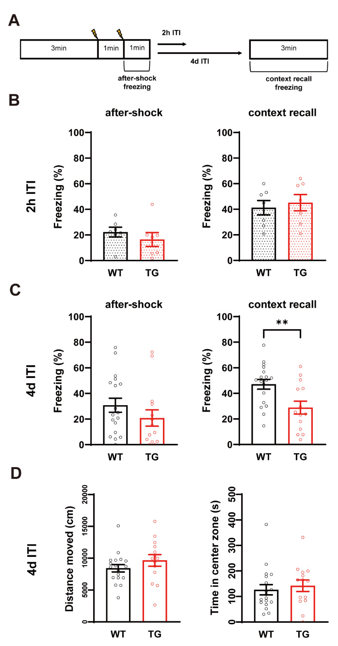
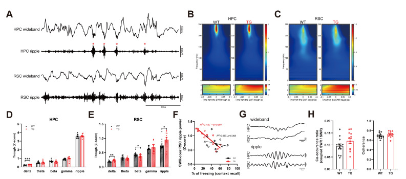
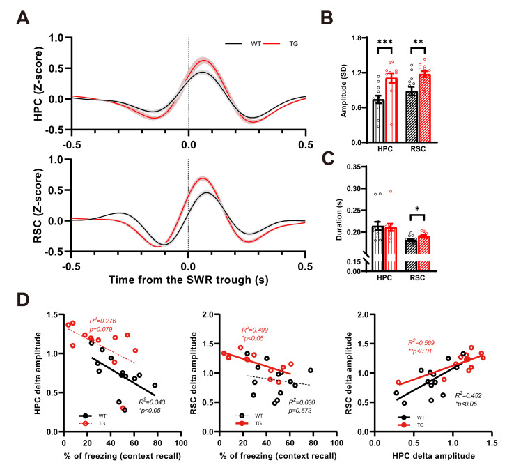
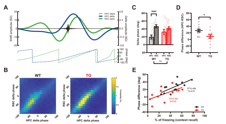
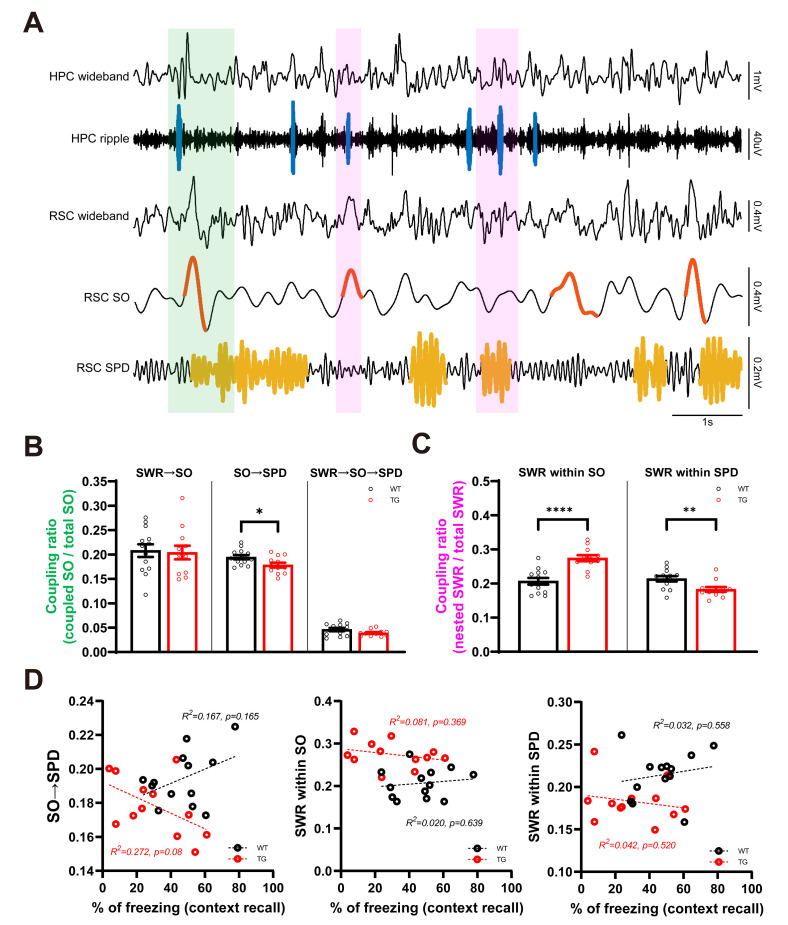
Alzheimer's disease (AD) is the most common cause of dementia, characterized by prominent episodic memory dysfunction. Recent studies have suggested that there is a sequential mechanism in the memory deficit, with long-term ones preceding short-term ones. However, there is lack of explanation for these symptoms. Interaction between the hippocampus and retrosplenial cortex (RSC) during slow-wave sleep (SWS) is a crucial step for successful long-term memory formation. In particular, sharp-wave ripple (SWR) is a principal hippocampus oscillation that coordinates with RSC activity. To determine the relationship between memory dysfunction and SWR-related oscillation changes in AD, we implanted local field potential electrodes in the hippocampus and RSC of AD model mice (APP/PS1). We found that the SWR-coupled ripple wave increased in the RSC, while the amplitude of the SWR was preserved. In addition, the corresponding delta power in hippocampus and RSC was elevated, together with altered delta synchrony in AD mice. All these findings showed a significant correlation with long-term memory deficits measured in contextual fear conditions. Our study suggests that altered SWR-coupled oscillations are a possible underlying mechanism of episodic memory dysfunction in AD mice.

Keywords: Alzheimer disease; Brain waves; Episodic memory; Slow-wave sleep.


Figures

Fig. 1
Fig. 1
Long-term memory deficit in 12-month-old AD mice. (A) Schematic representation of CFC protocol. There were distinct batches with different conditioning to context recall intertrial time interval (ITI, 2 h versus 4 days). (B) No difference in after-shock and context recall freezing between 2h ITI WT (n=7) and TG (n=7). (C) In 4d ITI experiment, TG (n=14) mice show significant decrease in the context recall freezing compared to WT (n=18) (**p<0.01, Student’s t-test). (D) No difference in total traveled distance and time spent in center zone during the open field test, between WT and TG of 4d ITI group.
Fig. 2
Fig. 2
Increased amplitude of SWR coordinated RSC ripple oscillation in AD mice. (A) Example traces (wideband: 1~250 Hz, ripple: 100~250 Hz) of HPC and RSC LFPs during SWS. Sharp wave ripples (SWRs) are marked on ripple filtered HPC trace (red asterisk). Note the RSC also has a prominent amplitude change in ripple band oscillation like as HPC. (B) Grand average spectrogram of HPC LFP around SWRs. Prominent ripple band power (top panels) and delta frequency power (bottom panels) changes are detected in both of WT (n=13) and TG (n=12) mice. (C) Grand average spectrogram of coordinated RSC LFP around HPC SWR events. Similar with HPC spectrogram, RSC LFPs also show high amplitude in ripple and delta oscillations around SWRs. Color bar in panel B & C indicates the z-scored amplitude. (D) Individually quantified peak band power in HPC channel (delta: 1~4 Hz, theta: 5~10 Hz, beta: 12~30 Hz, gamma: 31~100 Hz, ripple: 100~250 Hz). Transgenic mice show significantly increased delta power compared to WT (***p<0.001, Student’s t-test). (E) Individually quantified peak band power in RSC channel. Significantly increased in ripple and delta amplitude is detected in TG mice (*p<0.05 and **p<0.01 respectively, Student’s t-test), together with reduced beta oscillation power. (F) Sharp wave ripple (SWR) corresponding RSC ripple amplitude is correlated with context recall freezing in TG mice (WT: p=0.393, TG: ***p<0.001, Pearson’s correlation). (G) Example HPC LFP trace with cortical ripple in RSC (top two traces, wideband). Retrosplenial cortex (RSC) show large ripple oscillation respond to HPC SWR (bottom two traces, ripple filtered). (H) Co-occurrence rate of HPC SWR and RSC ripple (occurred within 50 ms from the SWR trough) is equivalent between WT and TG mice (left). Coherence between SWR and RSC ripple is not changed in TG mice (right).
Fig. 3
Fig. 3
Increased amplitude of SWR coordinated RSC delta oscillation in AD mice. (A) Grand average of raw delta trace around SWRs. Shaded area indicates SEM of averaged delta wave. Note that, both HPC (top panel) and RSC (bottom panel) show the negative and positive peaks before and after the SWR trough respectively. (B) Amplitude (negative peak to positive peak) of SWR related delta oscillation is higher in TG mice in both HPC and RSC (***p<0.001, Mann-Whitney test, **p<0.05, Student’s t-test). (C) Duration (from negative peak to positive peak) of delta wave is significantly increased at the RSC of TG mice **p<0.05, Student’s t-test). (D) HPC (left panel) show negative correlation with freezing level in context recall session, although TG only shows marginal p-value (WT: *p<0.05, TG: p=0.08, Pearson’s correlation). Only TG mice showed significant correlation between RSC delta power (middle panel) and recall freezing level (WT: p=0.573, TG: *p<0.05, Pearson’s correlation). Delta amplitude of HPC and RSC closely associated regardless of genotype (right panel, WT: *p<0.05, TG: **p<0.01, Pearson’s correlation).
Fig. 4
Fig. 4
Reduced difference of SWR coordinated delta phases in AD mice. (A) Example of SWR trace (black) along with delta oscillation from HPC (blue) and RSC (green). Corresponding delta phase change is represented in below. (B) Group averaged SWR nesting delta phase in scaled matrix. The phase location of each SWR is analyzed and divided by total SWR number of each mouse. (C) Median delta phase of HPC and RSC in individual. There is no group difference in regional phase, but the regional relationship is changed (WT HPC-RSC, ####p<0.0001, paired t-test). (D) Delta phase difference between HPC and RSC is decreased in TG mice (*p<0.05, Student’s t-test). (E) Phase difference is positively correlated with context recall freezing level (WT: *p<0.05, TG: **p<0.01, Pearson’s correlation).
Fig. 5
Fig. 5
Altered sleep oscillation coupling between HPC and RSC in AD mice. (A) Example traces of HPC and RSC during SWS. Sharp wave ripple is marked on HPC ripple band (100~250 Hz) filtered trace with blue line. Cortical slow oscillation (SO) and spindle (SPD) events are labeled with red and yellow line respectively on filtered RSC channel (RSC SO : RSC 0.5~4 Hz filtered trace, RSC SPD : RSC 7~15 Hz filtered trace). Note the serial event of SWR-SO-SPD (green box) and co-occurrence of SWR within SO and SPD (magenta box). (B) Sequential appearance of SWR-SO and SWR-SO-SPD is unchanged, but SO-SPD pair event is reduced in TG mice (*p<0.05, Student’s t-test). (C) Co-occurrence ratio of SWR-SO and SWR-SPD is altered in TG mice (**p<0.01, **p<0.0001, Student’s t-test). (D) No correlation between changed HPC-RSC sleep oscillation coupling properties and freezing level in context recall.

References

    1. Knopman DS, Amieva H, Petersen RC, Chételat G, Holtzman DM, Hyman BT, Nixon RA, Jones DT. Alzheimer disease. Nat Rev Dis Primers. 2021;7:33. doi: 10.1038/s41572-021-00269-y. - DOI - PMC - PubMed
    1. Ballard C, Gauthier S, Corbett A, Brayne C, Aarsland D, Jones E. Alzheimer's disease. Lancet. 2011;377:1019–1031. doi: 10.1016/S0140-6736(10)61349-9. - DOI - PubMed
    1. Weintraub S, Wicklund AH, Salmon DP. The neuropsychological profile of Alzheimer disease. Cold Spring Harb Perspect Med. 2012;2:a006171. doi: 10.1101/cshperspect.a006171. - DOI - PMC - PubMed
    1. Pause BM, Zlomuzica A, Kinugawa K, Mariani J, Pietrowsky R, Dere E. Perspectives on episodic-like and episodic memory. Front Behav Neurosci. 2013;7:33. doi: 10.3389/fnbeh.2013.00033. - DOI - PMC - PubMed
    1. Tromp D, Dufour A, Lithfous S, Pebayle T, Després O. Episodic memory in normal aging and Alzheimer disease: insights from imaging and behavioral studies. Ageing Res Rev. 2015;24(Pt B):232–262. doi: 10.1016/j.arr.2015.08.006. - DOI - PubMed