. 2021 Dec;11(4):120-132.
doi: 10.4236/ojbd.2021.114012. Epub 2021 Dec 2.

Primary Cardiac Lymphoma: Three Case Reports and a Review of the Literature

Affiliations

Primary Cardiac Lymphoma: Three Case Reports and a Review of the Literature

Clifford Michael Csizmar et al. Open J Blood Dis. 2021 Dec.

Abstract

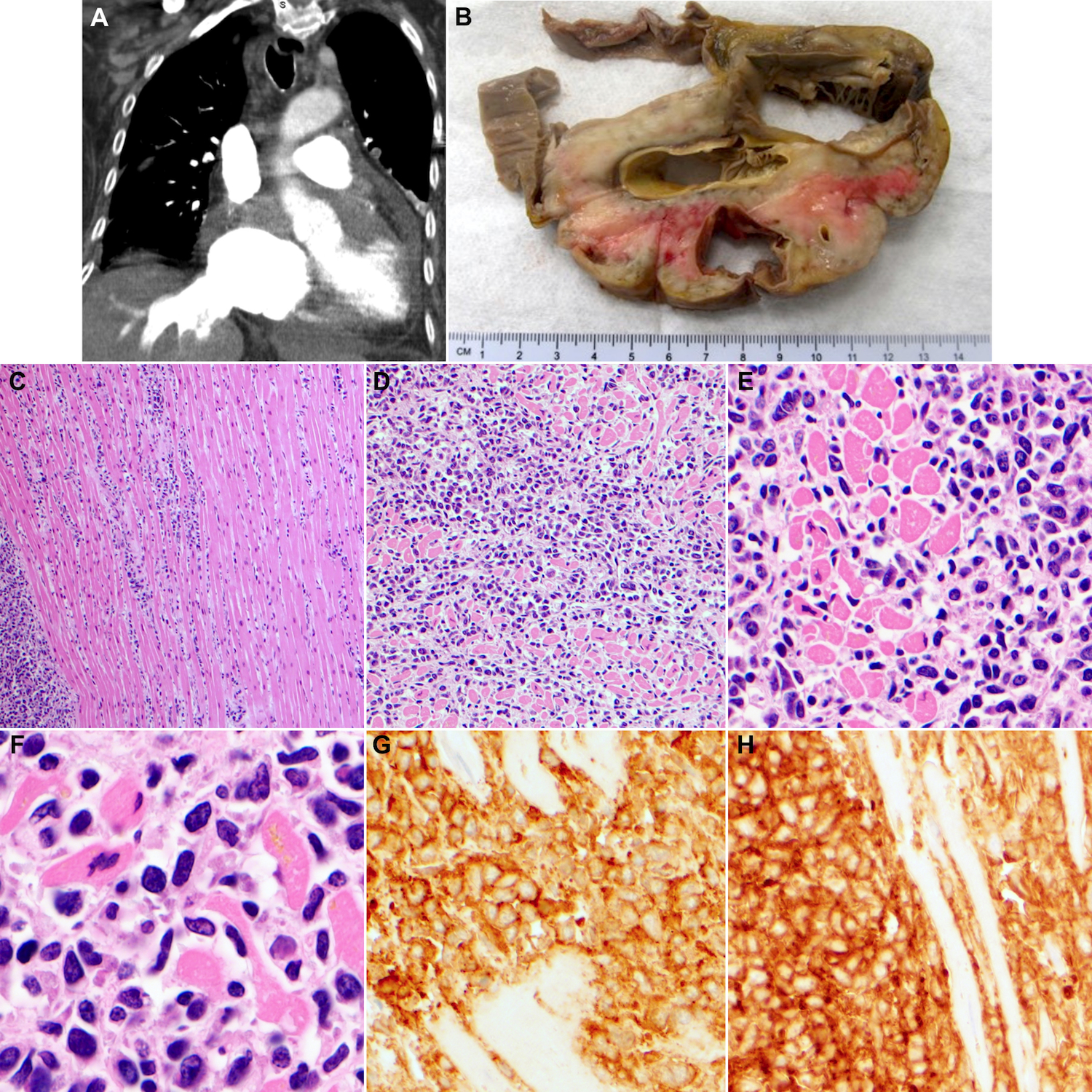
Primary cardiac lymphoma (PCL) is a rare entity that comprises only 1-2% of all cardiac tumors. Due to their scarcity and variable clinical presentation, early diagnosis is challenging. In this series, three cases of PCL from a single institution are described, which highlight the spectrum of presenting features and emphasize common principles. In the first case, a 73-year-old male who presented with dyspnea was found to have a 12.1 cm mass in the right ventricle. Biopsy via cardiac catheterization revealed diffuse large B cell lymphoma (DLBCL). He was treated with chemoimmunotherapy and survived for two months. The second case describes a 55-year-old female who presented with chest pain. Imaging revealed a 3.1 cm right atrial mass and bilateral pleural effusions, with cytology from the latter demonstrating DLBCL. She was lost to follow up after three cycles of chemoimmunotherapy. In the last case, an 80-year-old female presented with weakness. A 4.0 cm mass was discovered in the right atrium and the patient expired shortly after admission. Autopsy confirmed the diagnosis of DLBCL. These case summaries are followed by a review of the clinical presentation, diagnostic approach, and treatment outcomes of PCL.

Keywords: Primary cardiac lymphoma; cardiac tumors; diffuse large B cell lymphoma.


Conflict of interest statement

CONFLICTS OF INTEREST The authors have no conflicts of interest associated with this work.

Figures

Figure 1.
Figure 1.. Cardiac imaging and histology for case one.
Cardiac MRI (A) and chest CT (B) imaging depicting a large right ventricular mass. (C) Hematoxylin and eosin (H&E) staining of biopsied tissue from cardiac mass depicting diffuse infiltration of large, malignant, lymphoid cells; 500x magnification.
Figure 2.
Figure 2.. Cardiac imaging, histology, and flow cytometry for case two.
(A) Axial and (B) coronal sections of PET/CT imaging depicting a large hypermetabolic mass centered in the right atrium with involvement of the right ventricle. (C) H&E staining of biopsied tissue from cardiac mass depicting diffuse infiltration by large lymphoid cells with extensive necrosis. IHC staining of the mass demonstrating expression of (D) CD20, (D) CD45, and (F) CD99. Histologic images all depicted at 500x magnification. (G-I) Flow cytometry of pleural fluid. The three gated dotplots show all B cells that coexpress CD19 and CD20. The population of interest is gated in black, with non-neoplastic B cells in magenta and aqua. Large cells are gated based on size (high FSC) in panel G. They are kappa monotypic (panel H), and they express CD20 and dim CD5 (panel I).
Figure 3.
Figure 3.. Cardiac imaging, autopsy findings, and histology for case three.
(A) Coronal section of chest CT imaging depicting an enlarged cardiac silhouette with irregular thickening of the right atrium and ventricle. (B) Gross cross-section of the cardiac mass demonstrating invasion into the myometrium and encasement of the aorta and pulmonic trunk. H&E staining of biopsied tissue from cardiac mass depicting large lymphoid cells dissecting the myocardial fascicles at (C) 100x, (D) 200x, (E) 500x, and (F) 1,000x magnification. IHC staining of the mass demonstrating expression of (G) CD20 and (H) CD45, both at 500x magnification.

References

    1. Miller DV, Mookadam F, Mookadam M, Edwards WD and Macon WR (2007) Primary cardiac plasmablastic (diffuse large B-cell) lymphoma mimicking left ventricular aneurysm with mural thrombus. Cardiovascular Pathology, 16, 111–114. 10.1016/j.carpath.2006.09.003 - DOI - PubMed
    1. Aimanan K, Kumar KS, Mohd Arif MN, Noor Zuraini M, Ramdzan MJ and Hamdan L (2016) Rare Intracardiac Tumor: Primary Cardiac Lymphoma Presenting as Atypical Angina. Case Reports in Medicine, 2016, 4125295. 10.1155/2016/4125295 - DOI - PMC - PubMed
    1. Gordon MJ, Danilova O, Spurgeon S and Danilov AV (2016) Cardiac non-Hodgkin’s lymphoma: clinical characteristics and trends in survival. European Journal of Haematology, 97, 445–452. 10.1111/ejh.12751 - DOI - PubMed
    1. Carras S, Berger F, Chalabreysse L, Callet-Bauchut E, Cordier JF, Salles G, et al. (2017) Primary cardiac lymphoma: diagnosis, treatment and outcome in a modern series. Hematological Oncology, 35, 510–519. 10.1002/hon.2301 - DOI - PubMed
    1. Patel J, Melly L and Sheppard MN (2010) Primary cardiac lymphoma: B- and T-cell cases at a specialist UK centre. Annals of Oncology, 21, 1041–1045. 10.1093/annonc/mdp424 - DOI - PubMed

LinkOut - more resources