

Metabolic correlates of olfactory dysfunction in COVID-19 and Parkinson's disease (PD) do not overlap
- PMID: 34984501
- PMCID: PMC8727173
- DOI: 10.1007/s00259-021-05666-9
Metabolic correlates of olfactory dysfunction in COVID-19 and Parkinson's disease (PD) do not overlap
Abstract
Purpose: Hyposmia is a common feature of COVID-19 and Parkinson's disease (PD). As parkinsonism has been reported after COVID-19, a link has been hypothesized between SARS-CoV2 infection and PD. We aimed to evaluate brain metabolic correlates of isolated persistent hyposmia after mild-to-moderate COVID-19 and to compare them with metabolic signature of hyposmia in drug-naïve PD patients.
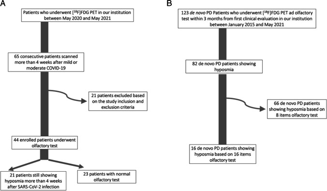
Methods: Forty-four patients who experienced hyposmia after SARS-COV2 infection underwent brain [18F]-FDG PET in the first 6 months after recovery. Olfaction was assessed by means of the 16-item "Sniffin' Sticks" test and patients were classified as with or without persistent hyposmia (COVID-hyposmia and COVID-no-hyposmia respectively). Brain [18F]-FDG PET of post-COVID subgroups were compared in SPM12. COVID-hyposmia patients were also compared with eighty-two drug-naïve PD patients with hyposmia. Multiple regression analysis was used to identify correlations between olfactory test scores and brain metabolism in patients' subgroups.
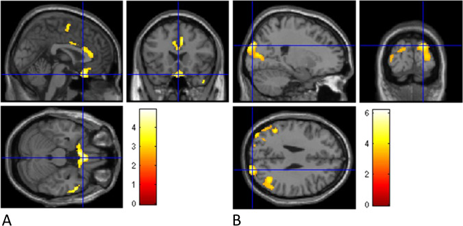
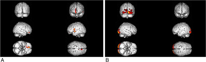
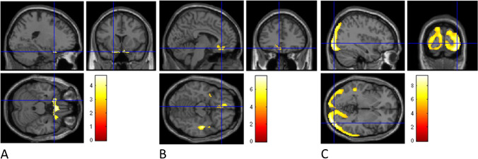
Results: COVID-hyposmia patients (n = 21) exhibited significant hypometabolism in the bilateral gyrus rectus and orbitofrontal cortex with respect to COVID-non-hyposmia (n = 23) (p < 0.002) and in middle and superior temporal gyri, medial/middle frontal gyri, and right insula with respect to PD-hyposmia (p < 0.012). With respect to COVID-hyposmia, PD-hyposmia patients showed hypometabolism in inferior/middle occipital gyri and cuneus bilaterally. Olfactory test scores were directly correlated with metabolism in bilateral rectus and medial frontal gyri and in the right middle temporal and anterior cingulate gyri in COVID-hyposmia patients (p < 0.006) and with bilateral cuneus/precuneus and left lateral occipital cortex in PD-hyposmia patients (p < 0.004).
Conclusion: Metabolic signature of persistent hyposmia after COVID-19 encompasses cortical regions involved in olfactory perception and does not overlap metabolic correlates of hyposmia in PD.
Keywords: Anosmia; Brain PET; COVID-19; Parkinson’s disease; [18F]FDG.
© 2022. The Author(s), under exclusive licence to Springer-Verlag GmbH Germany, part of Springer Nature.
Conflict of interest statement
Silvia Morbelli received speaking honoraria from GE Healthcare and AAA. Flavio Nobili received fees from BIAL for consultation, from GE Healthcare for teaching talks, and from Roche for board participation. Dario Arnaldi received fees from Fidia for lectures and board participation. Matteo Pardini receives research support from Novartis and Nutricia and fees from Novartis, Merck, and Biogen. All other authors declare no competing interests.
Figures
References
-
- Lechien JR, Chiesa-Estomba CM, De Siati DR, Horoi M, Le Bon SD, Rodriguez A, et al. Olfactory and gustatory dysfunctions as a clinical presentation of mild-to-moderate forms of the coronavirus disease (COVID-19): a multicenter European study. Eur Arch Otorhinolaryngol. 2020;277:2251–2261. doi: 10.1007/s00405-020-05965-1. - DOI - PMC - PubMed
MeSH terms
Substances
LinkOut - more resources
Full Text Sources
Medical
Research Materials
Miscellaneous
