. 2022 Jan 4;79(1):23.
doi: 10.1007/s00018-021-04099-9.

Transient nuclear lamin A/C accretion aids in recovery from vapor nanobubble-induced permeabilisation of the plasma membrane

Affiliations

Transient nuclear lamin A/C accretion aids in recovery from vapor nanobubble-induced permeabilisation of the plasma membrane

Gaëlle Houthaeve et al. Cell Mol Life Sci. .

Abstract

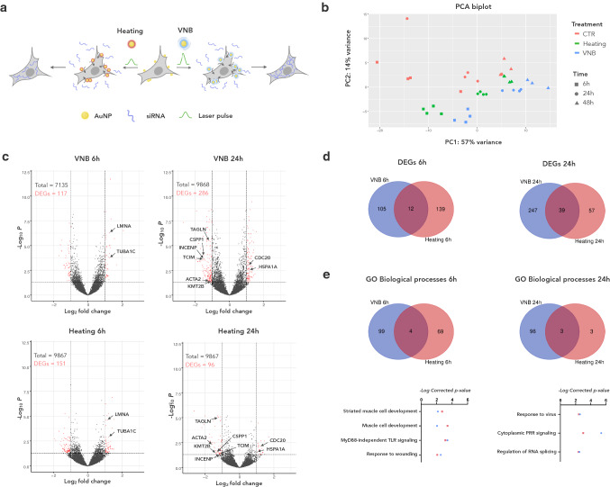
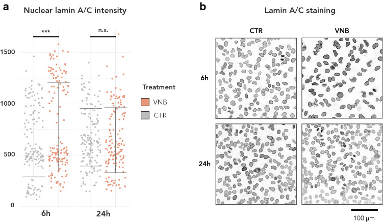
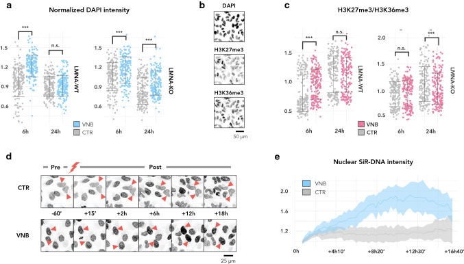
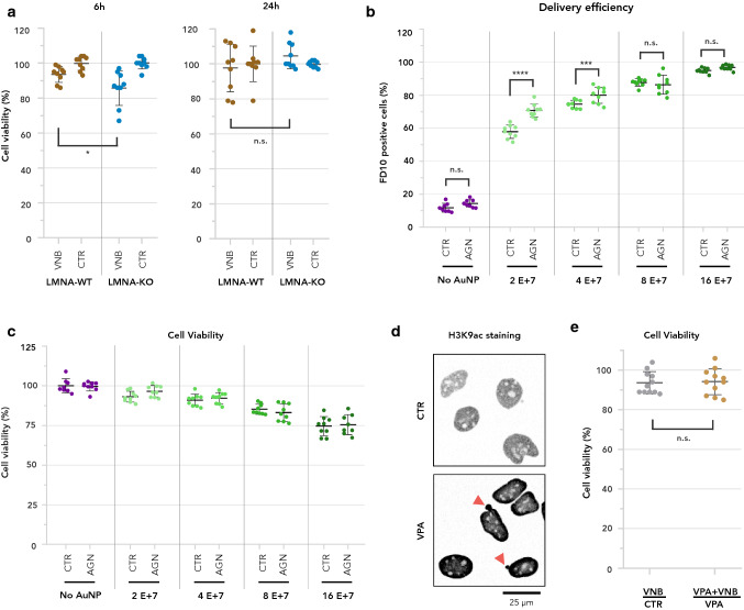
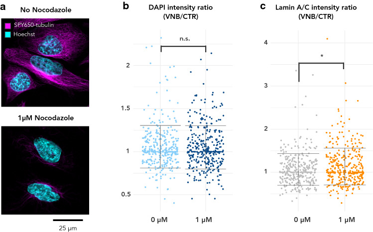
Vapor nanobubble (VNB) photoporation is a physical method for intracellular delivery that has gained significant interest in the past decade. It has successfully been used to introduce molecular cargo of diverse nature into different cell types with high throughput and minimal cytotoxicity. For translational purposes, it is important to understand whether and how photoporation affects cell homeostasis. To obtain a comprehensive view on the transcriptional rewiring that takes place after VNB photoporation, we performed a longitudinal shotgun RNA-sequencing experiment. Six hours after photoporation, we found a marked upregulation of LMNA transcripts as well as their protein products, the A-type lamins. At the same time point, we observed a significant increase in several heterochromatin marks, suggesting a global stiffening of the nucleus. These molecular features vanished 24 h after photoporation. Since VNB-induced chromatin condensation was prolonged in LMNA knockout cells, A-type lamins may be required for restoring the nucleus to its original state. Selective depletion of A-type lamins reduced cell viability after VNB photoporation, while pharmacological stimulation of LMNA transcription increased the percentage of successfully transfected cells that survived after photoporation. Therefore, our results suggest that cells respond to VNB photoporation by temporary upregulation of A-type lamins to facilitate their recovery.

Keywords: A-type lamins; Chromatin; Gold nanoparticles; Photoporation; Plasma membrane; Vapor nanobubbles.


Conflict of interest statement

The authors declare that they have no conflict of interest.

Figures

Fig. 1
Fig. 1
The transcriptional response to VNB photoporation and photothermal heating is limited and is both time- and modality-dependent. a Schematic illustration of the experimental procedure in which HeLa cells were either subjected to VNB or heating using non-targeting siRNA as cargo; b Principal component analysis (PCA) of the normalised gene counts (expressed as fragments per kilobase million, FPKM) for VNB, heating and CTR conditions at three time points; c Volcano plots show the fold change and the adjusted p value of individual differentially expressed genes (DEGs) 6 h (left) and 24 h (right) after VNB (top) and after heating (down). DEGs with greater than twofold change and adjusted p value < 0.05 are color-coded in red; d Venn diagrams illustrating the number of unique and shared DEGs when comparing VNB and heating 6 h pp (left) and 24 h pp (right). e Venn diagram showing the number of unique and shared gene ontology (GO) biological processes when comparing VNB and heating at 6 h pp (left) and 24 h pp (right). Underneath the Venn diagrams, the significance of enrichment in the list of DEGs is plotted for shared GO biological processes for VNB (blue) and heating (red)
Fig. 2
Fig. 2
Quantitative immunofluorescence reveals nuclear accumulation of A-type lamins after VNB photoporation. a Lamin A/C intensity in nuclei ROIs as determined by quantitative immunofluorescence (IF) in VNB versus CTR cells for 6 h and 24 h pp (***P < 0.001); b Representative images of IF staining for lamin A/C. The grayscale range has been inverted for clarity (darker signal equals stronger intensity)
Fig. 3
Fig. 3
VNB photoporation causes a transient chromatin compaction, which is prolonged in LMNA-KO cells. a Normalized nuclear DAPI intensity plotted per timepoint for LMNA-WT (left) and LMNA-KO cells (right) (***P < 0.001); b Representative example of a staining with DAPI, anti-H3K27me3, and anti-H3K36me3 on CTR cells at 6 h. c Ratio of heterochromatin (H3K27me3) to euchromatin (H3K36me3) plotted per timepoint for LMNA-WT (left) and LMNA-KO (right) (***P < 0.001); d Montage of SiR-DNA stained LMNA-WT cells before photoporation treatment (− 60’) and at several timepoints after photoporation treatment (+ 15’, + 2 h, + 6 h, + 12 h and + 18 h). Control cells were irradiated in the absence of AuNPs; e Line graphs of the mean SiR-DNA signal (± standard error) in VNB-treated cells (VNB) and control cells that were irradiated in the absence of AuNPs (CTR). Per treatment, SiR-DNA signal was normalised to the mean intensity of the first timepoint (+ 15’) post laser irradiation (n = 4 cells per treatment)
Fig. 4
Fig. 4
Upregulation of A-type lamins allows cells to cope with VNB photoporation. a Percentage of viability compared to untreated cells is plotted either 6 h or 24 h after VNB, for LMNA-WT cells and for LMNA-KO cells. A two-tailed unpaired student’s T test was performed to determine statistical differences (*P < 0.05); b Percentage of cells positive for FD10 2 h after VNB as measured by quantitative flow cytometry for photoporated cells that were pre-incubated with AGN (AGN) or were untreated (CTR). FD10 delivery efficiency was determined for different concentrations of AuNP (0 = No AuNP, 2E + 7, 4E + 7, 8E + 7, 16E + 7). Sidak’s multiple comparisons test was performed to determine statistical differences (****P < 0.0001; ***P = 0.0002); c Percentage of viability 2 h after VNB compared to untreated cells is plotted for cells that were incubated with AGN (AGN) and cells that did not receive AGN (CTR). Sidak’s multiple comparisons test was performed to determine statistical differences; d Representative images of anti-H3K9ac immunofluorescence staining in control cells (CTR) and cells treated with VPA (VPA). Grayscale images were inverted for clarity; e Ratio of viability (%) for cells treated with VNB versus untreated control cells (VNB/CTR) and for cells treated with VNB in the presence of VPA versus cells treated with VPA only (VNB + VPA/VPA). A Mann–Whitney U test was performed to determine statistical differences (n.s.: P > 0.05)
Fig. 5
Fig. 5
Microtubule polymerisation is not required for chromatin compaction and tempers lamin A/C accrual after VNB photoporation. a Staining of microtubules with the vital stain SPY650-tubulin of untreated control cells and cells treated with 1 mM Nocodazole for 2 h; b Normalized DAPI intensity in VNB-treated vs. CTR cells treated with 0 µM (DMSO) or 1 µM Nocodazole. c Normalized nuclear lamin A/C intensity in VNB-treated vs. CTR cells treated with 0 µM (DMSO) or 1 µM Nocodazole

Similar articles

Cited by

References

    1. Morshedi Rad D, et al. A comprehensive review on intracellular delivery. Adv Mater. 2021;33(13):e2005363. - PubMed
    1. Stewart MP, Langer R, Jensen KF. Intracellular delivery by membrane disruption: mechanisms, strategies, and concepts. Chem Rev. 2018;118(16):7409–7531. - PMC - PubMed
    1. Howarth JL, Lee YB, Uney JB. Using viral vectors as gene transfer tools (Cell Biology and Toxicology Special ssue: ETCS-UK 1 day meeting on genetic manipulation of cells) Cell Biol Toxicol. 2010;26(1):1–20. - PMC - PubMed
    1. Thomas CE, Ehrhardt A, Kay MA. Progress and problems with the use of viral vectors for gene therapy. Nat Rev Genet. 2003;4(5):346–358. - PubMed
    1. Meacham JM, et al. Physical methods for intracellular delivery: practical aspects from laboratory use to industrial-scale processing. J Lab Autom. 2014;19(1):1–18. - PMC - PubMed

LinkOut - more resources