

Production and application of mouse monoclonal antibodies targeting linear epitopes in pB602L of African swine fever virus
- PMID: 34984562
- PMCID: PMC8843907
- DOI: 10.1007/s00705-021-05335-0
Production and application of mouse monoclonal antibodies targeting linear epitopes in pB602L of African swine fever virus
Abstract
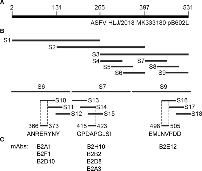
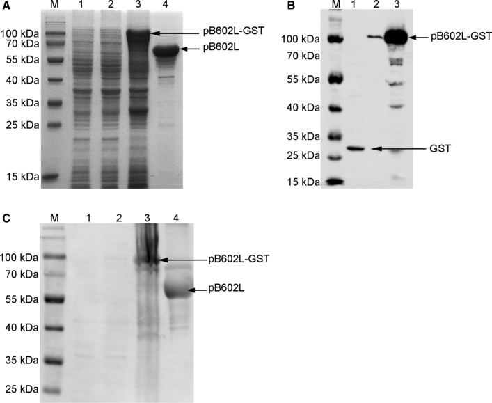
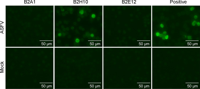
African swine fever (ASF) is an acute hemorrhagic disease of domestic pigs. The causative agent of ASF, ASF virus (ASFV), is a double-stranded DNA virus, the sole member in the family Asfarviridae. The non-structural protein pB602L of ASFV is a molecular chaperone of the major capsid protein p72 and plays a key role in icosahedral capsid assembly. This protein is antigenic and is a target for developing diagnostic tools for ASF. To generate monoclonal antibodies (mAbs) against pB602L, a prokaryotically expressed recombinant pB602L protein was produced, purified, and used as an antigen to immunize mice. A total of eight mouse mAbs were obtained, and their binding epitopes were screened by Western blot using an overlapping set of polypeptides from pB602L. Three linear epitopes were identified and designated epitope 1 (366ANRERYNY373), epitope 2 (415GPDAPGLSI423), and epitope 3 (498EMLNVPDD505). Based on the epitope recognized, the eight mAbs were placed into three groups: group 1 (B2A1, B2F1, and B2D10), group 2 (B2H10, B2B2, B2D8, and B2A3), and group 3 (B2E12). The mAbs B2A1, B2H10, and B2E12, each representing one of the groups, were used to detect pB602L in ASFV-infected porcine alveolar macrophages (PAMs) and pig tissues, using an indirect fluorescence assay (IFA) and immunohistochemical staining, respectively. The results showed that pB602L was detectable with all three mAbs in immunohistochemical staining, but only B2H10 was suitable for detecting the proteins in ASFV-infected PAMs by IFA. In summary, we developed eight anti-pB602L mouse mAbs recognizing three linear epitopes in the protein, which can be used as reagents for basic and applied research on ASFV.
© 2021. The Author(s).
Conflict of interest statement
The authors declared that they have no conflict of interest.
Figures
Similar articles
-
A novel conserved B-cell epitope in pB602L of African swine fever virus.Appl Microbiol Biotechnol. 2024 Dec;108(1):78. doi: 10.1007/s00253-023-12921-6. Epub 2024 Jan 9. Appl Microbiol Biotechnol. 2024. PMID: 38194141 Free PMC article.
-
Linear epitopes in African swine fever virus p72 recognized by monoclonal antibodies prepared against baculovirus-expressed antigen.J Vet Diagn Invest. 2018 May;30(3):406-412. doi: 10.1177/1040638717753966. Epub 2018 Jan 12. J Vet Diagn Invest. 2018. PMID: 29327672 Free PMC article.
-
Epitope mapping and establishment of a blocking ELISA for mAb targeting the p72 protein of African swine fever virus.Appl Microbiol Biotechnol. 2024 May 29;108(1):350. doi: 10.1007/s00253-024-13146-x. Appl Microbiol Biotechnol. 2024. PMID: 38809284 Free PMC article.
-
Structure of African Swine Fever Virus and Associated Molecular Mechanisms Underlying Infection and Immunosuppression: A Review.Front Immunol. 2021 Sep 6;12:715582. doi: 10.3389/fimmu.2021.715582. eCollection 2021. Front Immunol. 2021. PMID: 34552586 Free PMC article. Review.
-
African Swine Fever Virus: A Review.Viruses. 2017 May 10;9(5):103. doi: 10.3390/v9050103. Viruses. 2017. PMID: 28489063 Free PMC article. Review.
Cited by
-
Detection of African swine fever virus antibodies in serum using a pB602L protein-based indirect ELISA.Front Vet Sci. 2022 Sep 23;9:971841. doi: 10.3389/fvets.2022.971841. eCollection 2022. Front Vet Sci. 2022. PMID: 36213400 Free PMC article.
-
Establishment of a Dual-Antigen Indirect ELISA Based on p30 and pB602L to Detect Antibodies against African Swine Fever Virus.Viruses. 2023 Aug 30;15(9):1845. doi: 10.3390/v15091845. Viruses. 2023. PMID: 37766252 Free PMC article.
-
Screening and identification of linear B-cell epitopes on structural proteins of African Swine Fever Virus.Virus Res. 2024 Dec;350:199465. doi: 10.1016/j.virusres.2024.199465. Epub 2024 Sep 25. Virus Res. 2024. PMID: 39306245 Free PMC article.
-
B602L-Fc fusion protein enhances the immunogenicity of the B602L protein of the African swine fever virus.Front Immunol. 2023 Jun 22;14:1186299. doi: 10.3389/fimmu.2023.1186299. eCollection 2023. Front Immunol. 2023. PMID: 37426672 Free PMC article.
-
Lactobacillus plantarum Surface-Displayed ASFV (p14.5) Can Stimulate Immune Responses in Mice.Vaccines (Basel). 2022 Feb 24;10(3):355. doi: 10.3390/vaccines10030355. Vaccines (Basel). 2022. PMID: 35334986 Free PMC article.
References
-
- Chen W, Zhao D, He X, Liu R, Wang Z, Zhang X, Li F, Shan D, Chen H, Zhang J, Wang L, Wen Z, Wang X, Guan Y, Liu J, Bu Z. A seven-gene-deleted African swine fever virus is safe and effective as a live attenuated vaccine in pigs. Sci China Life Sci. 2020;63(5):623–634. doi: 10.1007/s11427-020-1657-9. - DOI - PMC - PubMed
MeSH terms
Substances
Grants and funding
- 2016YFD0500705/national basic research program of china (973 program)
- 2017YFD0500105/national basic research program of china (973 program)
- 2017YFC1200502/national basic research program of china (973 program)
- Y2017LM08/central public-interest scientific institution basal research fund, chinese academy of fishery sciences
LinkOut - more resources
Full Text Sources
Molecular Biology Databases
