. 2022 Jan;19(1):274-288.
doi: 10.1007/s13311-021-01159-7. Epub 2022 Jan 4.

Dendrimer-2PMPA Delays Muscle Function Loss and Denervation in a Murine Model of Amyotrophic Lateral Sclerosis

Affiliations

Dendrimer-2PMPA Delays Muscle Function Loss and Denervation in a Murine Model of Amyotrophic Lateral Sclerosis

Carolyn Tallon et al. Neurotherapeutics. 2022 Jan.

Abstract

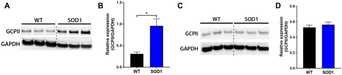
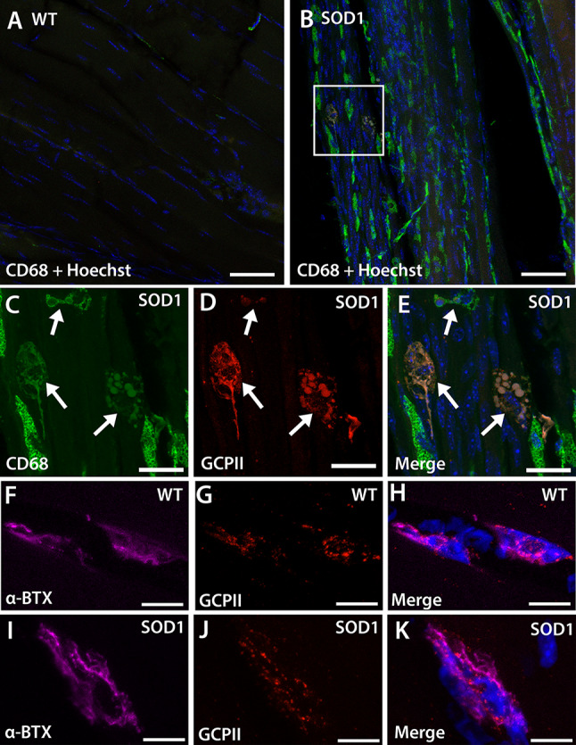
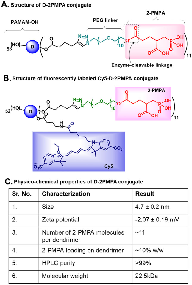
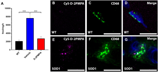
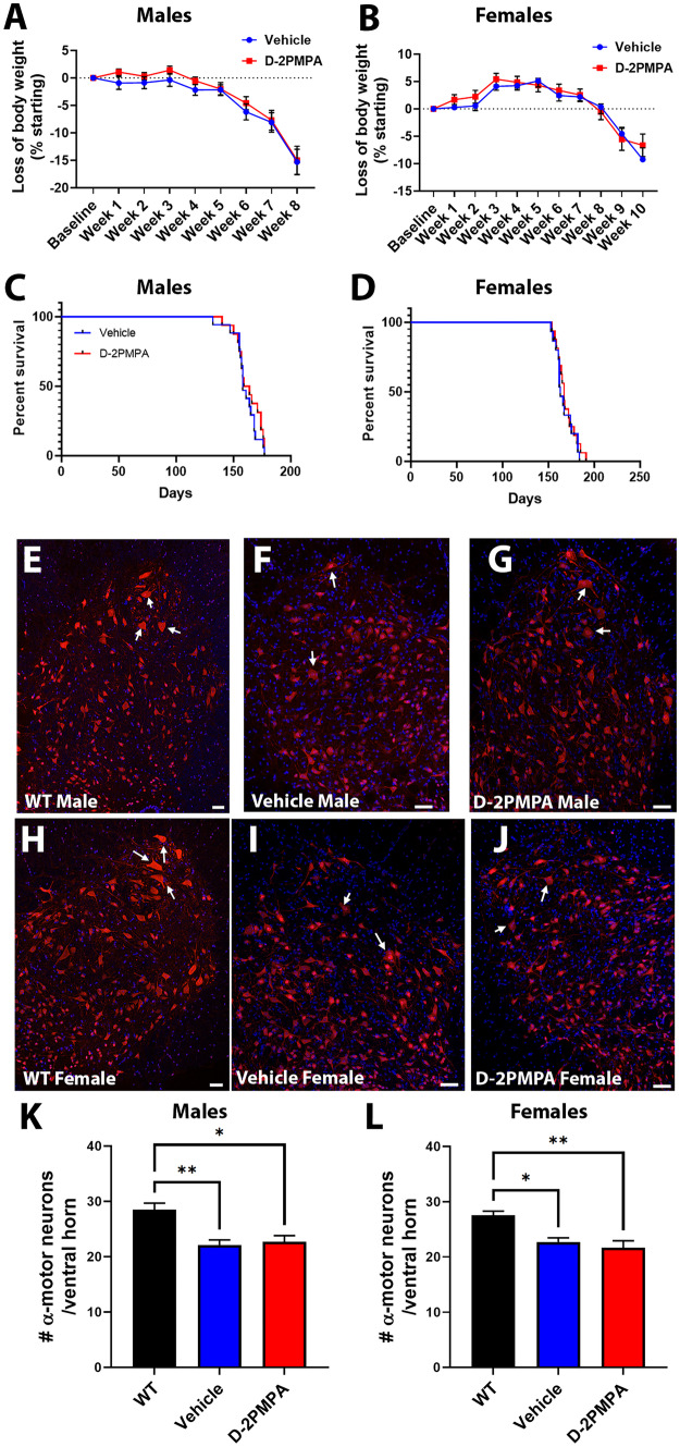
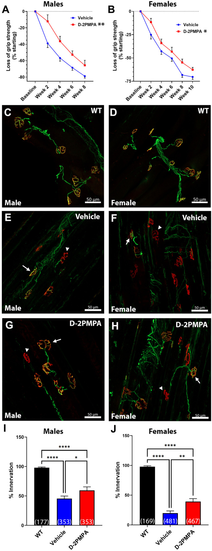
Amyotrophic lateral sclerosis (ALS) is a devastating neurodegenerative disease where muscle weakness and neuromuscular junction (NMJ) denervation precede motor neuron cell death. Although acetylcholine is the canonical neurotransmitter at the mammalian NMJ synapse, glutamate has recently been identified as a critical neurotransmitter for NMJ development and maintenance. One source of glutamate is through the catabolism of N-acetyl-aspartyl-glutamate (NAAG), which is found in mM concentrations in mammalian motoneurons, where it is released upon stimulation and hydrolyzed to glutamate by the glial enzyme glutamate carboxypeptidase II (GCPII). Using the SOD1G93A model of ALS, we found an almost fourfold elevation of GCPII enzymatic activity in SOD1G93A versus WT muscle and a robust increase in GCPII expression which was specifically associated with activated macrophages infiltrating the muscle. 2-(Phosphonomethyl)pentanedioic acid (2PMPA) is a potent GCPII inhibitor which robustly blocks glutamate release from NAAG but is highly polar with limited tissue penetration. To improve this, we covalently attached 2PMPA to a hydroxyl polyamidoamine (PAMAM-G4-OH) dendrimer delivery system (D-2PMPA) which is known to target activated macrophages in affected tissues. Systemic D-2PMPA therapy (20 mg/kg 2PMPA equivalent; IP 2 × /week) was found to localize in muscle macrophages in SOD1G93A mice and completely normalize the enhanced GCPII activity. Although no changes in body weight or survival were observed, D-2PMPA significantly improved grip strength and inhibited the loss of NMJ innervation in the gastrocnemius muscles. Our finding that inhibiting elevated GCPII activity in SOD1G93A muscle can prolong muscle function and delay NMJ denervation may have early therapeutic implications for ALS patients.

Keywords: Amyotrophic lateral sclerosis; Dendrimer; Glutamate; Glutamate carboxypeptidase II (GCPII); N-acetyl-aspartyl-glutamate (NAAG); NMDA; Neuroinflammation; Neuromuscular junction (NJM); mGluR3.


Figures

Fig. 1
Fig. 1
GCPII expression is elevated in SOD1G93A muscle. A Western blot from WT and SOD1G93A gastrocnemius muscles. GAPDH shown as a loading control. B Quantification of the blot shown in A. n = 3/group. C Western blot from the lumbar region of the spinal cord from WT and SOD1G93A mice. GAPDH is shown as a loading control. D Quantification of the blot from C. n = 3/group. Bars represent mean ± SEM. Black bars represent WT values. Blue bars represent SOD1G93A values. *p < 0.05
Fig. 2
Fig. 2
GCPII expression is localized to macrophages in SOD1G93A gastrocnemius muscle. A, B Representative images demonstrating that WT muscle (A) has limited CD68 + macrophage staining while SOD1G93A muscle (B) has a large influx of CD68 + macrophages. Scale bar, 50 µm. CE High magnification of the boxed area from SOD1G93A muscle showing CD68 + macrophages (C, arrows) with GCPII expression (D, arrows). Merged image with Hoechst-stained nuclei (blue) shown in E. Scale bar, 20 µm. F, H WT NMJs stained with α-bungarotoxin (F and H, α-BTX, purple) have GCPII staining colocalization (G and H, red). IK SOD1G93A NMJs stained with α-bungarotoxin (I and K, α-BTX, purple) also have GCPII staining colocalization (J and K, red). Nuclei stained blue. Scale bar, 10 µm
Fig. 3
Fig. 3
The structures and physicochemical characterization of dendrimer-2PMPA conjugates. A The structure of D-2PMPA conjugate having 2PMPA covalently attached to the dendrimer surface through PEG linkers via enzyme-sensitive ester linkages. B The structure of fluorescently labeled Cy5-D-2PMPA conjugate with the attachment of both the drug and the fluorophore for imaging purposes. C Table showing the physicochemical characteristics of D-2PMPA
Fig. 4
Fig. 4
Cy5-labeled D-2PMPA is targeted to macrophages in SOD1G93A gastrocnemius muscle. A GCPII activity in gastrocnemius muscles excised from WT and SOD1G93A mice treated with vehicle or D-2PMPA for 2 weeks. Bars represent mean ± SEM. Black bars represent WT values. Blue bars represent SOD1G93A vehicle–treated mouse values. Red bars represent SOD1G93A D-2PMPA–treated mouse values. ***p < 0.001. n = 5/group. B, D Representative images from gastrocnemius muscles in WT mice showing no Cy5-D-2PMPA staining (B) localizing with CD68 + macrophages (C). Merged image shown in D with nuclei stained blue. E, G Representative images from gastrocnemius muscles in SOD1G93A mice showing Cy5-D-2PMPA staining (B) localizing with CD68 + macrophages (C). Merged image shown in D with nuclei stained blue. Scale bar, 10 µm
Fig. 5
Fig. 5
D-2PMPA treatment does not improve disease outcome in SOD1G93A mice. A, B Weekly body weight is not changed in male mice (A) or female mice (B) following treatment. Points represent mean ± SEM. C, D Overall survival is not altered in male mice (C) or female mice (D) following treatment. Blue points represent SOD1G93A vehicle–treated mouse values. Red points represent SOD1G93A D-2PMPA–treated mouse values. E, J Representative images of lumbar spinal cord ventral horns. Male WT mice (E) have more dense neuronal staining than vehicle (F) or D-2PMPA mice (G). Female WT mice (H) also have more dense neuronal staining than vehicle (I) or D-2PMPA mice (J). Neurons labeled red with NeuN and nuclei labeled blue with Hoechst. Arrows pointing to examples of α motor neurons. Scale bar, 100 µm. K, L Quantification of the number of α motor neurons (cell body area > 250 µm2) per ventral horn from males (K) and females (L). Bars represent mean ± SEM. Black bars represent WT values. Blue bars represent SOD1G93A vehicle–treated mouse values. Red bars represent SOD1G93A D-2PMPA–treated mouse values. AD Male vehicle n = 20, D-2PMPA n = 21; female vehicle n = 18, D-2PMPA n = 19. I Male WT n = 4; female WT n = 4; male vehicle n = 5; male D-2PMPA n = 6; female vehicle n = 4; female D-2PMPA n = 4. *p < 0.05. **p < 0.01
Fig. 6
Fig. 6
D-2PMPA treatment selectively improves muscle function and innervation in SOD1G93A mice. A, B Grip strength data from male (A) and female (B) mice. D-2PMPA–treated mice have significantly higher grip strength over 8 weeks or 10 weeks, respectively. Vehicle male n = 20, female n = 18; D-2PMPA male n = 21, female n = 19. Points represent mean ± SEM. Blue points represent SOD1G93A vehicle–treated mouse values. Red points represent SOD1G93A D-2PMPA–treated mouse values. C, H Representative images of WT (male C; female D), vehicle (male E; female F), and D-2PMPA (male G; female H)–treated SOD1G93A gastrocnemius muscle after 4 weeks (male) or 6 weeks (female). Axons stained green. NMJs stained red. Scale bar, 50 µm. Arrows indicate innervated NMJ. Arrowheads indicate denervated NMJs. I, J Quantification of the percentage of innervated NMJs from male (I) and female (J) mice. Bars represent mean ± SEM. Black bars represent WT values. Blue bars represent SOD1G93A vehicle–treated mouse values. Red bars represent SOD1G93A D-2PMPA–treated mouse values. Numbers in parenthesis indicate the total number of NMJs. N = 6/group. *p < 0.05. **p < 0.01. ****p < 0.0001

References

    1. Cleveland DW, Rothstein JD. From charcot to lou gehrig: deciphering selective motor neuron death in als. Nature Reviews Neuroscience. 2001;2(11):806–819. doi: 10.1038/35097565. - DOI - PubMed
    1. Oskarsson B, Gendron TF, Staff NP. Amyotrophic Lateral Sclerosis: An Update for 2018. Mayo Clin Proc. 2018;93(11):1617–1628. doi: 10.1016/j.mayocp.2018.04.007. - DOI - PubMed
    1. Azzouz M, Leclerc N, Gurney M, Warter J-M, Poindron P, Borg J. Progressive motor neuron impairment in an animal model of familial amyotrophic lateral sclerosis. Muscle & Nerve. 1997;20(1):45–51. doi: 10.1002/(SICI)1097-4598(199701)20:1<45::AID-MUS6>3.0.CO;2-H. - DOI - PubMed
    1. Fischer LR, Culver DG, Tennant P, Davis AA, Wang M, Castellano-Sanchez A, et al. Amyotrophic lateral sclerosis is a distal axonopathy: evidence in mice and man. Experimental Neurology. 2004;185(2):232–240. doi: 10.1016/j.expneurol.2003.10.004. - DOI - PubMed
    1. Hegedus J, Putman CT, Gordon T. Time course of preferential motor unit loss in the SOD1G93A mouse model of amyotrophic lateral sclerosis. Neurobiology of Disease. 2007;28(2):154–164. doi: 10.1016/j.nbd.2007.07.003. - DOI - PubMed

Publication types