. 2022 Apr;119(4):1129-1141.
doi: 10.1002/bit.28030. Epub 2022 Jan 25.

On-chip magnetophoretic capture in a model of malaria-infected red blood cells

Affiliations

On-chip magnetophoretic capture in a model of malaria-infected red blood cells

Marco Giacometti et al. Biotechnol Bioeng. 2022 Apr.

Abstract

The search for new rapid diagnostic tests for malaria is a priority for developing an efficient strategy to fight this endemic disease, which affects more than 3 billion people worldwide. In this study, we characterize systematically an easy-to-operate lab-on-chip, designed for the magnetophoretic capture of malaria-infected red blood cells (RBCs). The method relies on the positive magnetic susceptibility of infected RBCs with respect to blood plasma. A matrix of nickel posts fabricated in a silicon chip placed face down is aimed at attracting infected cells, while healthy cells sediment on a glass slide under the action of gravity. Using a model of infected RBCs, that is, erythrocytes with methemoglobin, we obtained a capture efficiency of about 70% after 10 min in static conditions. By proper agitation, the capture efficiency reached 85% after just 5 min. Sample preparation requires only a 1:10 volume dilution of whole blood, previously treated with heparin, in a phosphate-buffered solution. Nonspecific attraction of untreated RBCs was not observed in the same time interval.

Keywords: lab-on-a-chip; magnetorphoretic separation; malaria.


Figures

Figure 1
Figure 1
Concept of the method for the magnetophoretic separation of malaria‐infected red blood cells. Under the action of the magnetic field gradient produced by Ni microposts on a microchip in the external field H e, infected RBC are captured by the Ni posts, while healthy RBCs sediment on a glass slide
Figure 2
Figure 2
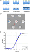
(a) Process flow for the fabrication of the microchip. (b) Chip layout (top view); D is post diameter, S is post spacing in the hcp lattice; scale bar is 20 µm. (c) Magnetization loops from arrays of Ni posts for applied field in the plane of the chip (IP) or perpendicular to it (OOP)
Figure 3
Figure 3
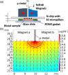
(a) Cross section of the setup for the experiments of magnetophoretic capture. (b) Simulated spatial distribution of the gradient of the H 2 field produced by the assembly of the external magnets. Arrows indicate the direction of ∇H 2, that is, of the magnetophoretic force, while its intensity is given by the color plot
Figure 4
Figure 4
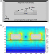
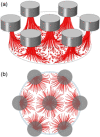
(a) Geometry adopted for the finite element simulations of the magnetic field gradient generated by the Ni pillars and t‐RBC trajectories using Comsol 5.3a. (b) Simulation of the magnetic field gradient generated by the (40–80) Ni pillars geometry. The color plot represents log 10(∇H 2) intensity; red arrows indicate its direction
Figure 5
Figure 5
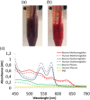
Appearance of (a) a t‐RBC suspension immediately after NaNO2 treatment versus (b) that of a fully oxygenated RBC suspension; (c) absorbance spectra of bovine (solid lines) and human (dotted lines) methemoglobin (red), oxyhemoglobin (blue) and plasma (green); the spectrum for PBS (yellow) is also shown
Figure 6
Figure 6
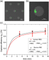
Illustration of the method for the determination of the capture efficiency. (a, b) Optical microscopy images of green fluorescent t‐RBCs captured on pillars. The images are the result of the merge of a green channel fluorescence image and a phase‐contrast channel. (c) Capture efficiency versus time for bovine and human blood samples made of mixtures of untreated RBCs and RBCs treated with NaNO 2. The error bars correspond to the FWHM of the distribution of capture efficiencies measured in repeated experiments on the same sample
Figure 7
Figure 7
Finite element simulations of t‐RBCs capture trajectory on a 40‐µm diameter and 80‐µm spacing pillars geometry using Comsol 5.3a. (a) side view, (b) top view
Figure 8
Figure 8
Capture efficiency curves for different combinations of experimental parameters for MSs with 0.02% volume concentration of t‐RBCs. (a) Magnet M1, different geometries, 20 µm gasket, (b) small magnet, different geometries 40 µm gasket, (c) magnet M2, 40 µm gasket, (d) COMSOL simulations, conditions corresponding to (c)
Figure 9
Figure 9
Effect of the matrix of u‐RBC on the magnetophoretic capture of t‐RBCs in IBMSs. (a) Total hematocrit of 0.4% and volumetric fraction of fluorescent t‐RBCs of 0.02%, (b) total hematocrit of 4% and volumetric fraction of fluorescent t‐RBCs of 0.02%
Figure 10
Figure 10
Capture efficiency of stained t‐RBCs and u‐RBCs, corresponding to the 0.5% of the total number of RBCs in the sample (0.02% volumetric fraction over the whole sample), suspended in whole blood diluted (1:10) in PBS and heparin to obtain a 4% hematocrit

Similar articles

Cited by

References

    1. Bertacco, R. , Petti, D. , Ferrari, G. , Albisetti, E. , & Giacometti, M. (2017). Device and method for the quantification of cellular and non‐cellular blood components. Ital. Pat. n. 102017000082112.
    1. Bertacco, R. , Fiore, G. B. , Ferrari, G. , Giacometti, M. , Milesi, F. , Coppadoro, L. P. & Giuliani, E. (2019). Apparato per la quantificazione di componenti biologiche disperse in un fluido. Ital. Pat. n. 102019000000821.
    1. Blue Martin, A. , Wu, W.‐T. , Kameneva, M. V. , & Antaki, J. F. (2017). Development of a high‐throughput magnetic separation device for malaria‐infected erythrocytes. Annual Review of Biomedical Engineering, 45, 2888–2898. http://link.springer.com/10.1007/s10439-017-1925-2 - DOI - PMC - PubMed
    1. Bottiger, L. E. , & Svedberg, C. A. (1967). Normal erythrocyte sedimentation rate and age. BMJ, 2, 85–87. https://www.bmj.com/lookup/doi/10.1136/bmj.2.5544.85 - DOI - PMC - PubMed
    1. Cohen, J. M. , Smith, D. L. , Cotter, C. , Ward, A. , Yamey, G. , Sabot, O. J. , & Moonen, B. (2012). Malaria resurgence: A systematic review and assessment of its causes. Malaria Journal, 11, 122. https://malariajournal.biomedcentral.com/articles/10.1186/1475-2875-11-122 - DOI - PMC - PubMed

Publication types