. 2022 Mar 9;96(5):e0175221.
doi: 10.1128/JVI.01752-21. Epub 2022 Jan 5.

A Pseudorabies Virus Serine/Threonine Kinase, US3, Promotes Retrograde Transport in Axons via Akt/mToRC1

Affiliations

A Pseudorabies Virus Serine/Threonine Kinase, US3, Promotes Retrograde Transport in Axons via Akt/mToRC1

Andrew D Esteves et al. J Virol. .

Abstract

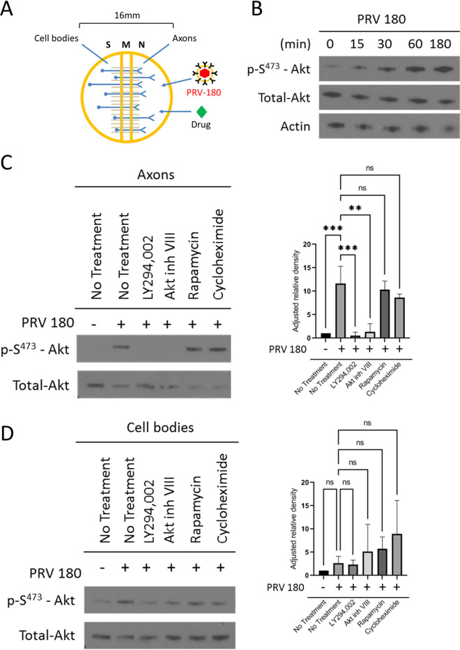
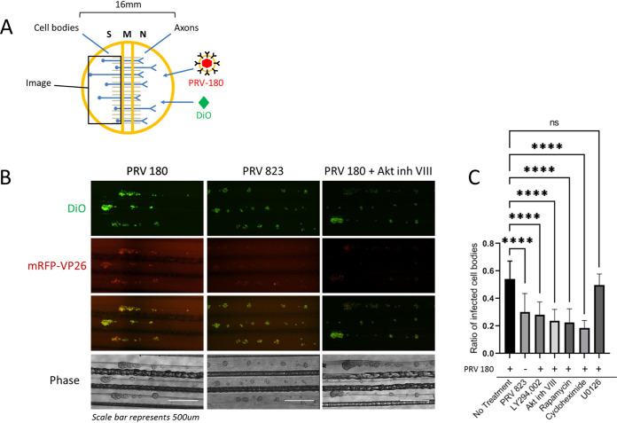
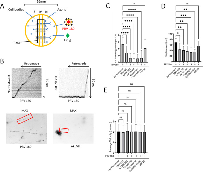
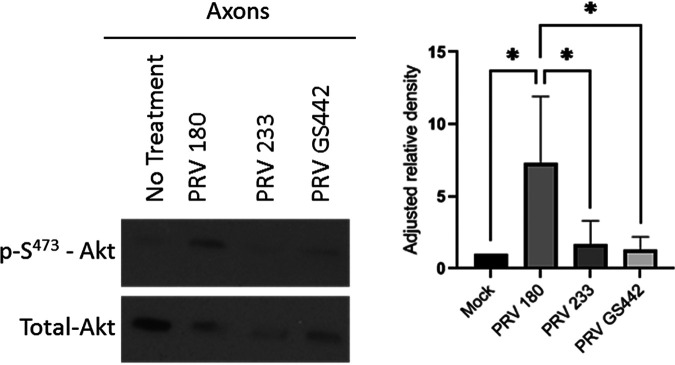
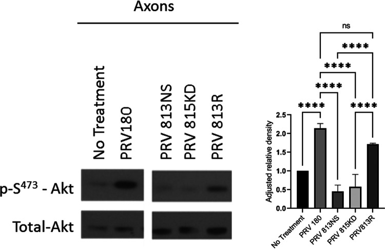
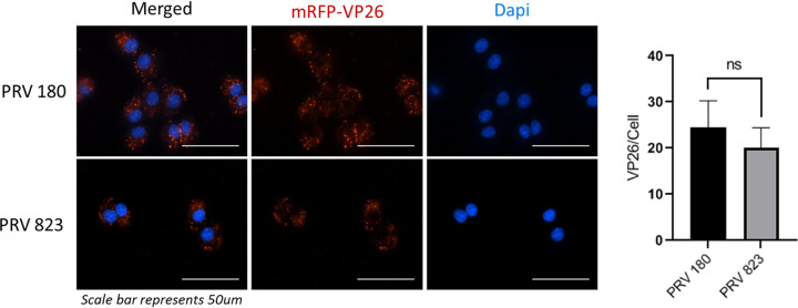
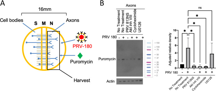
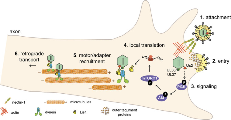
Infection of peripheral axons by alpha herpesviruses (AHVs) is a critical stage in establishing a lifelong infection in the host. Upon entering the cytoplasm of axons, AHV nucleocapsids and associated inner-tegument proteins must engage the cellular retrograde transport machinery to promote the long-distance movement of virion components to the nucleus. The current model outlining this process is incomplete, and further investigation is required to discover all viral and cellular determinants involved as well as the temporality of the events. Using a modified trichamber system, we have discovered a novel role of the pseudorabies virus (PRV) serine/threonine kinase US3 in promoting efficient retrograde transport of nucleocapsids. We discovered that transporting nucleocapsids move at similar velocities in both the presence and absence of a functional US3 kinase; however, fewer nucleocapsids are moving when US3 is absent, and they move for shorter periods of time before stopping, suggesting that US3 is required for efficient nucleocapsid engagement with the retrograde transport machinery. This led to fewer nucleocapsids reaching the cell bodies to produce a productive infection 12 h later. Furthermore, US3 was responsible for the induction of local translation in axons as early as 1 h postinfection (hpi) through the stimulation of a phosphatidylinositol 3-kinase (PI3K)/Akt-mToRC1 pathway. These data describe a novel role for US3 in the induction of local translation in axons during AHV infection, a critical step in transport of nucleocapsids to the cell body. IMPORTANCE Neurons are highly polarized cells with axons that can reach centimeters in length. Communication between axons at the periphery and the distant cell body is a relatively slow process involving the active transport of chemical messengers. There is a need for axons to respond rapidly to extracellular stimuli. Translation of repressed mRNAs present within the axon occurs to enable rapid, localized responses independently of the cell body. AHVs have evolved a way to hijack local translation in the axons to promote their transport to the nucleus. We have determined the cellular mechanism and viral components involved in the induction of axonal translation. The US3 serine/threonine kinase of PRV activates Akt-mToRC1 signaling pathways early during infection to promote axonal translation. When US3 is not present, the number of moving nucleocapsids and their processivity are reduced, suggesting that US3 activity is required for efficient engagement of nucleocapsids with the retrograde transport machinery.

Keywords: Akt; PRV; US3; axon; intracellular transport; kinase; mToRC1; pseudorabies virus; retrograde transport; translation; viral entry.


Conflict of interest statement

The authors declare no conflict of interest.

We declare that we have no competing interests.

Figures

FIG 1
FIG 1
Akt is phosphorylated in axons during PRV infection. (A) A Campenot trichamber neuronal culture system divided into soma (S), middle (M), and neurite (N) compartments used to separate neuronal cell bodies from axons. Addition of virus or drug into the N compartment allows the study of axonal responses independent of the cell body. (B and C) Immunoblots of p-S473-Akt in axons infected with PRV 180 in the N compartment. (B) Infections continued for 0 min, 15 min, 30 min, 60 min, and 180 min. (C and D) N compartments were pretreated with LY294002, Akt inhibitor VIII, rapamycin, or cycloheximide prior to infection. (C) N compartments were harvested 1 hpi. (D) S compartments were harvested 1 hpi in the N compartment. (C and D) p-S473-Akt bands were normalized to total-Akt bands using a densitometry assay. Means and standard deviations (SD) for condition (n = 3) are plotted. **, P ≤ 0.01, and ***, P ≤ 0.001, using one-way analysis of variance (ANOVA); ns, not significant.
FIG 2
FIG 2
Disruption of Akt signaling pathways reduced PRV retrograde infection. (A) N-compartment axons were treated with FAST-DiO 12 h prior to infection. PRV 180 or PRV 823 was added to axons, and at 12 hpi, S-compartment cell bodies were tile imaged. For conditions involving inhibitor treatment, inhibitor was added to N compartments 1 h prior to infection, and at 6 hpi, unabsorbed virus inoculum and inhibitor were removed from the N compartment. (B) Tile images of neuron cell bodies in S compartments. Bar, 500 μm. (C) Quantification of primarily infected cells. The ratio of dual-colored to total green cell bodies for each condition was calculated. Means and SD for 10 chambers for each condition are plotted. ****, P ≤ 0.0001, using one-way ANOVA; ns, not significant.
FIG 3
FIG 3
Quantification of PRV transport kinetics in axons. (A) N compartments were infected with PRV 180 or PRV 823. At 2 hpi, 30-s videos of moving nucleocapsids in M compartments were recorded. Inhibitor was added to N compartments 1 h prior to infection when specified. (B) Maximum-intensity projections (bottom) were created from the videos to visualize nucleocapsid displacement. Moving nucleocapsids are represented as tracks in the image (red box). Kymographs (top) were made from the maximum-intensity projections to visualize nucleocapsid velocity throughout the recording process. Diagonal lines starting from the upper right corner represent retrograde movement. Horizontal lines represent stationary nucleocapsids. (C) Quantification of the number of moving nucleocapsids in M compartments. (D) The displacement of individual nucleocapsids over the 30-s recordings were measured for each condition. (E) The average velocity of the nucleocapsids moving in the retrograde direction was calculated by acquiring the mean of all instantaneous velocities of ≥1 μm/s. Data are means and SD for 7 chambers and 5 fields of view per chamber. ****, P ≤ 0.0001, ***, P ≤ 0.001, **, P ≤ 0.01, and *, P ≤ 0.05, using one-way ANOVA; ns, not significant.
FIG 4
FIG 4
Akt phosphorylation in axons occurred after entry of PRV into the cytoplasm. Immunoblot of p-S473-Akt in axons infected with PRV 180, PRV 233, or PRV GS442 in N compartments. p-S473-Akt bands were normalized to total-Akt bands using a densitometry assay. Means and SD (n = 3) for each condition are plotted. *, P ≤ 0.05, using one-way ANOVA.
FIG 5
FIG 5
US3 induced Akt phosphorylation in axons. Immunoblot of p-S473-Akt in axons infected with PRV 180, PRV 813NS, PRV 815KD, or PRV 813R in N compartments. p-S473-Akt bands were normalized to total-Akt bands using a densitometry assay. Means and SD (n = 3) for each condition are plotted. ****, P ≤ 0.0001, using one-way ANOVA. The image was produced from cropped portions of a single membrane.
FIG 6
FIG 6
US3 does not affect entry of PRV into cells. Fluorescence imaging of PRV 180 and PRV 823 nucleocapsids in Rat2 fibroblasts. A synchronized infection assay was used to allow entry of all bound virion particles to occur simultaneously. Nucleocapsids that entered the cytoplasm were manually counted. Data are means and SD, with 5 replicates and 2 to 5 cells per replicate for each condition. ns, not significant using an unpaired t test.
FIG 7
FIG 7
US3 and Akt phosphorylation are required for virus-induced translation in axons. (A) N compartments were infected with PRV 180 for 1 h prior to harvest. Puromycin was added to N compartments at 45 mpi to label nascent peptides. (B) Immunoblot of puromycin-incorporated peptides from axons in the N compartment. PRV 180 or PRV 813NS were added to N compartments for 1 h. Puromycin was added to N compartments 45 mpi. Inhibitor was added 1 h prior to infection when specified. A single band is visible for each condition in this blot, representing the most abundant peptide synthesized at the time of incubation. Other peptide bands become visible at higher exposure times. The molecular weight ladder shown is a Bio-Rad Precision Plus protein standard (1610374).
FIG 8
FIG 8
Model for PRV-induced translation in axons. (Step 1) PRV virions bind to nectin-1 receptors on the host cell membrane. (Step 2) Entry is mediated by fusion of the viral envelope with the cell’s plasma membrane, allowing nucleocapsid and tegument proteins to enter the cytoplasm. Inner tegument proteins (US3, UL36, and UL37) stay bound to nucleocapsids. (Step 3) US3 stimulates a PI3K/Akt-mToRC1 signaling pathway either directly or indirectly (the dashed arrow indicates that the direct kinase target is still unknown) to induce translation of axonal mRNAs leading to Lis1 expression (step 4). (Steps 5 and 6) Nucleocapsid engagement with the retrograde transport machinery is mediated by the inner tegument and Lis1 (step 5), and subsequent retrograde transport through the axon occurs (step 6).

Similar articles

Cited by

References

    1. Pomeranz LE, Reynolds AE, Hengartner CJ. 2005. Molecular biology of pseudorabies virus: impact on neurovirology and veterinary medicine. Microbiol Mol Biol Rev 69:462–500. 10.1128/MMBR.69.3.462-500.2005. - DOI - PMC - PubMed
    1. Döhner K, Wolfstein A, Prank U, Echeverri C, Dujardin D, Vallee R, Sodeik B. 2002. Function of dynein and dynactin in herpes simplex virus capsid transport. Mol Biol Cell 13:2795–2809. 10.1091/mbc.01-07-0348. - DOI - PMC - PubMed
    1. Sodeik B, Ebersold M, Helenius A. 1997. Microtubule-mediated transport of incoming herpes simplex virus 1 capsids to the nucleus. J Cell Biol 136:1007–1021. 10.1083/jcb.136.5.1007. - DOI - PMC - PubMed
    1. Richards AL, Sollars PJ, Pitts JD, Stults AM, Heldwein EE, Pickard GE, Smith GA. 2017. The pUL37 tegument protein guides alpha-herpesvirus retrograde axonal transport to promote neuroinvasion. PLoS Pathog 13:e1006741. 10.1371/journal.ppat.1006741. - DOI - PMC - PubMed
    1. Sekine E, Schmidt N, Gaboriau D, O'Hare P. 2017. Spatiotemporal dynamics of HSV genome nuclear entry and compaction state transitions using bioorthogonal chemistry and super-resolution microscopy. PLoS Pathog 13:e1006721. 10.1371/journal.ppat.1006721. - DOI - PMC - PubMed

Publication types

MeSH terms

Substances