. 2022 Mar 9;96(5):e0167521.
doi: 10.1128/JVI.01675-21. Epub 2022 Jan 5.

Virus-Like Particles Containing the E2 Core Domain of Hepatitis C Virus Generate Broadly Neutralizing Antibodies in Guinea Pigs

Affiliations

Virus-Like Particles Containing the E2 Core Domain of Hepatitis C Virus Generate Broadly Neutralizing Antibodies in Guinea Pigs

Joey McGregor et al. J Virol. .

Abstract

A vaccine to prevent hepatitis C virus (HCV) infection is urgently needed for use alongside direct-acting antiviral drugs to achieve elimination targets. We have previously shown that a soluble recombinant form of the glycoprotein E2 ectodomain (residues 384 to 661) that lacks three variable regions (Δ123) is able to elicit a higher titer of broadly neutralizing antibodies (bNAbs) than the parental form (receptor-binding domain [RBD]). In this study, we engineered a viral nanoparticle that displays HCV glycoprotein E2 on a duck hepatitis B virus (DHBV) small surface antigen (S) scaffold. Four variants of E2-S virus-like particles (VLPs) were constructed: Δ123-S, RBD-S, Δ123A7-S, and RBDA7-S; in the last two, 7 cysteines were replaced with alanines. While all four E2-S variant VLPs display E2 as a surface antigen, the Δ123A7-S and RBDA7-S VLPs were the most efficiently secreted from transfected mammalian cells and displayed epitopes recognized by cross-genotype broadly neutralizing monoclonal antibodies (bNMAbs). Both Δ123A7-S and RBDA7-S VLPs were immunogenic in guinea pigs, generating high titers of antibodies reactive to native E2 and able to prevent the interaction between E2 and the cellular receptor CD81. Four out of eight animals immunized with Δ123A7-S elicited neutralizing antibodies (NAbs), with three of those animals generating bNAbs against 7 genotypes. Immune serum generated by animals with NAbs mapped to major neutralization epitopes located at residues 412 to 420 (epitope I) and antigenic region 3. VLPs that display E2 glycoproteins represent a promising vaccine platform for HCV and could be adapted to large-scale manufacturing in yeast systems. IMPORTANCE There is currently no vaccine to prevent hepatitis C virus infection, which affects more than 71 million people globally and is a leading cause of progressive liver disease, including cirrhosis and cancer. Broadly neutralizing antibodies that recognize the E2 envelope glycoprotein can protect against heterologous viral infection and correlate with viral clearance in humans. However, broadly neutralizing antibodies are difficult to generate due to conformational flexibility of the E2 protein and epitope occlusion. Here, we show that a VLP vaccine using the duck hepatitis B virus S antigen fused to HCV glycoprotein E2 assembles into virus-like particles that display epitopes recognized by broadly neutralizing antibodies and elicit such antibodies in guinea pigs. This platform represents a novel HCV vaccine candidate amenable to large-scale manufacture at low cost.

Keywords: glycoprotein E2; hepatitis C virus; vaccine; virus-like particle.


Conflict of interest statement

The authors declare no conflict of interest.

Figures

FIG 1
FIG 1
Schematic representation of constructs used in this study. (A) Soluble proteins RBD, Δ123, RBDA7, and Δ123A7. Scale is a representation of the amino acid numbering according to the H77c prototype. Variable regions (red), variable regions replaced by GSSG linkers (dotted lines), epitope I, II, and III regions (thick gray lines), N-linked glycans (brown trees), cysteine residues (yellow lines), Cys-Ala mutations (green lines), and 6×His tag (HHHHHH) are indicated. (B) E2-S constructs where E2 is fused at the C terminus to the N terminus of S (light blue). Regions within the E2 area are as described for panel A.
FIG 2
FIG 2
Western blot analysis of E2-DHBVS VLPs showing the expression of DHBVS alone and 4 variants of E2-S VLPs. HEK293T cells were cotransfected with E2-S and DHBVS and VLPs purified by ultracentrifugation through a 20% sucrose cushion. Reducing SDS-PAGE was performed, followed by Western blotting with anti-E2 bNMAb HCV1 (green) and anti-S MAb 7C12 (red). Purified soluble protein Δ123 and DHBVS were included as controls. (B) Immunoblot of Δ123A7-VLPs sedimented through an iodixanol gradient. VLP-containing fractions were resolved by reducing SDS-PAGE followed by Western blotting with anti-E2 bNMAb HCV1 (green) and anti-S MAb 7C12 (red). (C) Immunoblot of VLPs after E2-S and DHBVS-containing fractions were pooled, pelleted, and resuspended in PBS. (D) Deglycosylation of Δ123A7-S or Δ123A7 soluble protein with either PNGase F or Endo-H. Untreated, no enzyme; control, no enzyme or DTT. Analysis was done as described for panel A. (E) Transmission electron microscopy of purified E2Δ123A7-S VLPs negatively stained with 1% (wt/vol) uranyl acetate. (F) Immunoelectron microscopy of purified E2Δ123A7-S VLPs incubated with human MAb HC84.27 (anti-E2), mouse MAb 7C12 (anti-S), 5-nm gold-conjugated anti-human immunoglobulin, and 10-nm gold-conjugated anti-mouse immunoglobulin, followed by staining with 2% (wt/vol) uranyl acetate. Scale bars (E and F) = 50 nm.
FIG 3
FIG 3
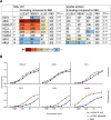
Antigenic characterization of E2-S VLPs and E2 soluble protein. (A) Binding of MAbs HCV1, AR3C, HC84.27, HC11, HC84.1, MAb44, 2A12, and AR1A to plate-bound chimeric E2-S VLPs (RBD-S, RBDA7-S, Δ123-S, or Δ123A7-S) at a single dilution of 0.5 μg/mL and soluble protein (RBD, RBDA7, Δ123, or Δ123A7) at a single dilution of 5 μg/ml. Specific binding was detected with the appropriate HRP-conjugated secondary antibody. MAb binding was calculated as a percentage compared to RBD binding, and mean percentages calculated from 4 separate experiments are shown. Binding: >400%, maroon; >300%, orange; >200%, light orange; >130%, yellow; and <80%, blue. Mean RBD binding absorbance values for VLP and soluble protein are indicated. (B) Comparison of the antigenicity of Δ123A7-S VLP and soluble monomeric Δ123A7 protein. Neutralizing MAbs HC84.27, AR3C, and HC11 and non-NMAbs 2A12, AR1A, and MAb26 were added to ELISA plates coated with either VLPs or soluble protein to assess reactivity. Specific binding was detected with the appropriate HRP-conjugated secondary antibody. Curves were fitted using nonlinear regression in GraphPad Prism.
FIG 4
FIG 4
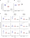
Reactivity of immune sera toward monomeric Δ123A7 protein (A) and S VLPs (B). Dilutions of immune sera were added to plates coated with monomeric Δ123A7 protein or S VLPs. Antibody titers were calculated as the reciprocal dilution of serum required for a 10-fold increase over binding to BSA (background binding). (C) Reactivity of immune sera toward continuous NAb epitopes. Homologous genotype 1a H77c peptides and heterologous genotype 2a J6 and genotype 3a S52 peptides (peptide 410-428, peptide 430-451, and peptide 523-549) were used to capture antibodies present in the immune serum. Antibody titers were calculated as the reciprocal dilution of serum required for a 10-fold increase over binding to BSA. The dotted line in panels A to C represents the lower limit of detection for the assay. Statistical analysis for panels A to C was performed using a Kruskal-Wallis test followed by Dunn’s multiple-comparison test in GraphPad Prism (v. 9). *, P ≤ 0.05; **, P ≤ 0.01; ***, P ≤ 0.001; ****, P ≤ 0.0001. The horizontal bars indicate the mean titers.
FIG 5
FIG 5
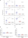
Analysis of antibody specificity. (A) Competitive ELISA to determine specificity of the antibody response. Serial dilutions of immune sera were mixed with a constant amount of MAb prior to incubation with plate-bound Δ123. The reciprocal dilutions of serum required for 50% inhibition (ID50) relative to MAb binding in the absence of sera are shown. (B) Reactivity of serum to HVR1 peptide. Plate-bound genotype 1a H77c HVR1 peptide was used to capture antibodies present in the immune serum. Antibody titers were calculated as the reciprocal dilution of serum required for a 10-fold increase over background binding. (C) Competitive ELISA to determine specificity of the antibody response to HVR1 MAb36. Serial dilutions of immune serum were added to a constant amount of MAb36 prior to incubation with plate-bound Δ123. The reciprocal dilution of serum required for 50% inhibition was calculated using binding of MAb in the absence of serum as 100% binding. The dotted line in panels A to C represents the lower limit of detection for the assay. Statistical analysis for panels A to C was performed using a Kruskal-Wallis test followed by Dunn’s multiple-comparison test in GraphPad Prism (v. 9). *, P ≤ 0.05; **, P ≤ 0.01; ***, P ≤ 0.001; ****, P ≤ 0.0001. The horizontal bars indicate the mean titers.
FIG 6
FIG 6
(A) Ability of immune sera to inhibit the binding of homologous H77c RBD to CD81-LEL. Immune sera were precleared of interfering antibodies and incubated with H77c RBD before being added to CD81-LEL-coated plates. RBD binding was detected by E2-specific MAb H53 and HRP-conjugated secondary antibody, and the reciprocal dilution of serum required to inhibit E2-CD81 interaction by 50% and 80% was calculated. (B) Ability of immune serum to neutralize homologous genotype 1a pp virus. Serial dilutions of Δ123A7-S VLP, RBDA7-S VLP, or S VLP precleared sera were mixed with HCVpp (H77c, genotype 1a). Luciferase activity was measured, and the reciprocal dilution of serum required to achieve 50% and 80% neutralization was calculated. Statistical analysis for panels A and B was performed using a Kruskal-Wallis test followed by Dunn’s multiple-comparison test in GraphPad Prism (v. 9). *, P ≤ 0.05; **, P ≤ 0.01; ***, P ≤ 0.001. The horizontal bars indicate the mean titers. (C) Ability of sera to neutralize diverse HCV genotypes. Serial dilutions of precleared Δ123A7-S VLP, RBDA7-S VLP, or S VLP sera or MAb24 miniperm (7 μg/mL) were mixed with either HCVpp (genotype 1a) or HCVcc (genotypes 2 to 7). Luciferase activity was measured, and the reciprocal dilution of serum required to achieve 50% neutralization was calculated. The data were derived from three independent experiments performed in triplicate, with bars representing the means and error bars showing SEM. In panels A to C, the dotted line represents the lower limit of detection of the assay.
FIG 7
FIG 7
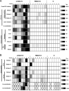
Binding and neutralizing titers for individual animals vaccinated with either Δ123A7-S VLPs, RBDA7-S VLPs, or S VLPs. Titers generated against homologous genotype 1a (H77c) (A) and heterologous genotype 2a (J6) and genotype 3a (S52) (B) were plotted in a heat map. Higher titers are represented by increasingly darker tone. A white box indicates activity below the level of detection for that assay. An “X” indicates that the experiment was not conducted.

References

    1. Thrift AP, El-Serag HB, Kanwal F. 2017. Global epidemiology and burden of HCV infection and HCV-related disease. Nat Rev Gastroenterol Hepatol 14:122–132. 10.1038/nrgastro.2016.176. - DOI - PubMed
    1. Pawlotsky JM. 2014. New hepatitis C therapies: the toolbox, strategies, and challenges. Gastroenterology 146:1176–1192. 10.1053/j.gastro.2014.03.003. - DOI - PubMed
    1. Falade-Nwulia O, Suarez-Cuervo C, Nelson DR, Fried MW, Segal JB, Sulkowski MS. 2017. Oral direct-acting agent therapy for hepatitis C virus infection: a systematic review. Ann Intern Med 166:637–648. 10.7326/M16-2575. - DOI - PMC - PubMed
    1. Falade-Nwulia O, Sulkowski MS, Merkow A, Latkin C, Mehta SH. 2018. Understanding and addressing hepatitis C reinfection in the oral direct-acting antiviral era. J Viral Hepat 25:220–227. 10.1111/jvh.12859. - DOI - PMC - PubMed
    1. Cox AL. 2015. Global control of hepatitis C virus. Science 349:790–791. 10.1126/science.aad1302. - DOI - PubMed

Publication types

MeSH terms