. 2022 Jan 5;18(1):e1010182.
doi: 10.1371/journal.ppat.1010182. eCollection 2022 Jan.

Structure of a bacterial Rhs effector exported by the type VI secretion system

Affiliations

Structure of a bacterial Rhs effector exported by the type VI secretion system

Patrick Günther et al. PLoS Pathog. .

Abstract

The type VI secretion system (T6SS) is a widespread protein export apparatus found in Gram-negative bacteria. The majority of T6SSs deliver toxic effector proteins into competitor bacteria. Yet, the structure, function, and activation of many of these effectors remains poorly understood. Here, we present the structures of the T6SS effector RhsA from Pseudomonas protegens and its cognate T6SS spike protein, VgrG1, at 3.3 Å resolution. The structures reveal that the rearrangement hotspot (Rhs) repeats of RhsA assemble into a closed anticlockwise β-barrel spiral similar to that found in bacterial insecticidal Tc toxins and in metazoan teneurin proteins. We find that the C-terminal toxin domain of RhsA is autoproteolytically cleaved but remains inside the Rhs 'cocoon' where, with the exception of three ordered structural elements, most of the toxin is disordered. The N-terminal 'plug' domain is unique to T6SS Rhs proteins and resembles a champagne cork that seals the Rhs cocoon at one end while also mediating interactions with VgrG1. Interestingly, this domain is also autoproteolytically cleaved inside the cocoon but remains associated with it. We propose that mechanical force is required to remove the cleaved part of the plug, resulting in the release of the toxin domain as it is delivered into a susceptible bacterial cell by the T6SS.


Conflict of interest statement

The authors have declared that no competing interests exist.

Figures

Fig 1
Fig 1. High-resolution structure of the T6SS effector RhsA.
(A) Genomic context of rhsA (PFL_6096, blue) in Pseudomonas protegens Pf-5. Upstream genes encoding the cognate VgrG protein, vgrG1 (PFL_6094, green) and RhsA-specific chaperone, eagR1 (PFL_6095, light brown) are shown. Self-protection against RhsA is conferred via expression of the downstream immunity-encoding gene, rhsI (dark brown). (B) Representative cryo-EM 2-D class averages of the assembled pre-firing complex composed of VgrG1, RhsA and EagR1 (left) and a schematic representation of each of the components that comprise this complex (right). Scale bar, 10 nm. (C) Full-length RhsA contains a prePAAR motif and a TMD comprising two transmembrane helices upstream of its PAAR domain. The RhsAΔTMD truncation of RhsA was used in this study to determine the high-resolution structure of the RhsA cocoon. (D) Cryo-EM density of RhsAΔTMD displayed perpendicular to the central symmetry axis of the barrel and rotated 90° clockwise (map postprocessed with DeepEMhancer). (E) Cartoon representation of the atomic model of RhsA colored in rainbow from N-terminus (blue) to C-terminus (red).
Fig 2
Fig 2. Autoproteolysis of RhsA occurs at its N- and C-terminus.
(A) RhsA is autoproteolytically cleaved at its C-terminus at position W1350. The end of the connected density is indicated with an asterisk. Catalytic aspartates D1324 and D1346, are shown in stick representation. (B) Western blot analysis shows proteolytic cleavage of the C-terminal toxin domain. Mutation of either D1324 or D1346 to asparagine prevents autoproteolytic cleavage of the C-terminal RhsA toxin domain. Blots were performed against the Rhs barrel (α-RhsA) and against a C-terminal VSV-G epitope tag (α-VSV-G). (C) Outcome of intraspecific growth competition assays between the indicated P. protegens donor and recipient strains. Donor strains were competed against recipient strains that either contain (light grey) or lack the rhsA-rhsI effector-immunity pair (dark grey). The recipient strains also lack pppA to stimulate type VI secretion in donors [14]. Data are mean ± s.d. for n = 3 biological replicates and are representative of two independent experiments. (D) The toxin domain of RhsA is encapsulated by its cocoon-shaped Rhs repeat-containing domain. Difference map of the encapsulated toxin at extremely low-density threshold and low pass-filtered to 20 Å (green) is shown. The Rhs cocoon is depicted using a transparent space-filling representation at normal map threshold. (E) Regions of the toxin domain are stabilized inside the cocoon through interactions with the C-terminal autoprotease domain. The densities appear at the same density threshold as the rest of the map. The atomic models are shown in stick representation. (F) RhsA undergoes N-terminal cleavage at proline 305. The end of the density is indicated with an asterisk. (G) N-terminal cleavage occurs in RhsA and a mutant lacking the prePAAR-TMD (RhsAΔTMD, residues 74-CT) but not in a mutant lacking the entire N-terminal region (RhsAΔN, residues 297-CT). The indicated RhsA constructs were purified from E. coli and subject to Western blot and detected using an N-terminal His6-tag antibody (α-his6).
Fig 3
Fig 3. A unique plug domain seals the Rhs barrel of RhsA.
(A) Surface representation of the cork domain of RhsA. The molecular surface is colored according to hydrophobicity where ochre and white indicate hydrophobic and hydrophilic regions, respectively. The Rhs barrel is shown as a cartoon. (B) Enlarged view of the cork domain of RhsA. The hydrophobic surface spirals around the domain as indicated by the black dashed line. (C) The upper Rhs repeats of RhsA possess complementary hydrophobic patches to those found on its plug domain (shown in cartoon representation, red). (D) The cocoon structure of RhsA is closed off by an N-terminal plug comprised of a ‘cork’ domain (orange, density representation), a ‘seal’ peptide (mesh), and an anchor helix (E, F). The seal and the cork density together form a cap structure. (E) The seal peptide, which also functions as the linker to the unmodelled PAAR domain, not only complements the shape of the cork but is also the entry point of the N-terminal part of the protein into the inside of the cocoon. The cocoon remains stably bound to the cleaved N-terminal region, including the PAAR domain, due to the anchor helix inside the cocoon. (F) The anchor helix of RhsA is stabilized by hydrophobic interactions with the inner wall of the Rhs repeats. (G) Cartoon representation of the cork region of the RhsA plug domain and a comparison with the homologous N-terminal plug domain of Photorhabdus luminescens TcdB2 (grey, PDB ID: 6H6G) and the unique FN-plug domain of human teneurin2 (green, PDB ID: 6FB3). The lower part of each depicted plug domain inserts into each of their respective Rhs barrels. The cork domain of RhsA is colored in rainbow.
Fig 4
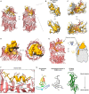
Fig 4. Structural comparison of Rhs repeat containing proteins.
The C-terminal autoproteolysis domain (left and middle) or YD-shell plug (right) is structurally conserved among diverse Rhs proteins (blue). RhsA and BC components of Tc toxin complexes encapsulate their toxic effector domains (red) whereas in teneurin proteins the toxin domain is appended to the outside of the Rhs barrel (red). A distinguishing feature of these Rhs proteins is the unique N-terminal plug domain for each protein family (orange). RhsA is capped by a cork-like plug domain that seals the Rhs barrel (orange). In Tc toxins, the homologous plug domain acts as constriction site and the cocoon is sealed off by a β-propeller domain (green). Teneurin proteins are capped with a non-homologous FN-plug (orange) that is stabilized by an NHL domain (green). The lower row shows schematic representations of the domain organizations.
Fig 5
Fig 5. High-resolution structure of P. protegens VgrG1.
(A) The RhsA barrel of the assembled pre-firing complex (PFC) displays high positional flexibility relative to VgrG1. Representative cryo-EM 2-D class averages depicting flexibility are shown. Scale bar, 10 nm. (B) Cryo-EM density and (C) ribbon representation of the molecular model of the P. protegens VgrG1 trimer viewed along the long axis of the protein. Each protomer is colored differently to highlight their positions within the homotrimeric VgrG1 spike.
Fig 6
Fig 6. Model of T6SS-dependent delivery of RhsA into the cytoplasm of a susceptible bacterial cell.
(1–3) RhsA undergoes N- and C-terminal autoproteolytic processing. The prePAAR motif ‘completes’ the PAAR domain fold. The EagR1 chaperone solubilizes the two transmembrane helices of RhsA to facilitate loading onto its cognate cytoplasmic VgrG1 (via the prePAAR + PAAR domain). (4) The secretion competent RhsA effector is loaded onto the VgrG1 spike. (5) During a firing event the EagR1 chaperone is dissociated from the complex and the T6SS injects the PFC into target cells where it crosses the peptidoglycan (PG) layer and inserts into the inner membrane (IM) (5A). Alternatively, the tip of the VgrG1 spike could be directly delivered into the cytosol along with RhsA (5B). (6–8) Two different scenarios (6A or 6B,C) are proposed as possible mechanisms for toxin domain release from the cocoon into the cytosol of the target cell. Either the toxin domain alone (mechanism 6A) or the entire RhsA barrel is translocated across the inner membrane (mechanism 6B). In both cases, the seal of the cocoon is likely removed by translocation-induced pulling and the energy required for release and translocation of the toxin domain out of the Rhs cage is probably driven by its spontaneous refolding in the prey bacterium’s cytosol.

References

    1. Mougous JD, Cuff ME, Raunser S, Shen A, Zhou M, Gifford CA, et al.. A virulence locus of Pseudomonas aeruginosa encodes a protein secretion apparatus. Science (80-). 2006;312: 1526–1530. doi: 10.1126/science.1128393 - DOI - PMC - PubMed
    1. Pukatzki S, Ma AT, Sturtevant D, Krastins B, Sarracino D, Nelson WC, et al.. Identification of a conserved bacterial protein secretion system in Vibrio cholerae using the Dictyostelium host model system. Proc Natl Acad Sci U S A. 2006;103: 1528–1533. doi: 10.1073/pnas.0510322103 - DOI - PMC - PubMed
    1. Russell AB, Hood RD, Bui NK, Leroux M, Vollmer W, Mougous JD. Type VI secretion delivers bacteriolytic effectors to target cells. Nature. 2011;475: 343–349. doi: 10.1038/nature10244 - DOI - PMC - PubMed
    1. Chang Y, Rettberg LA, Ortega DR, Jensen GJ. In vivo structures of an intact type VI secretion system revealed by electron cryotomography. EMBO Rep. 2017;18: 1090–1099. doi: 10.15252/embr.201744072 - DOI - PMC - PubMed
    1. Silverman JM, Brunet YR, Cascales E, Mougous JD. Structure and regulation of the type VI secretion system. Annu Rev Microbiol. 2012;66: 453–472. doi: 10.1146/annurev-micro-121809-151619 - DOI - PMC - PubMed

Publication types

Substances

Supplementary concepts