. 2022 Jan 19;13(2):245-256.
doi: 10.1021/acschemneuro.1c00653. Epub 2022 Jan 5.

Distinct Dibasic Cleavage Specificities of Neuropeptide-Producing Cathepsin L and Cathepsin V Cysteine Proteases Compared to PC1/3 and PC2 Serine Proteases

Affiliations

Distinct Dibasic Cleavage Specificities of Neuropeptide-Producing Cathepsin L and Cathepsin V Cysteine Proteases Compared to PC1/3 and PC2 Serine Proteases

Michael C Yoon et al. ACS Chem Neurosci. .

Abstract

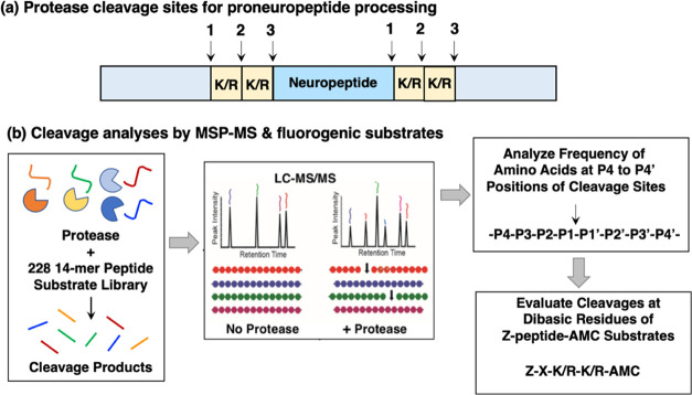
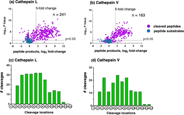
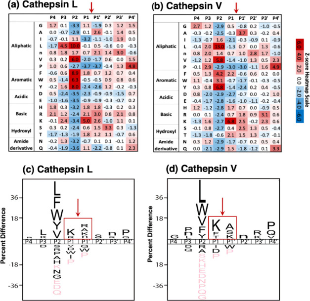
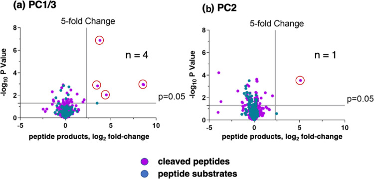
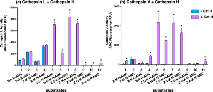
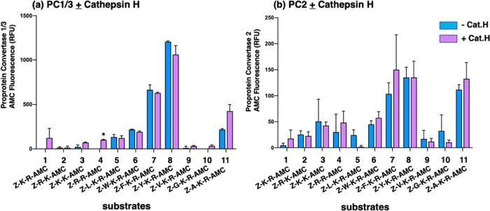
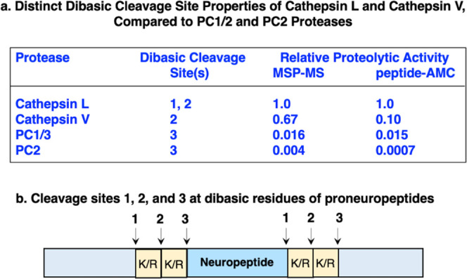
Neuropeptides, functioning as peptide neurotransmitters and hormones, are generated from proneuropeptide precursors by proteolytic processing at dibasic residue sites (i.e., KR, RK, KK, RR). The cysteine proteases cathepsin L and cathepsin V, combined with the serine proteases proprotein convertases 1 and 2 (PC1/3 and PC2), participate in proneuropeptide processing to generate active neuropeptides. To compare the dibasic cleavage properties of these proteases, this study conducted global, unbiased substrate profiling of these processing proteases using a diverse peptide library in multiplex substrate profiling by mass spectrometry (MSP-MS) assays. MSP-MS utilizes a library of 228 14-mer peptides designed to contain all possible protease cleavage sites, including the dibasic residue sites of KR, RK, KK, and RR. The comprehensive MSP-MS analyses demonstrated that cathepsin L and cathepsin V cleave at the N-terminal side and between the dibasic residues (e.g., ↓K↓R, ↓R↓K, and K↓K), with a preference for hydrophobic residues at the P2 position of the cleavage site. In contrast, the serine proteases PC1/3 and PC2 displayed cleavage at the C-terminal side of dibasic residues of a few peptide substrates. Further analyses with a series of dipeptide-AMC and tripeptide-AMC substrates containing variant dibasic sites with hydrophobic P2 residues indicated the preferences of cathepsin L and cathepsin V to cleave between dibasic residue sites with preferences for flanking hydrophobic residues at the P2 position consisting of Leu, Trp, Phe, and Tyr. Such hydrophobic amino acids reside in numerous proneuropeptides such as pro-NPY and proenkephalin that are known to be processed by cathepsin L. Notably, cathepsin L displayed the highest specific activity that was 10-, 64-, and 1268-fold greater than cathepsin V, PC1/3, and PC2, respectively. Peptide-AMC substrates with dibasic residues confirmed that PC1/3 and P2 cleaved almost exclusively at the C-terminal side of dibasic residues. These data demonstrate distinct dibasic cleavage site properties and a broad range of proteolytic activities of cathepsin L and cathepsin V, compared to PC1/3 and PC2, which participate in producing neuropeptides for cell-cell communication.

Keywords: cathepsin; mass spectrometry; neuropeptide; peptidomics; proprotein convertase; protease.


Conflict of interest statement

The authors declare no competing financial interest.

Figures

Figure 1
Figure 1
Scheme for the cleavage profiling of the proneuropeptide processing proteases cathepsin L, cathepsin V, PC1/3, ad PC2, by MSP-MS and analyses by fluorogenic peptide substrates. (a) Proneuropeptides undergo proteolytic processing at dibasic residue sites. Neuropeptides are generated from proneuropeptide precursors that require proteolytic processing at dibasic sites (K/R-K/R) to generate active neuropeptides. (b) Strategy for the cleavage profiling of cathepsin L, cathepsin V, PC1/3, and PC2 processing enzymes by MSP-MS and fluorogenic substrates. The cleavage profile properties of cathepsin L and cathepsin V cysteine proteases, combined with PC1/3 and PC2 serine proteases, were evaluated by global, unbiased multiplex substrate profiling by mass spectrometry (MSP-MS) and fluorogenic peptide-AMC substrates containing variant dibasic residue sequences. For MSP-MS, the 228 peptide library was incubated with each of the processing proteases (as described in the Methods section), and peptide cleavage products were subjected to nano-LC–MS/MS tandem mass spectrometry for identification and quantification. Peptide cleavage products were analyzed for the frequency of each of the different amino acid residues at positions P4–P4′ and at the cleaved P1↓P1′ cleavage site. Based on MSP-MS results, peptide-AMC substrates were designed to further assess the dibasic cleavage site preferences of these proteases.
Figure 2
Figure 2
Cathepsin L and cathepsin V peptide cleavage profiling analyzed by multiplex substrate profiling by mass spectrometry (MSP-MS). (a,b) Volcano plots of cleaved peptides generated by cathepsin L (a) and cathepsin V (b). The log2 ratios of relative quantities of peptide products generated by cathepsin L or cathepsin V (60 min incubation at pH 5.5) compared to no enzyme activity controls are illustrated with −log10p values. Peptide products generated with at least a 5-fold change above no enzyme activity controls and with p < 0.05 numbered 241 and 163 peptides in panels “a” and “b”, respectively, representing 8.1 and 7.9%, respectively, of the entire number of 2964 cleavage sites among the 228 peptides of the library. Peptide sequences were analyzed for the frequencies of amino acid residues at the P4–P4′ positions of the P1–↓P1′cleavage site. (c,d) Cleavage positions of 14-mer peptide substrates for cathepsin L (c) and cathepsin V (d). The number of cleavages by cathepsin L and cathepsin V at each of the peptide bonds of the 14-mer peptide substrates are illustrated.
Figure 3
Figure 3
Cathepsin L and cathepsin V preferences for P4–P4′ residues of peptide cleavage sites in MSP-MS analyses. (a, b) Heat maps of amino acids preferred at cleavages sites. The peptide library cleavage data for cathepsin L (panel a) and cathepsin V (panel b) shows the frequencies of amino acid residues at each of the P4 to the P4′ positions of cleaved peptides, shown as the heat maps of Z-scores (explained in the Methods section) that compare protease cleavages with that of the reference peptide library. (c, d) IceLogo of cathepsin L and cathepsin V for preferred cleavages at P4–P4′ residues. Cathepsin L (panel c) and cathepsin V (panel d) cleavage data is illustrated by iceLogo. IceLogo shows the relative frequency of the preferred residues at the P1–↓P1′ cleavage site and at the P4–P4′ residues. Black letters above the line of P4–P4′ positions indicate preferred amino acid residues of the protease with p < 0.05, compared to the reference (negative data) of all possible residues at each position. Pink letters indicate residues that were never found at the indicated cleavage position.
Figure 4
Figure 4
PC1/3 and PC2 cleavage profiling analyzed by MSP-MS. Volcano plots of PC1/3 (panel a) and PC2 (panel b) peptide cleavages from MSP-MS data show the log2 ratios of relative quantities of peptide products generated by PC1/3 and PC2 (60 min incubation at pH 5.5) compared to no enzyme activity controls, illustrated by −log10p values. Peptide products generated with at least a 5-fold change above controls and with p < 0.05 were analyzed for the frequencies of amino acid residues at the P4–P4′ positions of the P1–↓P1′ cleavage site.
Figure 5
Figure 5
Cathepsin L and cathepsin V cleavage specificities at dibasic residue sites assessed with variant dipeptide-AMC and tripeptide-AMC substrates. Cathepsin L (panel a) and cathepsin V (panel b) were evaluated for the cleavage of dipeptide-AMC substrates containing the four dibasic variant cleavage sites KR, RK, KK, and RR and compared to the cleavage of tripeptide-AMC substrates containing the K-R with adjacent hydrophobic residues (Leu, Trp, Phe, Tyr, Val) or nonpolar residues (Gly, Ala) at the N-terminal side of the dibasic K-R site. Cathepsin L and cathepsin V were incubated with each of these substrates at 37 °C for 60 min. Then, the aminopeptidase cathepsin H or control buffer was added and incubation continued at 37 °C for another 30 min to allow conversion of basic residue-extended AMC products to free AMC for fluorometric measurement (conducted as described in the Methods section, with example shown in the Supporting Information, Figure S1). Controls included incubation of cathepsin H alone with each of the substrates, which resulted in no fluorescence, indicating that cathepsin H does not remove the blocked N-terminal residues of Z-peptide-AMC substrates. Comparison of fluorescence observed in the absence and presence of cathepsin H is illustrated, with significant differences with p < 0.05 (student’s t-test, n = 3) indicated by asterisks.
Figure 6
Figure 6
PC1/3 and PC2 cleavage properties examined with variant dipeptide-AMC and tripeptide-AMC substrates containing dibasic residue sites. PC1/3 (panel a) and PC2 (panel b) were evaluated for the cleavage of dipeptide-AMC substrates containing the four dibasic variant cleavage sites KR, RK, KK, and RR and compared to the cleavage of tripeptide-AMC substrates containing the K-R with hydrophobic residues (Leu, Trp, Phe, Tyr, Val) or nonpolar residues (Gly, Ala) at the N-terminal side of the dibasic K-R site. After incubation of PC1/3 or PC2 with each of these substrates at 37 °C for 120 min, control buffer or the aminopeptidase cathepsin H was added and incubation at 37 °C continued for another 30 min (37 °C) to allow conversion of basic residue-extended AMC products to free AMC for fluorometric measurement. Comparison of fluorescence observed in the absence and presence of cathepsin H is illustrated and included evaluation of significant differences with p < 0.05 (Student’s t-test, n = 3).
Figure 7
Figure 7
Distinct dibasic cleavage properties of cathepsin L and cathepsin V cysteine proteases compared to PC1/3 and PC2 serine proteases involved in proneuropeptide processing. (a) Information is shown for the relative proteolytic activity by the MSP-MS analyses of peptide library substrates and relative proteolytic activity observed in peptide-AMC assays using standard substrates for cathepsin L and cathepsin V (Z-F-R-AMC), and PC1/3 and PC2 (pERTKR-AMC). (b) Locations of dibasic cleavage sites (#1, 2, and 3) for each of the proneuropeptide processing proteases cathepsin L, cathepsin V, PC1/3, and PC2 are indicated.

References

    1. Hook V.; Funkelstein L.; Lu D.; Bark S.; Wegrzyn J.; Hwang S. R. Proteases for processing proneuropeptides into peptide neurotransmitters and hormones. Annu. Rev. Pharmacol. Toxicol. 2008, 48, 393–423. 10.1146/annurev.pharmtox.48.113006.094812. - DOI - PMC - PubMed
    1. Hook V.; Lietz C. B.; Podvin S.; Cajka T.; Fiehn O. Diversity of neuropeptide cell-cell signaling molecules generated by proteolytic processing revealed by neuropeptidomics mass spectrometry. J. Am. Soc. Mass Spectrom. 2018, 29, 807–816. 10.1007/s13361-018-1914-1. - DOI - PMC - PubMed
    1. Seidah N. G.; Sadr M. S.; Chrétien M.; Mbikay M. The multifaceted proprotein convertases: their unique, redundant, complementary, and opposite functions. J. Biol. Chem. 2013, 288, 21473–21481. 10.1074/jbc.R113.481549. - DOI - PMC - PubMed
    1. Felig P.; Baxter J. D.; Frohman L. A.. Endocrinology and Metabolism, 3rd ed.; McGraw Hill, Inc.: New York, 1981.
    1. Inturrisi C. E. Clinical pharmacology of opioids for pain. Clin. J. Pain 2002, 18, S3–S13. 10.1097/00002508-200207001-00002. - DOI - PubMed

Publication types