. 2022 Jan 4;38(1):110187.
doi: 10.1016/j.celrep.2021.110187.

Calcium-dependent ESCRT recruitment and lysosome exocytosis maintain epithelial integrity during Candida albicans invasion

Affiliations

Calcium-dependent ESCRT recruitment and lysosome exocytosis maintain epithelial integrity during Candida albicans invasion

Johannes Westman et al. Cell Rep. .

Abstract

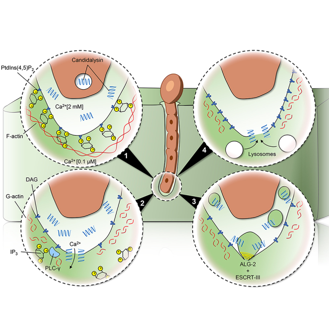
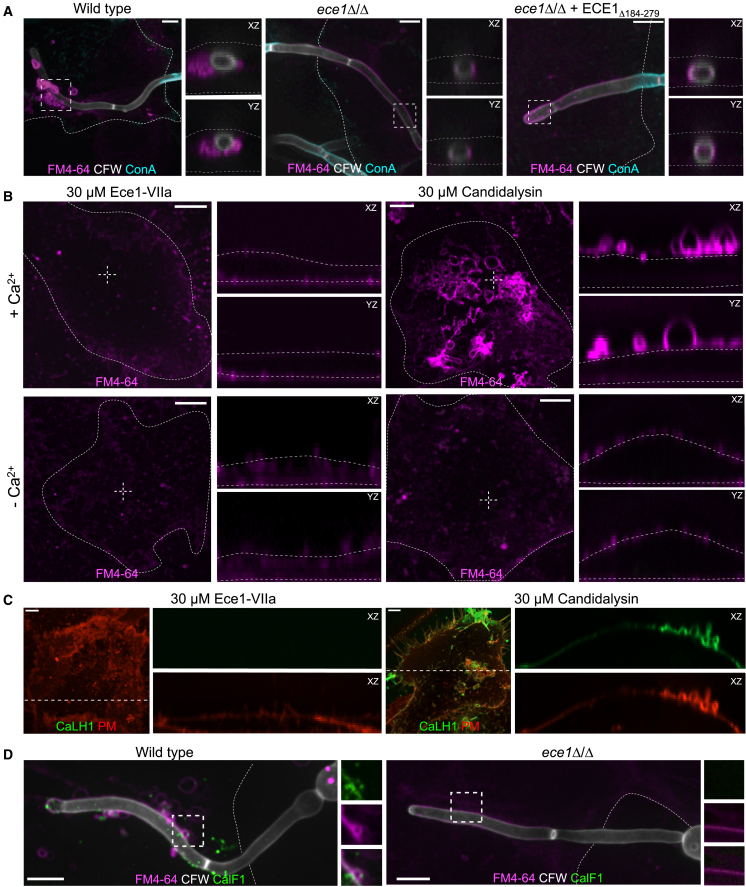
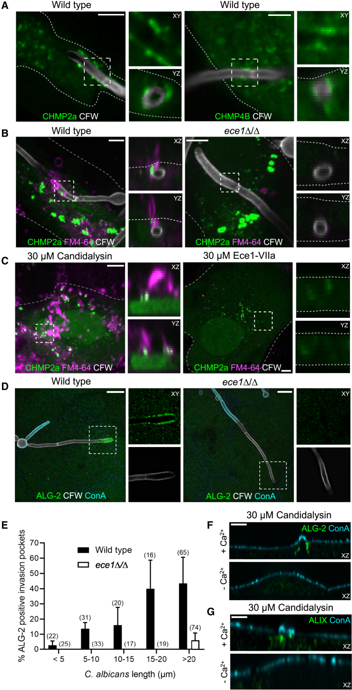
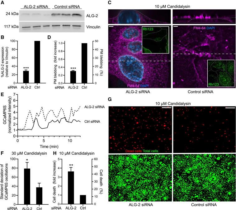
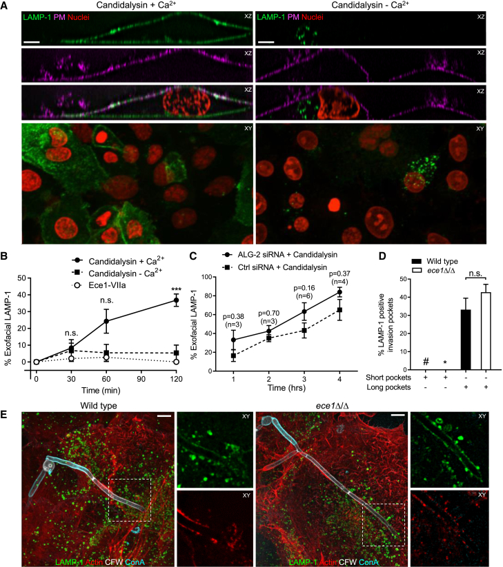
Candida albicans is both a commensal and an opportunistic fungal pathogen. Invading hyphae of C. albicans secrete candidalysin, a pore-forming peptide toxin. To prevent cell death, epithelial cells must protect themselves from direct damage induced by candidalysin and by the mechanical forces exerted by expanding hyphae. We identify two key Ca2+-dependent repair mechanisms employed by epithelial cells to withstand candidalysin-producing hyphae. Using camelid nanobodies, we demonstrate candidalysin secretion directly into the invasion pockets induced by elongating C. albicans hyphae. The toxin induces oscillatory increases in cytosolic [Ca2+], which cause hydrolysis of PtdIns(4,5)P2 and loss of cortical actin. Epithelial cells dispose of damaged membrane regions containing candidalysin by an Alg-2/Alix/ESCRT-III-dependent blebbing process. At later stages, plasmalemmal tears induced mechanically by invading hyphae are repaired by exocytic insertion of lysosomal membranes. These two repair mechanisms maintain epithelial integrity and prevent mucosal damage during both commensal growth and infection by C. albicans.

Keywords: ALG-2; Candida albicans; ESCRT; calcium; candidalysin; epithelia; lysosome; membrane damage; plasma membrane; repair.


Conflict of interest statement

Declaration of interests The authors declare no competing interests.

Figures

None
Graphical abstract
Figure 1
Figure 1
Characterization of the C. albicans-containing invasion pocket (A–C) Confocal micrographs of short TR146 invasion pockets induced by wild-type C. albicans. In these and subsequent images the entire C. albicans was stained with calcofluor white (CFW) and those portions of the fungus outside the invasion pocket with concanavalin A (ConA). The PM was identified by expression of PM-RFP. Here and in all subsequent images, actin refers specifically to F-actin, which was stained with phalloidin (A); LAMP-1 immunostaining (B); PtdIns(3)P detected by expression of PX-GFP (C). (D–F) Micrographs of RAW264.7 dectin-1 macrophages with frustrated phagosomes containing C. albicans. Micrographs show F-actin (D); LAMP-1 (E); PtdIns(3)P (F). (G–I) Micrographs of long TR146 invasion pockets containing wild-type C. albicans. Micrographs show F-actin (G); LAMP-1 (H); PtdIns(3)P (I). Images are representative of ≥3 experiments of each type. All scale bars represent 5 μm.
Figure 2
Figure 2
Damage to the invasion pocket leads to influx of extracellular Ca2+, hydrolysis of PtdIns(4,5)P2, and loss of cortical actin (A) Confocal micrograph of short invasion pockets containing C. albicans. TR146 cells were transfected with the PtdIns(4,5)P2-binding probe PLCδ-GFP and stained as in Figure 1. (B) Micrograph of a long invasion pocket. TR146 cells transfected with the PtdIns(4)P-binding probe 2xP4M-GFP. (C) Micrograph of PLCδ-GFP-transfected TR146 cells before (top) and after (bottom) ionomycin treatment. (D) Immunofluorescence staining of candidalysin using the CaLF1 nanobody in invasion pockets formed by wild-type C. albicans (left) or by the ece1Δ/Δ mutant (right). Images representative of ≥30 observations in 3 independent experiments. (E) Micrographs of PLCδ-GFP-transfected TR146 cells before (top left) and after treatment with the control peptide Ece1-VIIa (top middle), and after treatment with 30 μM candidalysin (top right) in the presence of extracellular Ca2+. Bottom panels show a similar experiment in the absence of extracellular Ca2+. (F) Quantification of PM/cytosolic ratio of PtdIns(4,5)P2 from experiments like those in (E). Data are means ± SEM of ≥3 individual experiments of each type. (G) Micrograph of pockets induced by wild-type C. albicans (left) or the ece1Δ/Δ mutant (right). TR146 cells were transfected with the PtdIns(4,5)P2-binding probe PLCδ-GFP. Inset in left panel shows the outline of the hypha. Images are representative of ≥ 30 cells from 3 experiments of each type. All scale bars represent 5 μm.
Figure 3
Figure 3
Candidalysin causes [Ca2+]c oscillations (A–C) TR146 cells were transfected with GCaMP6s-GFP and incubated with 30 μM candidalysin in the presence (A, top row, and B) or absence of extracellular Ca2+ (A, bottom row, and C). GCaMP6s fluorescence was monitored every 20 s for 12 min, followed by addition of thapsigargin (TG, 500 nM) for an additional 3 min. [Ca2+]c changes, monitored using GCaMP6s, in 12 representative cells in response to candidalysin treatment in the presence of extracellular Ca2+, followed by TG. (C) [Ca2+]c changes of 12 representative cells after candidalysin treatment in the absence of extracellular Ca2+, followed by addition of TG. Images and graphs are representative of at least 3 experiments of each type. Scale bars represent 20 μm.
Figure 4
Figure 4
Epithelial cells respond to rupture by the formation of PM-derived blebs (A) Representative micrographs of TR146 invasion pockets induced by wild-type, ece1Δ/Δ, and ece1Δ/Δ+ECE1Δ184-279 strains of C. albicans. PM bleb formation was assessed by staining non-permeabilized cells with 10 μM FM4-64. Side panels show the XZ and YZ slices corresponding to areas marked by dashed boxes. (B) Micrographs of TR146 cells treated with 30 μM Ece1-VIIa peptide or candidalysin in the presence or absence of extracellular Ca2+. PM bleb formation was assessed by staining non-permeabilized cells with 10 μM FM4-64. Outlines of TR146 cells indicated by dotted lines. Side panels show the XZ and YZ slices corresponding to the areas marked by the white dashed crosshairs. (C) Micrographs of TR146 cells transfected with PM-RFP and treated with either 30 μM Ece1-VIIa peptide or candidalysin. Localization of candidalysin was assessed by staining non-permeabilized cells with CaLH1 nanobody. Side panels show XZ slices of individual channels, corresponding to the dashed white lines. Outlines of cells are indicated by dotted lines. (D) Representative confocal micrographs of TR146 invasion pockets induced by wild-type and ece1Δ/Δ strains of C. albicans. Bleb formation and candidalysin were visualized by staining non-permeabilized cells with 10 μM FM4-64 and CaLF1 nanobody, respectively. Side panels are XY slices showing CalF1 (green), FM4-64 (magenta), and merged channels, for areas marked by dotted boxes. Scale bars represent 5 μm. Images representative of ≥3 experiments of each type. See also Figure S1.
Figure 5
Figure 5
PM damage is followed by recruitment of ALG-2 and ESCRT-III leading to PM repair and shedding of the damaged membrane (A) Micrograph of invasion pockets containing C. albicans. TR146 cells were transfected with CHMP2a-GFP (left), CHMP4B-mCherry (right). Side panels show the XY and YZ slices corresponding to areas marked by the dotted boxes. (B) Micrograph of pockets containing wild-type or ece1Δ/Δ C. albicans. TR146 cells were transfected with CHMP2a-GFP (left), CHMP4B-mCherry (right), and PM bleb formation was assessed staining non-permeabilized cells with 10 μM FM4-64. Side panels show the XZ and YZ slices corresponding to the areas marked by the dotted boxes. (C) Micrographs of TR146 cells treated with 30 μM Ece1-VIIa peptide or candidalysin. Cells were transfected with CHMP2a-GFP and PM bleb formation assessed by staining non-permeabilized cells with 10 μM FM4-64. (A–C) Outlines of TR146 cells indicated by dotted lines. Scale bars represent 5 μm. (D) Confocal micrographs of TR146 cells infected with wild-type (left panel) or ece1Δ/Δ mutant C. albicans (right panel). ALG-2 was detected by immunofluorescence. Scale bar represents 10 μm. (E) Quantification of ALG-2 accumulation as a function of invasion pocket length, from experiments like those in (D). Data are means ± SEM of ≥3 individual experiments. (F and G) Micrographs of TR146 cells treated with 30 μM candidalysin. ALG-2 (F) and ALIX (G) detected by immunofluorescence and PM blebs detected staining the PM with ConA. Images are representative of ≥3 experiments of each type. See also Figure S2.
Figure 6
Figure 6
Silencing ALG-2 prevents ESCRT-III-mediated membrane repair and curtails epithelial viability TR146 cells were treated with ALG-2 siRNA or scrambled siRNA (control). (A and B) ALG-2 silencing verified by immunoblotting using vinculin as loading control. Data in B are means ± SEM of 6 separate determinations. (C) Representative micrographs of TR146 cells treated with 30 μM candidalysin. Membranes were stained with FM4-64 and PM rupture was detected by endomembrane staining and using DAPI. Insets: mitochondrial membrane potential assessed with rhodamine-123. Outlines of TR146 cells indicated by dotted lines. Scale bar represents 5 μm; dashed line indicates the position where the XZ image on top was constructed. (D) Quantification of PM blebbing from experiments like those in C. Data are means ± SEM of ≥3 individual experiments. (E) Representative time courses of [Ca2+]c determinations made using GCaMP6s following 30 μM candidalysin treatment in Ca2+-containing medium. TG (500 nM) was added where indicated. (F) Estimation of the [Ca2+]c changes induced by 10 μM candidalysin. Standard deviation of the [Ca2+]c changes induced by 10 μM candidalysin in cells treated with control or ALG-2 siRNA. Data are means ± SEM of at ≥3 individual experiments. (G) Representative micrographs of TR146 cells treated with 10 μM candidalysin and stained with propidium iodide to identify dead cells, then permeabilized with 0.2% Triton X-100 and counter-stained to visualize all nuclei using Sytox Green. Scale bar represents 200 μm. (H) Quantification of cell death from experiments like those in (C). Data are means ± SEM of ≥3 individual experiments. See also Figure S3.
Figure 7
Figure 7
Lysosome exocytosis contributes to repair PM damage during C. albicans infection (A) Representative micrographs of TR146 cells treated with 30 μM candidalysin in the presence or absence of extracellular Ca2+. Lysosome exocytosis assessed detecting exofacial LAMP-1 staining in non-permeabilized cells with an antibody directed to a luminal epitope. The PM was stained with ConA and nuclei were stained with propidium iodide (PI) to visualize all cells. Scale bars represent 5 μm in XZ panels and 15 μm in XY panels. (B) Quantification of exofacial LAMP1 from experiments like (A). Data are means ± SEM of ≥3 individual experiments of each type. (C) Quantification of exofacial LAMP-1 in TR146 cells treated with ALG-2 siRNA or control siRNA. Data are means ± SEM. p value and experimental number of determinations (n) shown above each data point. (D) Quantification of LAMP-1 insertion in invasion pockets formed in TR146 cells by wild-type or ece1Δ/Δ mutant C. albicans. Data are means ± SEM of ≥3 individual experiments. #, LAMP-1 not detected in >50 short pockets containing wild-type C. albicans. , LAMP-1 not detected in >50 short pockets containing ece1Δ/Δ C. albicans. (E) Representative micrographs of cells infected by wild-type (left panel) or ece1Δ/Δ mutant C. albicans (right panel). Scale bar represents 5 μm. See also Figure S4.

Similar articles

Cited by

References

    1. Aggor F.E.Y., Break T.J., Trevejo-Nuñez G., Whibley N., Coleman B.M., Bailey R.D., Kaplan D.H., Naglik J.R., Shan W., Shetty A.C., et al. Oral epithelial IL-22/STAT3 signaling licenses IL-17-mediated immunity to oral mucosal candidiasis. Sci. Immunol. 2020;5:eaba0570. - PMC - PubMed
    1. Allert S., Förster T.M., Svensson C.-M., Richardson J.P., Pawlik T., Hebecker B., Rudolphi S., Juraschitz M., Schaller M., Blagojevic M., et al. Candida albicans-Induced Epithelial Damage Mediates Translocation through Intestinal Barriers. MBio. 2018;9:e00915–e00918. - PMC - PubMed
    1. Babiychuk E.B., Monastyrskaya K., Potez S., Draeger A. Intracellular Ca(2+) operates a switch between repair and lysis of streptolysin O-perforated cells. Cell Death Differ. 2009;16:1126–1134. - PubMed
    1. Baracca A., Sgarbi G., Solaini G., Lenaz G. Rhodamine 123 as a probe of mitochondrial membrane potential: evaluation of proton flux through F(0) during ATP synthesis. Biochim. Biophys. Acta. 2003;1606:137–146. - PubMed
    1. Bichele R., Kärner J., Truusalu K., Smidt I., Mändar R., Conti H.R., Gaffen S.L., Peterson P., Laan M., Kisand K. IL-22 neutralizing autoantibodies impair fungal clearance in murine oropharyngeal candidiasis model. Eur. J. Immunol. 2018;48:464–470. - PMC - PubMed

Publication types

MeSH terms

Substances