

Volar versus combined dorsal and volar plate fixation of complex intraarticular distal radius fractures with small dorsoulnar fragment - a biomechanical study
- PMID: 34986819
- PMCID: PMC8734044
- DOI: 10.1186/s12891-021-04989-w
Volar versus combined dorsal and volar plate fixation of complex intraarticular distal radius fractures with small dorsoulnar fragment - a biomechanical study
Abstract
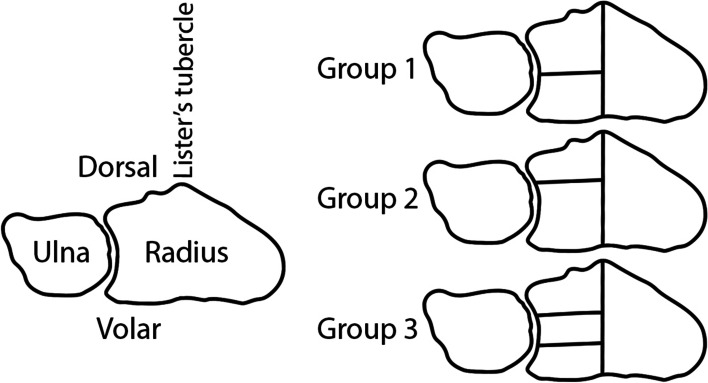
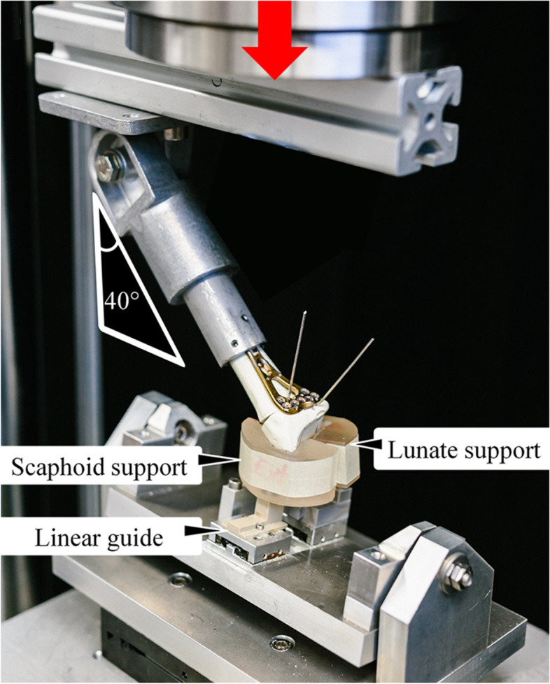
Complex intraarticular distal radius fractures (DRFs), commonly managed with volar locking plates, are challenging. Combined volar and dorsal plating is frequently applied for treatment, however, biomechanical investigations are scant. The aim of this biomechanical study was to investigate volar plating versus double plating in DRFs with different degrees of lunate facet comminution.Thirty artificial radii with simulated AO/OTA 23-C2.1 and C3.1 DRFs, including dorsal defect and lunate facet comminution, were assigned to 3 groups: Group 1 with two equally-sized lunate facet fragments; Group 2 with small dorsal and large volar fragment; Group 3 with three equally-sized fragments. The specimens underwent volar and double locked plating and non-destructive ramped loading in 0° neutral position, 40° flexion and 40° extension.In each tested position, stiffness: (1) did not significantly differ among groups with same fixation method (p ≥ 0.15); (2) increased significantly after supplemental dorsal plating in Group 2 and Group 3 (p ≤ 0.02).Interfragmentary displacements between styloid process and lunate facet in neutral position were below 0.5 mm, being not significantly different among groups and plating techniques (p ≥ 0.63).Following volar plating, angular displacement of the lunate facet to radius shaft was significantly lower in Group 1 versus both Group 2 and Group 3 (p < 0.01). It decreased significantly after supplemental dorsal plating in Group 2 and Group 3 (p < 0.01), but not in Group 1 (p ≥ 0.13), and did not differ significantly among the three groups after double plating (p ≥ 0.74).Comminution of the lunate facet within its dorsal third significantly affected the biomechanical outcomes related to complex intraarticular DRFs treated with volar and double locked plates.Double plating demonstrates superior stability versus volar plating only for lunate facet comminution within its dorsal third. In contrast, volar plating could achieve stability comparable with double plating when the dorsal third of the lunate facet is not separated by the fracture pattern. Both fixation methods indicated achievable absolute stability between the articular fragments.
Keywords: Biomechanical testing; Complex intraarticular distal radius fracture; Dorsoulnar fragment fixation; Double plating; Volar plate.
© 2022. The Author(s).
Conflict of interest statement
The authors declare that they have no affiliations with or involvement in any organization or entity with any financial interest (such as honoraria; educational grants; participation in speakers’ bureaus; membership, employment, consultancies, stock ownership, or other equity interest; and expert testimony or patent-licensing arrangements), or non-financial interest (such as personal or professional relationships, affiliations, knowledge or beliefs) in the subject matter or materials discussed in this manuscript
Figures


References
-
- Ikeda K, Osamura N, Tada K. Fixation of an ulnodorsal fragment when treating an intra-articular fracture in the distal radius. Hand Surg. 2014;19(1):139–144. - PubMed
-
- Miyashima Y, Kaneshiro Y, Yano K, Teraura H, Sakanaka H, Uemura T. Size and stabilization of the dorsoulnar fragment in AO C3-type distal radius fractures. Injury. 2019;50(11):2004–2008. - PubMed
MeSH terms
LinkOut - more resources
Full Text Sources
Medical
Miscellaneous
