. 2022 Jan;8(1):e001949.
doi: 10.1136/rmdopen-2021-001949.

Role of synovial fibroblast subsets across synovial pathotypes in rheumatoid arthritis: a deconvolution analysis

Affiliations

Role of synovial fibroblast subsets across synovial pathotypes in rheumatoid arthritis: a deconvolution analysis

Raphael Micheroli et al. RMD Open. 2022 Jan.

Abstract

Objectives: To integrate published single-cell RNA sequencing (scRNA-seq) data and assess the contribution of synovial fibroblast (SF) subsets to synovial pathotypes and respective clinical characteristics in treatment-naïve early arthritis.

Methods: In this in silico study, we integrated scRNA-seq data from published studies with additional unpublished in-house data. Standard Seurat, Harmony and Liger workflow was performed for integration and differential gene expression analysis. We estimated single cell type proportions in bulk RNA-seq data (deconvolution) from synovial tissue from 87 treatment-naïve early arthritis patients in the Pathobiology of Early Arthritis Cohort using MuSiC. SF proportions across synovial pathotypes (fibroid, lymphoid and myeloid) and relationship of disease activity measurements across different synovial pathotypes were assessed.

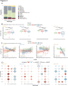
Results: We identified four SF clusters with respective marker genes: PRG4+ SF (CD55, MMP3, PRG4, THY1neg ); CXCL12+ SF (CXCL12, CCL2, ADAMTS1, THY1low ); POSTN+ SF (POSTN, collagen genes, THY1); CXCL14+ SF (CXCL14, C3, CD34, ASPN, THY1) that correspond to lining (PRG4+ SF) and sublining (CXCL12+ SF, POSTN+ + and CXCL14+ SF) SF subsets. CXCL12+ SF and POSTN+ + were most prominent in the fibroid while PRG4+ SF appeared highest in the myeloid pathotype. Corresponding, lining assessed by histology (assessed by Krenn-Score) was thicker in the myeloid, but also in the lymphoid pathotype + the fibroid pathotype. PRG4+ SF correlated positively with disease severity parameters in the fibroid, POSTN+ SF in the lymphoid pathotype whereas CXCL14+ SF showed negative association with disease severity in all pathotypes.

Conclusion: This study shows a so far unexplored association between distinct synovial pathologies and SF subtypes defined by scRNA-seq. The knowledge of the diverse interplay of SF with immune cells will advance opportunities for tailored targeted treatments.

Keywords: arthritis; fibroblasts; rheumatoid; synovitis.


Conflict of interest statement

Competing interests: None declared.

Figures

Figure 1
Figure 1
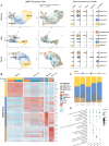
Individual synovial tissue datasets with respective used methods and number of synovial fibroblasts (SF), UMAP visualisation and SF positive (COL1A2) and negative (PTPRC, vWF) marker genes. Selected SF clusters in unsorted datasets are marked in grey in the UMAP plot. CER = Center of Experimental Rheumatology Zurich, UMAP = Uniform Manifold Approximation and Projection for Dimension Reduction. UMAP; Uniform Manifold Approximation and Projection for Dimension Reduction.
Figure 2
Figure 2
Identification of synovial fibroblast (SF) subsets (A) different integration methods with UMAP visualisation, grouping of cells according to datasets and violinplots showing the gene expression per cluster of CD55, THY1, CD34, POSTN and HLA-DRA. (B) Heatmap of the 10 most significant marker genes of each SF cluster (via Seurat). (C) Distribution of seurat clusters across the different datasets. (D) KEGG pathways enrichment analysis across SF clusters. Top 20 pathways are shown. UMAP; Uniform Manifold Approximation and Projection for Dimension Reduction.
Figure 3
Figure 3
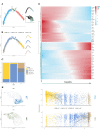
Pseudotime analysis of synovial fibroblast (SF) subsets. (A) Visualisation of the trajectory states of all SF. Top and middle right shows the pseudotime trajectory in the reduced dimension and UMAP visualisation. (B) Distribution of the SF cluster along the trajectory (right split by cluster). (C) Distribution of SF clusters in the main trajectory states 1, 3 and 5. (D) Most significant genes that covary across pseudotime split in two clusters (state one left, state five right). (E) Plot of gene expression levels of PRG4 and THY1 in the UMAP of the SF clusters on the left and along the pseudotime trajectory on the right. UMAP = Uniform Manifold Approximation and Projection for Dimension Reduction.
Figure 4
Figure 4
Synovial fibroblasts (SF) across synovial pathotypes and comparison with disease activity measurements. (A) Distribution of different cell types between the different synovial pathotypes. (B) Proportion of the different SF subtypes within the pathotypes. (C) Correlation between disease activity measurements and SF proportions across synovial pathotypes. (D) Dotplot with Pearson correlation of different clinical parameters with SF subtypes across pathotypes. DAS; disease activity score. ESR; erythrocyte sedimentation rate. FDR; false discovery rate. HAQ; health assessment questionnaire. SJC; swollen joint count. TJC; tender joint count. VAS; visual analog scale.

References

    1. Turner JD, Filer A. The role of the synovial fibroblast in rheumatoid arthritis pathogenesis. Curr Opin Rheumatol 2015;27:175–82. 10.1097/BOR.0000000000000148 - DOI - PubMed
    1. Humby F, Lewis M, Ramamoorthi N, et al. . Synovial cellular and molecular signatures stratify clinical response to csDMARD therapy and predict radiographic progression in early rheumatoid arthritis patients. Ann Rheum Dis 2019;78:761–72. 10.1136/annrheumdis-2018-214539 - DOI - PMC - PubMed
    1. Lewis MJ, Barnes MR, Blighe K, et al. . Molecular portraits of early rheumatoid arthritis identify clinical and treatment response phenotypes. Cell Rep 2019;28:2455–70. 10.1016/j.celrep.2019.07.091 - DOI - PMC - PubMed
    1. Stephenson W, Donlin LT, Butler A, et al. . Single-cell RNA-seq of rheumatoid arthritis synovial tissue using low-cost microfluidic instrumentation. Nat Commun 2018;9:791. 10.1038/s41467-017-02659-x - DOI - PMC - PubMed
    1. Mizoguchi F, Slowikowski K, Wei K, et al. . Functionally distinct disease-associated fibroblast subsets in rheumatoid arthritis. Nat Commun 2018;9:789. 10.1038/s41467-018-02892-y - DOI - PMC - PubMed