. 2021 Dec 20:12:801234.
doi: 10.3389/fphar.2021.801234. eCollection 2021.

mTORC1 Signaling Pathway Mediates Chronic Stress-Induced Synapse Loss in the Hippocampus

Affiliations

mTORC1 Signaling Pathway Mediates Chronic Stress-Induced Synapse Loss in the Hippocampus

Yu-Fei Luo et al. Front Pharmacol. .

Abstract

Background: The mechanistic target of rapamycin complex 1 (mTORC1) signaling has served as a promising target for therapeutic intervention of major depressive disorder (MDD), but the mTORC1 signaling underlying MDD has not been well elucidated. In the present study, we investigated whether mTORC1 signaling pathway mediates synapse loss induced by chronic stress in the hippocampus. Methods: Chronic restraint stress-induced depression-like behaviors were tested by behavior tests (sucrose preference test, forced swim test and tail suspension test). Synaptic proteins and alternations of phosphorylation levels of mTORC1 signaling-associated molecules were measured using Western blotting. In addition, mRNA changes of immediate early genes (IEGs) and glutamate receptors were measured by RT-PCR. Rapamycin was used to explore the role of mTORC1 signaling in the antidepressant effects of fluoxetine. Results: After successfully establishing the chronic restraint stress paradigm, we observed that the mRNA levels of some IEGs were significantly changed, indicating the activation of neurons and protein synthesis alterations. Then, there was a significant downregulation of glutamate receptors and postsynaptic density protein 95 at protein and mRNA levels. Additionally, synaptic fractionation assay revealed that chronic stress induced synapse loss in the dorsal and ventral hippocampus. Furthermore, these effects were associated with the mTORC1 signaling pathway-mediated protein synthesis, and subsequently the phosphorylation of associated downstream signaling targets was reduced after chronic stress. Finally, we found that intracerebroventricular infusion of rapamycin simulated depression-like behavior and also blocked the antidepressant effects of fluoxetine. Conclusion: Overall, our study suggests that mTORC1 signaling pathway plays a critical role in mediating synapse loss induced by chronic stress, and has part in the behavioral effects of antidepressant treatment.

Keywords: chronic restraint stress; depression; fluoxetine; mammalian target of rapamycin; postsynaptic density protein 95.


Conflict of interest statement

The authors declare that the research was conducted in the absence of any commercial or financial relationships that could be construed as a potential conflict of interest.

Figures
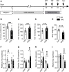
FIGURE 1
FIGURE 1
Effects of CRS on dorsal and ventral hippocampal IEG expression. (A) The schematic timeline of the experimental procedures. (B–E) Mice that exposed to CRS for 21 days displayed depression-like behavior, including reduced sucrose preference in the SPT (B), increased immobile time in the TST (C), increased floating time in the FST (D) and weight loss compared with the controls (E). (F–I) RT-qPCR assays found that the mRNA level of c-fos was selectively decreased in the dHIP, and not significantly changed in the vHIP (F). The mRNA level of △fos B was increased in the vHIP, without significant alteration in the dHIP (G). The mRNA level of Arc was upregulated in the dHIP but downregulated in the vHIP (H). The mRNA level of Egr was reduced in both the dHIP and vHIP (I). Data are presented as mean ± SEM. One-way ANOVA for all. n = 8-12 per group for (B–E); n = 5–7 per group for (F–I). **p < 0.01, ***p < 0.001 vs. Ctrl group.
FIGURE 2
FIGURE 2
Expression of glutamate receptors in the hippocampus of chronically stressed mice. (A,B) CRS selectively decreased the expression levels of PSD-95, GluA1, GluA3, GluN2A and GluN2B, while increased GluN1 expression in the dHIP. (C) The mRNA levels of PSD-95, GluN1 and GluN2A were reduced in the dHIP after CRS. (D,E) CRS selectively decreased the expression of PSD-95, GluA2, GluN2A and GluN2B, while increased GluN1 expression in the vHIP. (F) The mRNA levels of PSD-95, GluA3, GluA4, and GluN2B were reduced; while GluN1 mRNA level was upregulated in the vHIP after CRS. Data are presented as mean ± SEM. One-way ANOVA for all. n = 7–9 per group for (B,C,E,F). *p < 0.05, **p < 0.01, ***p < 0.001 vs. Ctrl group.
FIGURE 3
FIGURE 3
CRS mediates remarkable synapse losses in the hippocampus. (A–C) CRS decreased the expression of PSD-95 and synaptophysin in the CA1, DG, and CA3 regions of dHIP; representative Western blots (A), statistical analysis of PSD-95 (B) and synaptophysin (C). (D–F) CRS reduced the PSD-95 and synaptophysin expression in the CA1, DG, and CA3 of vHIP; representative Western blots (D), statistical analysis of PSD-95 (E) and synaptophysin (F). CRS induced synapse loss in the CA1, DG and CA3 of dHIP (G–I) and vHIP (J–L); representative Western blots (G, J), statistical analysis of PSD-95 (H, K) and synaptophysin (I, L). Data are presented as mean ± SEM. One-way ANOVA for all. n = 10–9 per group for (A–F), n = 8–9 per group for (G–L). *p < 0.05, **p < 0.01 vs. Ctrl group.
FIGURE 4
FIGURE 4
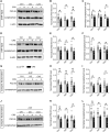
CRS mediates protein synthesis decreases by targeting mTORC1 signaling in the hippocampus. CRS significantly decreased the phosphorylation levels of the mTORC1 signaling pathway-associated targets, such as p-mTOR (A), p-Akt (B), p-ERK (C), p-p70S6K (D), p-4E-BP-1 (E) and p-CREB (F); while no effects on the expression of total mTOR, Akt, ERK, p70S6K, 4E-BP-1 and CREB were observed. Data are presented as mean ± SEM. One-way ANOVA for all. n = 7 per group for all. *p < 0.05, **p < 0.01, ***p < 0.001 vs. Ctrl group.
FIGURE 5
FIGURE 5
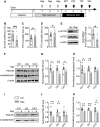
Treatment with rapamycin induces depression-like behaviors and synapse loss in the ventral hippocampus. (A) The schematic timeline of the experimental procedures. (B–D) Intracerebroventricular infusion of rapamycin simulated depression-like behaviors, including reduced sucrose preference in the SPT (B), increased immobile time in the TST (C), and increased floating time in the FST (D). (E–H) Rapamycin injection decreased mTOR activity in the vHIP (E), as well as the expression of PSD-95 and synaptophysin in the CA1, DG and CA3 of vHIP; representative Western blots (F), statistical analysis of PSD-95 (G) and synaptophysin (H). (I–K) Rapamycin treatment induced synapse loss in the CA1, DG and CA3 of vHIP; representative Western blots (I), statistical analysis of PSD-95 (J) and synaptophysin (K). Data are presented as mean ± SEM. One-way ANOVA for all. n = 11 per group for (B–D); n = 6 per group for (E, G,H, J,K). *p < 0.05, **p < 0.01, ***p < 0.001 vs. Ctrl group.
FIGURE 6
FIGURE 6
Rapamycin abolishes the effects of fluoxetine in chronically stressed mice. (A) The schematic timeline of the experimental procedures. (B–D) The antidepressant action of fluoxetine was abolished by rapamycin, including reduced sucrose preference in the SPT (B), increased immobile time in the TST (C), and increased floating time in the FST (D). (E) Schematic model for the role of mTORC1 signaling pathway in mediating synapse loss induced by chronic stress. CRS suppresses the mTORC1 signaling, followed by decreased phosphorylation levels of mTORC1 signaling-associated target proteins, which triggers the downregulation of protein synthesis and the synapse loss to contribute to depression-like behaviors. Data are presented as mean ± SEM. Two-way ANOVA for all. Respectively, n = 11, 9, 9 and 9 per group for (B–D). *p < 0.05, **p < 0.01 vs. Saline + Veh group; # p < 0.05, ## p < 0.01 vs. FLX + Veh group.

Similar articles

Cited by

References

    1. Abdallah C. G., Averill L. A., Gueorguieva R., Goktas S., Purohit P., Ranganathan M., et al. (2020). Modulation of the Antidepressant Effects of Ketamine by the mTORC1 Inhibitor Rapamycin. Neuropsychopharmacology 45 (6), 990–997. 10.1038/s41386-020-0644-9 - DOI - PMC - PubMed
    1. Abelaira H. M., Réus G. Z., Neotti M. V., Quevedo J. (2014). The Role of mTOR in Depression and Antidepressant Responses. Life Sci. 101 (1-2), 10–14. 10.1016/j.lfs.2014.02.014 - DOI - PubMed
    1. Admon R., Leykin D., Lubin G., Engert V., Andrews J., Pruessner J., et al. (2013). Stress-induced Reduction in Hippocampal Volume and Connectivity with the Ventromedial Prefrontal Cortex Are Related to Maladaptive Responses to Stressful Military Service. Hum. Brain Mapp. 34 (11), 2808–2816. 10.1002/hbm.22100 - DOI - PMC - PubMed
    1. Armbrecht E., Shah R., Poorman G. W., Luo L., Stephens J. M., Li B., et al. (2021). Economic and Humanistic Burden Associated with Depression and Anxiety Among Adults with Non-communicable Chronic Diseases (NCCDs) in the United States. J. Multidiscip Healthc. 14, 887–896. 10.2147/JMDH.S280200 - DOI - PMC - PubMed
    1. Autry A. E., Adachi M., Nosyreva E., Na E. S., Los M. F., Cheng P. F., et al. (2011). NMDA Receptor Blockade at Rest Triggers Rapid Behavioural Antidepressant Responses. Nature 475 (7354), 91–95. 10.1038/nature10130 - DOI - PMC - PubMed

LinkOut - more resources