. 2021 Dec 27:2021:4475184.
doi: 10.1155/2021/4475184. eCollection 2021.

Correlation Analysis of Synchronization Type and Degree in Respiratory Neural Network

Affiliations

Correlation Analysis of Synchronization Type and Degree in Respiratory Neural Network

Jieqiong Xu et al. Comput Intell Neurosci. .

Abstract

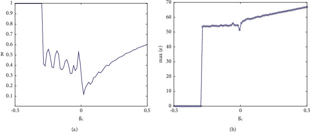
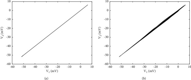
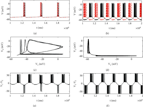
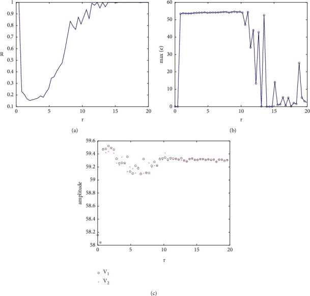
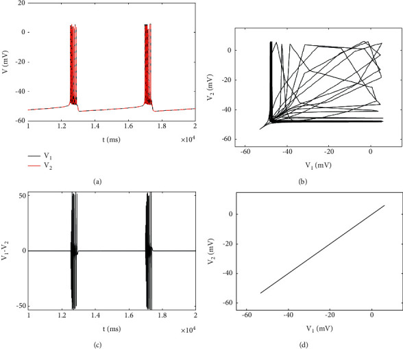
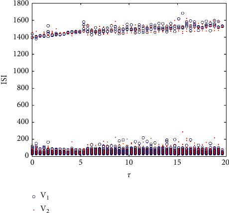
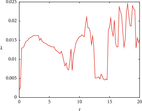
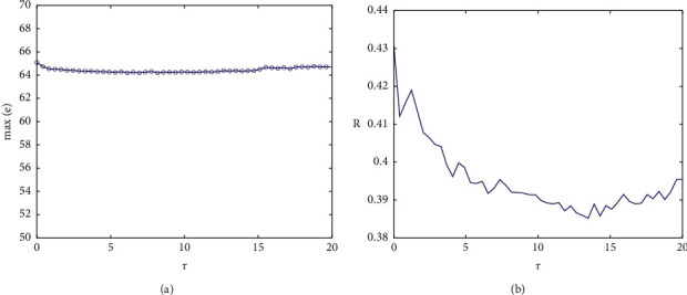
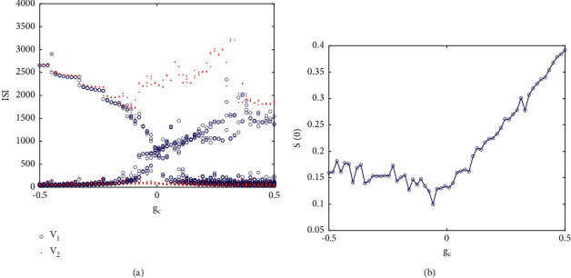
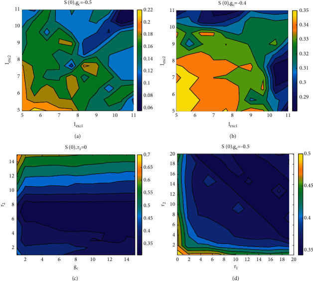
Pre-Bötzinger complex (PBC) is a necessary condition for the generation of respiratory rhythm. Due to the existence of synaptic gaps, delay plays a key role in the synchronous operation of coupled neurons. In this study, the relationship between synchronization and correlation degree is established for the first time by using ISI bifurcation and correlation coefficient, and the relationship between synchronization and correlation degree is discussed under the conditions of no delay, symmetric delay, and asymmetric delay. The results show that the phase synchronization of two coupling PBCs is closely related to the weak correlation, that is, the weak phase synchronization may occur under the condition of incomplete synchronization. Moreover, the time delay and coupling strength are controlled in the modified PBC network model, which not only reveals the law of PBC firing transition but also reveals the complex synchronization behavior in the coupled chaotic neurons. Especially, when the two coupled neurons are nonidentical, the complete synchronization will disappear. These results fully reveal the dynamic behavior of the PBC neural system, which is helpful to explore the signal transmission and coding of PBC neurons and provide theoretical value for further understanding respiratory rhythm.


Conflict of interest statement

The authors declare that they have no conflicts of interest.

Figures

Figure 1
Figure 1
ISI bifurcation diagram of single neuron with parameter Iexc.
Figure 2
Figure 2
(a) Time history diagram of membrane potential when Iexc=8.5. (b) The phase diagram corresponding to the plane (h, V).
Figure 3
Figure 3
Bifurcation diagram of a single PBC neuron with respect to parameter Iexc. The upper right corner shows the local enlarged view.
Figure 4
Figure 4
ISI bifurcation diagram of coupling strength gc.
Figure 5
Figure 5
(a) The variation between the correlation coefficient and coupling strength gc of two coupled neurons. (b) Change diagram between maximum synchronization difference and coupling strength gc.
Figure 6
Figure 6
The phase diagram of two coupled PBC neurons on the (V1, V2) plane when the coupling strength is different: (a) gc=−0.5 and (b) gc=−0.24.
Figure 7
Figure 7
The phase diagrams on different planes with different values of coupling strength, gc=−0.1 in the left column and gc=0.4 in the right column. (a) and (b) Time history diagram of the membrane potential of two neurons: black and red are neurons 1 and 2, respectively. (c) and (d) The phase diagram in planes (V1, V2). (e) and (f) Synchronization difference of membrane potential.
Figure 8
Figure 8
When gc=−0.5: (a) correlation coefficient of membrane potential of coupled neurons with time delay, (b) a graph showing the relationship between the maximum synchronization difference and the change of τ, and (c) the relationship between the amplitude of two PBC neurons and τ.
Figure 9
Figure 9
Phase plan diagram of two PBC neurons: (a) the time history diagram of coupled neurons when τ=5, (b) the phase diagram in the plane (V1, V2) when τ=5, (c) time history diagram of synchronization difference when τ=5, and (d) phase diagram in plane (V1, V2) when τ=17.
Figure 10
Figure 10
Bifurcation diagram of ISI and τ in two coupled neurons when gc=0.4.
Figure 11
Figure 11
The maximum Lyapunov exponent with time delay τ.
Figure 12
Figure 12
(a) The relationship between the maximum synchronization difference of the membrane potential of coupling neurons and τ. (b) The change diagram of correlation coefficient R and τ when gc=0.4.
Figure 13
Figure 13
Correlation coefficient and maximum synchronization difference of coupled neuron membrane potential in two-parameter space (τ, gc): (a) correlation coefficient, (b) maximum synchronization difference, and (c) similarity functions in the local neighborhood of graphs (a) and (b).
Figure 14
Figure 14
Correlation coefficient of asymmetric time-delay coupled neurons in two-parameter plane when gc=−0.5.
Figure 15
Figure 15
The phase difference between the two coupled neurons and τ2 when different τ1.
Figure 16
Figure 16
The relationship between period and amplitude with τ2 when different τ1.
Figure 17
Figure 17
(a) The relationship between the ISI sequence of two coupled neurons and the coupling strength in the case of nonidentical and asymmetric time-delay coupling. (b) The relationship between similarity function and coupling strength.
Figure 18
Figure 18
Similar functions on different two-parameter planes: (a) when gc=−0.5, the similar functions on the plane (Iexc1, Iexc2); (b) when gc=0.4, the similar functions on the plane (Iexc1, Iexc2); (c) when τ1=0, the similar functions on the plane (gc, τ2); and (d) when gc=−0.5, the similar functions on the plane (τ1, τ2).

Similar articles

Cited by

References

    1. Lestienne R. Spike timing, synchronization and information processing on the sensory side of the central nervous system. Progress in Neurobiology . 2001;65(6):545–591. doi: 10.1016/s0301-0082(01)00019-3. - DOI - PubMed
    1. Volgushev M., Chauvette S., Mukovski M., Timofeev I. Precise long-range synchronization of activity and silence in neocortical neurons during slow-wave sleep. Journal of Neuroscience . 2006;26(21):5665–5672. doi: 10.1523/jneurosci.0279-06.2006. - DOI - PMC - PubMed
    1. Codrons E., Bernardi N. F., Vandoni M., Bernardi L. Spontaneous group synchronization of movements and respiratory rhythms. PLoS One . 2014;9(9) doi: 10.1371/journal.pone.0107538.e107538 - DOI - PMC - PubMed
    1. Ramirez J. M., Baertsch N. A. The dynamic basis of respiratory rhythm generation: one breath at a time. Annual Review of Neuroscience . 2018;41:475–499. doi: 10.1146/annurev-neuro-080317-061756. - DOI - PMC - PubMed
    1. Hammond C., Bergman H., Brown P. Pathological synchronization in Parkinson’s disease: networks, models and treatments. Trends in neurosciences . 2007;30(7):357–364. doi: 10.1016/j.tins.2007.05.004. - DOI - PubMed

LinkOut - more resources