Case Reports
. 2021 Dec 26:2021:7697987.
doi: 10.1155/2021/7697987. eCollection 2021.

Tiger-Striped PASH: Recognition of a Unique Morphology Allows for a Zippered-Up Diagnosis of Pseudoangiomatous Stromal Hyperplasia of Breast

Affiliations
Case Reports

Tiger-Striped PASH: Recognition of a Unique Morphology Allows for a Zippered-Up Diagnosis of Pseudoangiomatous Stromal Hyperplasia of Breast

Mohamad Sakibuzzaman et al. Case Rep Pathol. .

Abstract

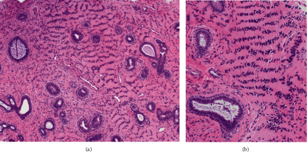
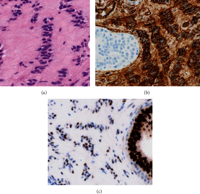
Pseudoangiomatous stromal hyperplasia (PASH) of the breast is histologically characterized by anastomosing and slit-like spaces invested by collagenous stroma and lined by flattened, spindle cells. These clear spaces that may mimic microscopic vascular channels do not contain red blood cells. Immunohistochemistry (IHC) studies may also help to confirm a diagnosis of PASH, with the spindled cells marking positively with CD34 and PR while demonstrating no reactivity with more specific endothelial antigens such as CD31 and ERG. In the current case, a 39-year-old female was diagnosed with cellular PASH of the right breast with unique histological patterns showing "tiger-striped" and "zippered" histologies. To our knowledge, this is the first report of these unique variant PASH morphologies.


Conflict of interest statement

All authors have no conflict of interests and no relevant financial disclosures.

Figures

Figure 1
Figure 1
(a) MRI of the right breast with IV gadolinium shows an oval enhancing mass in the upper inner right breast middle depth; (b) T2-weighted images show bright signal, though less bright than cysts seen elsewhere in the breast. Slit-like spaces are not definitely appreciated, possibly due to the small size of the finding. (c) Second-look ultrasound shows an oval, microlobulated, hypoechoic mass in the upper inner right breast, corresponding with that seen on MRI.
Figure 2
Figure 2
Low-power histologic assessment shows scattered nonneoplastic breast epithelial elements including scattered ducts and the edge of a terminal duct lobular unit with stromal expansion between and within these epithelial structures. Eosinophilic expanses of fibrous tissue are noted with linear arrangements of spindle cell nuclei resulting in a tiger-stripe-like pattern ((a) hematoxylin and eosin, 100X). At intermediate magnification, the nuclear arrangements with offset eosinophilic cytoplasmic bands emphasize the striped morphology ((b) hematoxylin and eosin, 200X).
Figure 3
Figure 3
High-power histologic assessment confirming a zippered morphology with interlacing lesional nuclei oriented perpendicular to the cleft space ((a) hematoxylin and eosin, 400X). The lesional spindled cells demonstrate diffuse and strong immunoreactivity with CD34 while the adjacent epithelium is nonreactive ((b) CD34 IHC, 600X). The lesional spindled cells also show immunoreactivity with progesterone receptor ((c) PR IHC, 600X).

References

    1. Naso J. R., Chiu C. G., Goecke M. E., Chang D., Shiau C. J. Benign spindle cell lesions of the breast: a diagnostic approach to solitary fibrous tumour, nodular pseudoangiomatous stromal hyperplasia and nodular fasciitis. Journal of Clinical Pathology . 2019;72(6):438–442. doi: 10.1136/jclinpath-2018-205561. - DOI - PubMed
    1. Layon D. R., Wang C., Roth S., Brooks A. D. Is surgical excision necessary in pseudoangiomatous stromal hyperplasia? The Breast Journal . 2016;22(5):595–596. doi: 10.1111/tbj.12643. - DOI - PubMed
    1. Yoon K. H., Koo B., Lee K. B., et al. Optimal treatment of pseudoangiomatous stromal hyperplasia of the breast. Asian Journal of Surgery . 2020;43(7):735–741. doi: 10.1016/j.asjsur.2019.09.008. - DOI - PubMed
    1. Vuitch M. F., Rosen P. P., Erlandson R. A. Pseudoangiomatous hyperplasia of mammary stroma. Human Pathology . 1986;17(2):185–191. doi: 10.1016/s0046-8177(86)80292-1. - DOI - PubMed
    1. Rosa G., Dawson A., Rowe J. J. Does identifying whether pseudoangiomatous stromal hyperplasia (PASH) is focal or diffuse on core biopsy correlate with a PASH nodule on excision? International Journal of Surgical Pathology . 2017;25(4):292–297. doi: 10.1177/1066896916676585. - DOI - PubMed

Publication types