

Case Reports
doi: 10.1016/j.hrcr.2021.09.009.
eCollection 2021 Dec.
Noninvasive electrocardiographic imaging of dynamic atrioventricular delay programming in a patient with left bundle branch block
Affiliations
- PMID: 34987974
- PMCID: PMC8695252
- DOI: 10.1016/j.hrcr.2021.09.009
Item in Clipboard
Case Reports
Noninvasive electrocardiographic imaging of dynamic atrioventricular delay programming in a patient with left bundle branch block
HeartRhythm Case Rep.
.
No abstract available
Keywords: Atrioventricular delay; CRT optimization; Electrocardiography imaging; MultiPoint Pacing; SyncAV.
Figures
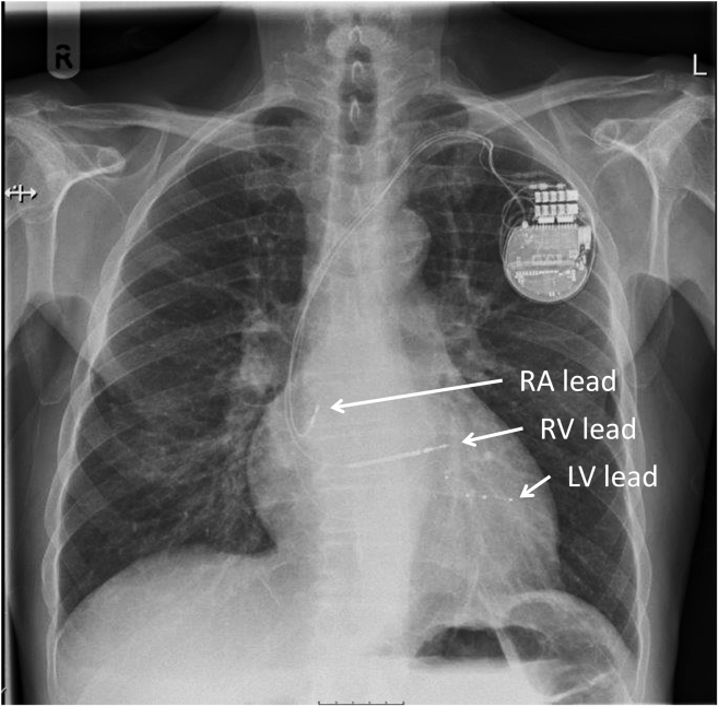
Chest radiograph displaying device and lead positions. This posteroanterior projection shows the final positions of the device and leads. Left ventricular (LV) lead was placed at the basal-mid posterolateral branch of the coronary sinus, right ventricular (RV) lead at the RV apex, and right atrial (RA) lead in the RA appendage.
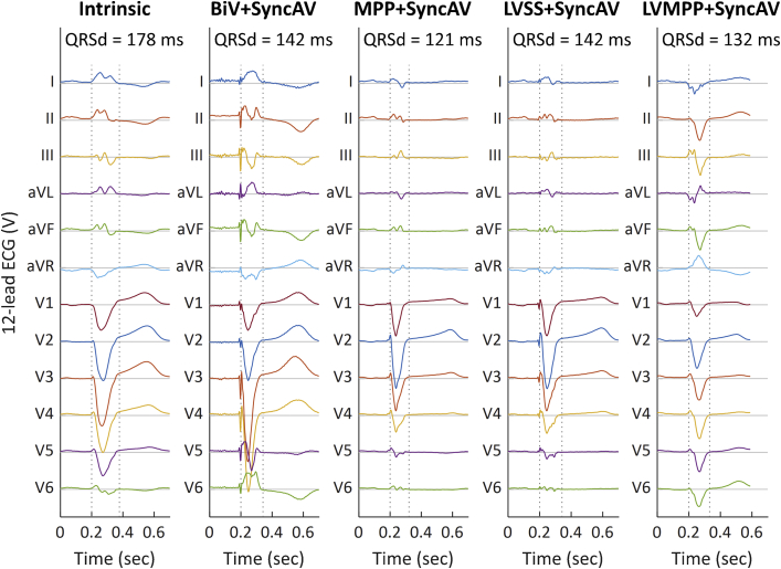
Twelve-lead surface electrocardiogram (ECG). The 12-lead surface ECG ensemble signals averaged over 3 successive beats are displayed for intrinsic conduction, biventricular (BiV) + SyncAV, Multi-Point Pacing (MPP) + SyncAV, left ventricle–only single-site pacing (LVSS) + SyncAV, and LV-only MPP (LVMPP) + SyncAV (left to right). QRS durations (QRSd) are displayed, with QRS start and end times shown as dashed vertical lines.
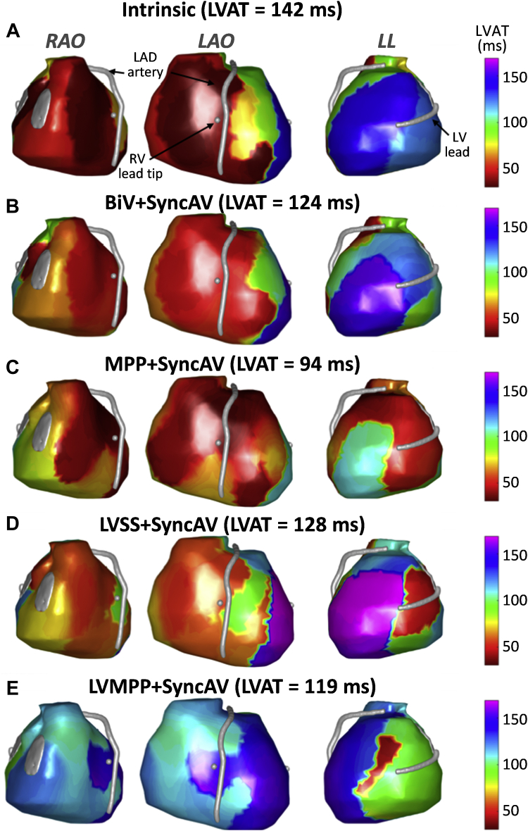
Electrocardiographic imaging activation maps. Epicardial ventricular surfaces of both ventricles are displayed in 3 views: from left to right, right anterior oblique (RAO), left anterior oblique (LAO), and left lateral (LL). The left anterior descending (LAD) artery, right ventricle (RV) lead tip, and left ventricle (LV) lead are depicted as silver surfaces. Color maps are projected onto the surface for activation time (AT, ms), ranging from early (red/orange) to late (blue/purple). Intrinsic conduction activation maps show the earliest-activating regions in the anterior RV (inclusive of septal activation) and RV free wall in keeping with intact right bundle conduction. The latest-activating LV segment is basal lateral, and a U-shaped line of activation discontinuity is present from the basal anterior-anterolateral LV spreading apically to the basal posterior LV. The impact of each pacing modality on activation pattern is displayed: A: intrinsic conduction; B: biventricular (BiV) + SyncAV; C: MultiPoint Pacing (MPP) + SyncAV; D: LV-only single-site pacing (LVSS) + SyncAV; E: LV-only MPP (LVMPP) + SyncAV.
References
-
- Arbelo E., Tolosana J.M., Trucco E., et al. Fusion-optimized intervals (FOI): a new method to achieve the narrowest QRS for optimization of the AV and VV intervals in patients undergoing cardiac resynchronization therapy. J Cardiovasc Electrophysiol. 2014;25:283–292. - PubMed
-
- Trucco E., Tolosana J.M., Arbelo E., et al. Improvement of reverse remodeling using electrocardiogram fusion-optimized intervals in cardiac resynchronization therapy: a randomized study. JACC Clin Electrophysiol. 2018;4:181–189. - PubMed
-
- Ellenbogen K.A., Gold M.R., Meyer T.E., et al. Primary results from the SmartDelay determined AV optimization: a comparison to other AV delay methods used in cardiac resynchronization therapy (SMART-AV) trial: a randomized trial comparing empirical, echocardiography-guided, and algorithmic atrioventricular delay programming in cardiac resynchronization therapy. Circulation. 2010;122:2660–2668. - PubMed
-
- Thibault B., Ritter P., Bode K., et al. Dynamic programming of atrioventricular delay improves electrical synchrony in a multicenter cardiac resynchronization therapy study. Heart Rhythm. 2019;16:1047–1056. - PubMed
Publication types
LinkOut - more resources
Full Text Sources
Research Materials
