Review
. 2021 Dec 20:9:798054.
doi: 10.3389/fcell.2021.798054. eCollection 2021.

Extracellular Vesicles: Potential Role in Remote Signaling and Inflammation in Trypanosoma cruzi-Triggered Disease

Affiliations
Review

Extracellular Vesicles: Potential Role in Remote Signaling and Inflammation in Trypanosoma cruzi-Triggered Disease

Luíza Dantas-Pereira et al. Front Cell Dev Biol. .

Abstract

Extracellular vesicles (EVs) act as cell communicators and immune response modulators and may be employed as disease biomarkers and drug delivery systems. In infectious diseases, EVs can be released by the pathogen itself or by the host cells (infected or uninfected), potentially impacting the outcome of the immune response and pathological processes. Chagas disease (CD) is caused by infection by the protozoan Trypanosoma cruzi and is the main cause of heart failure in endemic areas. This illness attracted worldwide attention due to the presence of symptomatic seropositive subjects in North America, Asia, Oceania, and Europe. In the acute phase of infection, nonspecific signs, and symptoms contribute to miss diagnosis and early etiological treatment. In this phase, the immune response is crucial for parasite control; however, parasite persistence, dysregulated immune response, and intrinsic tissue factors may contribute to the pathogenesis of chronic CD. Most seropositive subjects remain in the indeterminate chronic form, and from 30 to 40% of the subjects develop cardiac, digestive, or cardio-digestive manifestations. Identification of EVs containing T. cruzi antigens suggests that these vesicles may target host cells and regulate cellular processes and the immune response by molecular mechanisms that remain to be determined. Parasite-released EVs modulate the host-parasite interplay, stimulate intracellular parasite differentiation and survival, and promote a regulatory cytokine profile in experimental models of CD. EVs derived from the parasite-cell interaction inhibit complement-mediated parasite lysis, allowing evasion. EVs released by T. cruzi-infected cells also regulate surrounding cells, maintaining a proinflammatory profile. After a brief review of the basic features of EVs, the present study focuses on potential participation of T. cruzi-secreted EVs in cell infection and persistence of low-grade parasite load in the chronic phase of infection. We also discuss the role of EVs in shaping the host immune response and in pathogenesis and progression of CD.

Keywords: Chagas disease; Trypanosoma cruzi; extracellular vesicles; immune response; inflammation.


Conflict of interest statement

The authors declare that the research was conducted in the absence of any commercial or financial relationships that could be construed as a potential conflict of interest.

Figures

FIGURE 1
FIGURE 1
Timeline from the discovery of soluble exoantigens to description of the release of extracellular vesicles by Trypanosoma cruzi.
FIGURE 2
FIGURE 2
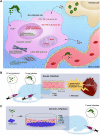
Main regulatory activities of EVs from various sources in a scenario of Trypanosoma cruzi infection analyzed in vitro (A), in vivo (B) and ex vivo (C). (A) EVs released by the parasite have been shown to promote increased transient intracellular calcium levels (Retana Moreira et al., 2019) and actin filament disorganization (Retana Moreira et al., 2019), modulate the expression of extracellular matrix components (Pinho et al., 2002; Retana Moreira et al., 2019), reduce the gene expression of proinflammatory cytokines (Cronemberger-Andrade et al., 2020) and reduce the release of NO, TNF, and IL-6 when in contact with uninfected cells (Nogueira et al., 2015; Lovo-Martins et al., 2018). The treatment with these EVs before infection with T. cruzi promotes an increase in parasite uptake in a target cell type-dependent manner (*) (Lovo-Martins et al., 2018). EVs released either by immune or nonimmune infected cells promote an increase in NO, TNF, IL-6, and IL-1β levels in uninfected cells (Choudhuri and Garg, 2020a). EVs from the blood sources obtained from CD patients promote an increase in NO and IL-1β release, increase mitochondrial ROS levels, and decrease mitochondrial membrane potential and inflammatory gene expression in EV-treated macrophages (Chowdhury et al., 2017). The treatment of macrophages with EVs obtained from the blood of mice with chronic infection increases TNF, IL-6, and IL-1β release (Choudhuri and Garg, 2020a). (B) BALB/c mice pretreated with EVs from T. cruzi and challenged by infection have reduced NO and TNF levels in the serum in the acute phase (Trocoli-Torrecilhas et al., 2009; Lovo-Martins et al., 2018), reduced iNOS expression (Trocoli-Torrecilhas et al., 2009), increased number of inflammatory infiltrates and amastigote nests in heart tissue (Trocoli-Torrecilhas et al., 2009; Lovo-Martins et al., 2018), and increased mortality (Trocoli-Torrecilhas et al., 2009). (C) Splenocytes obtained from chronically T. cruzi-infected C57Bl/6 mice treated with parasite EVs showed increased NO, TNF, IL-6, IFN-γ, and IL-10 release (Nogueira et al., 2015), contributing to the idea that EVs participate in CD pathogenesis.
FIGURE 3
FIGURE 3
Hypothetical integrative view of the effects of various sources of EV in the pathogenesis of CD. EVs of distinct origins may act in an autocrine, juxtracrine, and/or paracrine way, contributing to create inflammatory milieu (enriched in inflammatory cytokines and mediators), which may trigger tissue-specific injury mechanisms leading to heart dysfunction and behavioral and cognitive alterations. EVs from parasite and infected cells could cross the blood-brain barrier (BBB), interact with neurons/glial cells and stimulate the production of cytokines and co-stimulatory molecules already related to behavioral and cognitive alterations. Alternatively, parasites could cross the BBB and infect glial cells, which could release EVs and affect surrounding cells. In the heart, EVs from the parasite could, at first, reduce the production of molecules that would fight the parasite and favor the establishment of the infection. Posteriorly, the EVs from infected cells could sustain the inflammatory profile observed during chronic infection contributing to the heart dysfunction.

Similar articles

Cited by

References

    1. Abuin G., Colli W., Alves M. J. (1996). Turnover and Shedding of the Tc-85 Surface Glycoprotein of Trypanosoma Cruzi Trypomastigotes. Braz. J. Med. Biol. Res. 29 (3), 335–341. - PubMed
    1. Affranchino J., Ibañez C. F., Luquetti A. O., Rassi A., Reyes M. B., Macina R. A., et al. (1989). Identification of a Trypanosoma Cruzi Antigen that Is Shed during the Acute Phase of Chagas' Disease. Mol. Biochem. Parasitol. 34 (3), 221–228. 10.1016/0166-6851(89)90050-9 - DOI - PubMed
    1. Agusti R., Couto A. S., Alves M. J. M., Colli W., Lederkremer R. M. d. (2000). Lipids Shed into the Culture Medium by Trypomastigotes of Trypanosoma Cruzi. Mem. Inst. Oswaldo Cruz 95 (1), 97–102. 10.1590/s0074-02762000000100016 - DOI - PubMed
    1. Aliberti J. C., Cardoso M. A., Martins G. A., Gazzinelli R. T., Vieira L. Q., Silva J. S. (1996). Interleukin-12 Mediates Resistance to Trypanosoma Cruzi in Mice and Is Produced by Murine Macrophages in Response to Live Trypomastigotes. Infect. Immun. 64 (6), 1961–1967. 10.1128/iai.64.6.1961-1967.1996 - DOI - PMC - PubMed
    1. Almeida I. C., Camargo M. M., Procopio D. O., Silva L. S., Mehlert A., Travassos L. R., et al. (2000). Highly Purified Glycosylphosphatidylinositols from Trypanosoma Cruzi Are Potent Proinflammatory Agents. EMBO J. 19 (7), 1476–1485. 10.1093/emboj/19.7.1476 - DOI - PMC - PubMed

LinkOut - more resources