. 2022 Sep;25(3):745-755.
doi: 10.1007/s40477-021-00632-5. Epub 2022 Jan 6.

The role of ultrasound to evaluate the disorders of sex development: a pictorial essay

Affiliations

The role of ultrasound to evaluate the disorders of sex development: a pictorial essay

Takahiro Hosokawa et al. J Ultrasound. 2022 Sep.

Abstract

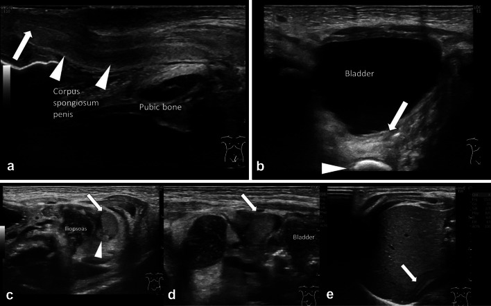
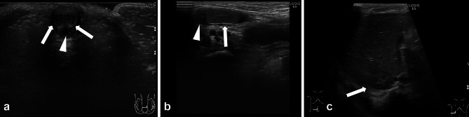
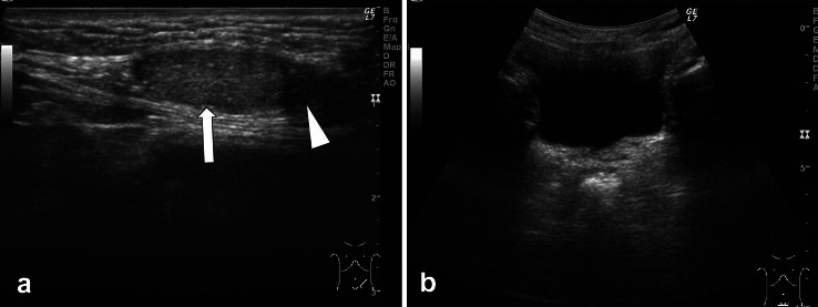
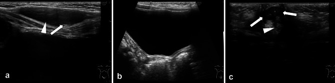
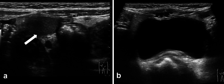
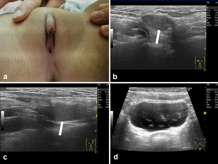
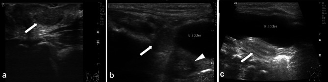
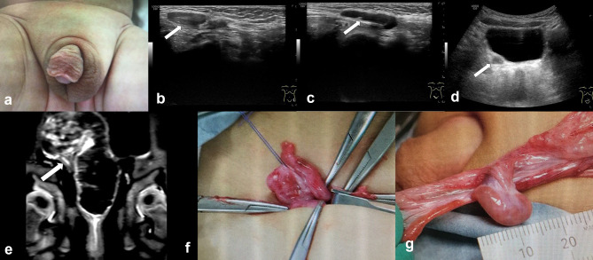
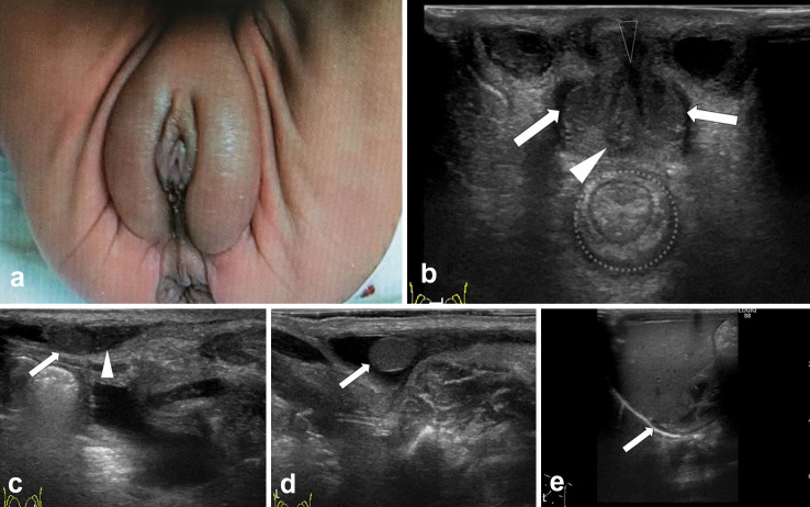
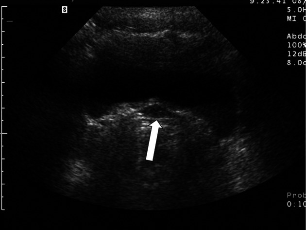
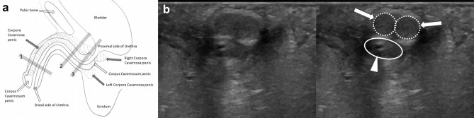
Ultrasonography is usually the first modality used to evaluate patients with disorders of sex development (DSD). To determine the sex in patients with DSD, the following four categories are carefully evaluated: chromosomal, gonadal, anatomical internal genitalia, and external genitalia. However, in the clinical setting, the only information that sonographers have prior to ultrasound examination is the appearance of the external genitalia. The following DSD presentations are commonly observed: (1) male external genitalia present at birth, without testis in the scrotum or with a small penis; (2) female external genitalia present at birth, with an inguinal hernia or clitoromegaly; (3) neonates with ambiguous genitalia at birth; and (4) female or male external genitalia without sexual maturity. In this retrospective study of several clinical cases, we demonstrated an ultrasound-based sex determination approach for these clinical presentations. We found that sonographers evaluated the external genitalia in relation to the distal urethra within the corpus spongiosum and corpus cavernosum and the presence or absence of follicles within the detected gonads to determine the sex of the patient.

Keywords: Diagnostic imaging; Disorders of sex development; Gonads; Transperineal sonogram.


Conflict of interest statement

T. H, Y. T, Y. S, M. H, and E. O declare that they have no financial or personal relationships that could lead to a conflict of interest.

Figures

Fig. 1
Fig. 1
Sonogram via the transperineal approach in neonates. a Image showing correct placement of the linear transducer in a sagittal orientation over the perineum in a neonate in the supine position. b Sonogram via the perineal approach in a normal female neonate showing the pubic symphysis (P), distal urethra (white-arrows), vagina (arrowheads), and rectum (open-arrows)
Fig. 2
Fig. 2
Male neonate with intra-abdominal testis. a A sagittal sonogram of the penis shows a normal corpus spongiosum penis (arrowhead), and the distal side of the urethra (arrow). The urethra is running within the corpus spongiosum. b Axial sonogram shows that the remnant Mullerian tissue cannot be detected as it is behind the bladder. The rectum (arrow) is visible between the bladder and sacrum (arrowhead). c Axial sonogram shows the right testis in the abdomen (arrow). The epididymis is also clearly visible on top of the testis (arrowhead). d Axial sonogram shows the left testis (arrow) located in the abdomen, next to the bladder. e Axial sonogram shows that the right adrenal gland (arrow) is of a normal size and positioned behind the liver
Fig. 3
Fig. 3
Male with DAX1 deficiency. a Axial sonogram of the penis shows the two columns of the corpus cavernosum penis (arrows) and the corpus spongiosum penis (arrowhead). b Axial sonogram shows the right testis located in the inguinal canal (arrow). The epididymis is visible on top of the testis (arrowhead). Axial sonogram shows a slightly smaller than normal adrenal gland (arrow) located behind the liver
Fig. 4
Fig. 4
One-year-old female with 46XY steroidogenic factor-1 deficiency. a Sonographic examination shows left testicular tissue (arrow) located in the left inguinal canal. Echogenicity is that of a normal testis. The epididymis is visible on top of the testis (arrowhead). b Neither a uterus nor a vagina can be visualized behind the bladder
Fig. 5
Fig. 5
Female with 46XY P450 oxidoreductase deficiency. a Axial sonogram shows the right testis in the right inguinal canal (arrow). The epididymis is visible (arrowhead). b The uterus and fallopian tubes are not detected behind the bladder. c The crus of the clitoris composed of two small corpus cavernosa (arrows) are visible in the enlarged clitoris. The distal side of the urethra (arrowhead) is located on the inferior surface of the clitoris
Fig. 6
Fig. 6
One-month-old female with androgen insensitivity syndrome. a Axial sonogram shows the right testis located in the inguinal canal (arrow). b The uterus and fallopian tubes are not detected behind the bladder
Fig. 7
Fig. 7
Nine-month-old female with 5 alpha-reductase deficiency. a External genitalia appear female with clitoromegaly. b Axial sonogram shows the right testis in the inguinal canal (arrow). Echogenicity is that of a normal testis. c On ultrasound, the left testis is detected in the left inguinal canal (arrow). d The uterus and fallopian tubes are not detected behind bladder
Fig. 8
Fig. 8
Neonate with 46XY/45X0 mixed gonadal dysgenesis. a Axial sonogram shows the left small testis (arrow) in the inguinal canal. b Axial sonogram shows the right testis located next to the bladder (arrow). The vagina is also present behind the bladder (arrowhead). c Sagittal sonogram shows a uterus (arrow) behind the bladder
Fig. 9
Fig. 9
Neonate with 45X0/47XYY. a External genitalia appear to be male because of the presence of a micro-penis and bifid scrotum. b Ultrasound shows the left testis (arrow) located in the inguinal canal. The echogenicity within that is of a normal testis, and no follicles are detected. c Ultrasound shows a cystic mass (arrow) in the right inguinal region. Gonadal tissue is not visible. A hypoplastic uterus (arrow) and vagina are detected behind the bladder. d Coronal T2 weighted magnetic resonance imaging shows right gonadal tissue (arrow) in the right uterine broad ligament. e During the operation. The right gonadal tissue is streaked like an ovary. f Left gonad located in the inguinal canal. Testicular tissue and epididymis are present
Fig. 10
Fig. 10
Neonate with congenital adrenal hyperplasia. a External genitalia show clitoromegaly and an opening on the distal side of the urethra not visible in the top of the penis. Pigmentation is evident. b Sagittal sonogram using the perineal approach shows that the distal urethra (arrow), vagina (arrowhead), and anus (vacant arrow) in separate and normal positions. c Axial sonogram shows gonadal tissue similar to an ovary with a follicle (arrow). d Ultrasound shows a normal uterus (arrow) behind the bladder. e Transverse sonogram demonstrates enlargement of the right adrenal gland cursors with maintained corticomedullary differentiation (arrow). f The left adrenal gland (arrow) is also enlarged, and both the left and right adrenal glands show a “cerebriform” appearance
Fig. 11
Fig. 11
Patient with severe hypospadias. a External genitalia are not identifiable as male or female. b Axial sonogram shows a very short corpus cavernosum penis (arrows) and corpus spongiosum penis with urethra (arrowhead). The urethra is running between the right and left corpus cavernosum penis. The proximal side of the urethra (arrowhead) and distal side (arrow) are vacant. No vagina is detected between the urethra and rectum (dotted circle). c Axial sonogram shows left testis (arrow) and epididymis (arrowhead) located in the inguinal canal. d Axial sonogram shows the right testis (arrow) located in the inguinal canal. e The right adrenal gland (arrow) behind the liver is of a normal size.
Fig. 12
Fig. 12
Eight-year-old with mosaic Turner syndrome. Ultrasound shows a small uterus (arrow) behind the bladder; however, no ovaries are detected
Fig. 13
Fig. 13
Normal male external genitalia. a Normal external genitalia in a male. Gray line 1 represents the angle of the axial sonogram of Fig. 3a. The corpus spongiosum penis is located just behind the two columns of the corpus cavernosum penis. Gray line 2 is representative of (b). The left and right corpus cavernosa are adjacent. Gray line 3 represents Fig. 11b. The left and right corpus cavernosum penises are not adjacent and the corpus spongiosum penis with the urethra is running between them. b The right panel is a schematic representation of the left panel. Two corpus cavernosa penises (arrows and dotted line) and the corpus spongiosum penis (arrowhead and continuous line) are shown. A catheter is inserted in the urethra
Fig. 14
Fig. 14
Normal female external genitalia. a The corpus clitoris is separated into a left and right cavernosum clitoris via the crus of clitoris. The urethra is not positioned around the cavernosum clitoris. Gray line 1 is representative of the sonogram of Fig. 5c. The two corpus cavernosa are adjacent. Gray line 2 shows the angle of the sonogram of (b). Two corpus cavernosum clitorises exist. b The right panel is a schematic presentation of the left panel. Two small corpus cavernosum clitorises (arrows) are visible. The urethra (arrowhead), vagina (dotted line), and anus (continuous line) are present

References

    1. Lambert SM, Vilain EJ, Kolon TF. A practical approach to ambiguous genitalia in the newborn period. Urol Clin North Am. 2010;37:195–205. doi: 10.1016/j.ucl.2010.03.014. - DOI - PubMed
    1. Kearsey I, Hutson JM. Disorders of sex development (DSD): not only babies with ambiguous genitalia. A practical guide for surgeons. Pediatr Surg Int. 2017;33:355–361. doi: 10.1007/s00383-016-4036-5. - DOI - PubMed
    1. Bennecke E, Bernstein S, Lee P, van de Grift TC, Nordenskjöld A, Rapp M, et al. Early genital surgery in disorders/differences of sex development: patients' perspectives. Arch Sex Behav. 2021;50:913–923. doi: 10.1007/s10508-021-01953-6. - DOI - PMC - PubMed
    1. Makiyan Z. Systematization of ambiguous genitalia. Organogenesis. 2016;12:169–182. doi: 10.1080/15476278.2016.1210749. - DOI - PMC - PubMed
    1. Chavhan GB, Parra DA, Oudjhane K, Miller SF, Babyn PS, Pippi Salle FL. Imaging of ambiguous genitalia: classification and diagnostic approach. Radiographics. 2008;28:1891–1904. doi: 10.1148/rg.287085034. - DOI - PubMed

LinkOut - more resources