

φYeO3-12 phage tail fiber Gp17 as a promising high specific tool for recognition of Yersinia enterocolitica pathogenic serotype O:3
- PMID: 34989907
- PMCID: PMC8739404
- DOI: 10.1186/s13568-021-01341-2
φYeO3-12 phage tail fiber Gp17 as a promising high specific tool for recognition of Yersinia enterocolitica pathogenic serotype O:3
Abstract
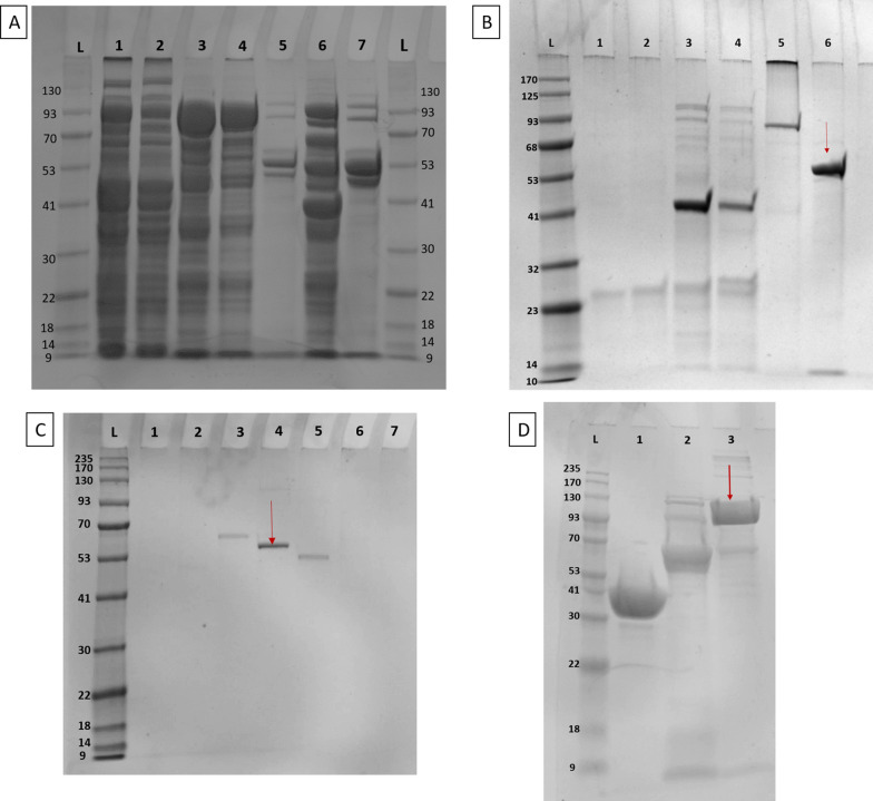
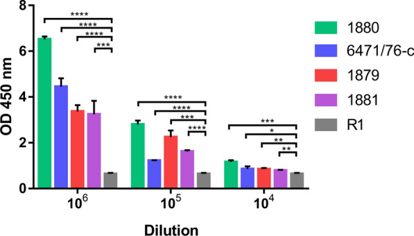
Yersiniosis is an infectious zoonotic disease caused by two enteropathogenic species of Gram-negative genus Yersinia: Yersinia enterocolitica and Yersinia pseudotuberculosis. Pigs and other wild and domestic animals are reservoirs for these bacteria. Infection is usually spread to humans by ingestion of contaminated food. Yersiniosis is considered a rare disease, but recent studies indicate that it is overlooked in the diagnostic process therefore the infections with this bacterium are not often identified. Reliable diagnosis of Yersiniosis by culturing is difficult due to the slow growth of the bacteria easily overgrown by other more rapidly growing microbes unless selec-tive growth media is used. Phage adhesins recognizing bacteria in a specific manner can be an excellent diagnostic tool, es-pecially in the diagnosis of pathogens difficult for culturing. In this study, it was shown that Gp17, the tail fiber protein (TFP) of phage φYeO3-12, specifically recognizes only the pathogenic Yersinia enterocolitica serotype O:3 (YeO:3) bacteria. The ELISA test used in this work confirmed the specific interaction of this protein with YeO:3 and demonstrated a promising tool for developing the pathogen recognition method based on phage adhesins.
Keywords: Diagnostic; ELISA; Phage; Phage adhesins; Tail fiber protein; Yersinia enterocolitica; Yersiniosis.
© 2022. The Author(s).
Conflict of interest statement
The authors declare that they have no competing interests.
Figures


Similar articles
-
Detection, seroprevalence and antimicrobial resistance of Yersinia enterocolitica and Yersinia pseudotuberculosis in pig tonsils in Northern Italy.Int J Food Microbiol. 2016 Oct 17;235:125-32. doi: 10.1016/j.ijfoodmicro.2016.07.033. Epub 2016 Jul 28. Int J Food Microbiol. 2016. PMID: 27500659
-
Yersiniosis in France: overview and potential sources of infection.Int J Infect Dis. 2016 May;46:1-7. doi: 10.1016/j.ijid.2016.03.008. Epub 2016 Mar 14. Int J Infect Dis. 2016. PMID: 26987478 Review.
-
Novel diagnostic ELISA test for discrimination between infections with Yersinia enterocolitica and Yersinia pseudotuberculosis.Eur J Clin Microbiol Infect Dis. 2018 Dec;37(12):2301-2306. doi: 10.1007/s10096-018-3373-9. Epub 2018 Sep 20. Eur J Clin Microbiol Infect Dis. 2018. PMID: 30238343
-
Nonessential genes of phage phiYeO3-12 include genes involved in adaptation to growth on Yersinia enterocolitica serotype O:3.J Bacteriol. 2005 Feb;187(4):1405-14. doi: 10.1128/JB.187.4.1405-1414.2005. J Bacteriol. 2005. PMID: 15687205 Free PMC article.
-
[Microbiology and epidemiology of Yersinia infections].Immun Infekt. 1990 Dec;18(6):178-85. Immun Infekt. 1990. PMID: 2076900 Review. German.
Cited by
-
Identification of receptor-binding protein and host receptor of non-lytic dsRNA phage phiNY.Microbiol Spectr. 2024 Oct 22;12(12):e0146724. doi: 10.1128/spectrum.01467-24. Online ahead of print. Microbiol Spectr. 2024. PMID: 39436121 Free PMC article.
-
The Novel Yersinia enterocolitica Telomere Phage vB_YenS_P840 Is Closely Related to PY54, but Reveals Some Striking Differences.Viruses. 2023 Sep 28;15(10):2019. doi: 10.3390/v15102019. Viruses. 2023. PMID: 37896796 Free PMC article.
-
The Biotechnological Application of Bacteriophages: What to Do and Where to Go in the Middle of the Post-Antibiotic Era.Microorganisms. 2023 Sep 13;11(9):2311. doi: 10.3390/microorganisms11092311. Microorganisms. 2023. PMID: 37764155 Free PMC article. Review.
-
Phage-derived proteins: Advancing food safety through biocontrol and detection of foodborne pathogens.Compr Rev Food Sci Food Saf. 2025 Mar;24(2):e70124. doi: 10.1111/1541-4337.70124. Compr Rev Food Sci Food Saf. 2025. PMID: 39898971 Free PMC article. Review.
-
Ultrasensitive Electrochemical Detection of Salmonella typhimurium in Food Matrices Using Surface-Modified Bacterial Cellulose with Immobilized Phage Particles.Biosensors (Basel). 2024 Oct 14;14(10):500. doi: 10.3390/bios14100500. Biosensors (Basel). 2024. PMID: 39451713 Free PMC article.
References
-
- Aziz M, Yalamanchili VS. Yersinia enterocolitica. Treasure Island: StatPearls Publishing; 2021. - PubMed
Grants and funding
LinkOut - more resources
Full Text Sources
