

Cerebral venous congestion exacerbates cerebral microhemorrhages in mice
- PMID: 34989944
- PMCID: PMC9135950
- DOI: 10.1007/s11357-021-00504-0
Cerebral venous congestion exacerbates cerebral microhemorrhages in mice
Abstract
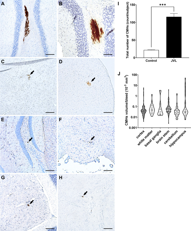
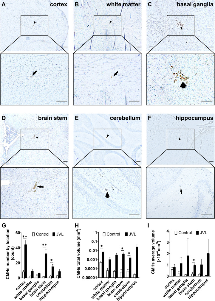
Cerebral microhemorrhages (CMHs; microbleeds), which are small focal intracerebral hemorrhages, importantly contribute to the pathogenesis of cognitive decline and dementia in older adults. Although recently it has been increasingly recognized that the venous side of the cerebral circulation likely plays a fundamental role in the pathogenesis of a wide spectrum of cerebrovascular and brain disorders, its role in the pathogenesis of CMHs has never been studied. The present study was designed to experimentally test the hypothesis that venous congestion can exacerbate the genesis of CMHs. Increased cerebral venous pressure was induced by internal and external jugular vein ligation (JVL) in C57BL/6 mice in which systemic hypertension was induced by treatment with angiotensin II plus L-NAME. Histological analysis (diaminobenzidine staining) showed that mice with JVL developed multiple CMHs. CMHs in mice with JVL were often localized adjacent to veins and venules and their morphology was consistent with venous origin of the bleeds. In brains of mice with JVL, a higher total count of CMHs was observed compared to control mice. CMHs were distributed widely in the brain of mice with JVL, including the cortical gray matter, brain stem, the basal ganglia, subcortical white matter, cerebellum, and the hippocampi. In mice with JVL, there were more CMHs predominantly in cerebral cortex, brain stem, and cerebellum than in control mice. CMH burden, defined as total CMH volume, also significantly increased in mice with JVL. Thus, cerebral venous congestion can exacerbate CMHs. These observations have relevance to the pathogenesis of cognitive impairment associated with right heart failure as well as elevated cerebral venous pressure due to jugular venous reflux in older adults.
Keywords: Cerebral circulation; Heart failure; ICH; Intracerebral hemorrhage; Microbleed; VCI; Vascular cognitive impairment; Vascular contributions to cognitive impairment and dementia (VCID); Vein; Venous congestion.
© 2022. The Author(s), under exclusive licence to American Aging Association.
Conflict of interest statement
Dr. Anna Csiszar serves as Associate Editor for The Journal of Gerontology, Series A: Biological Sciences and Medical Sciences and GeroScience. Dr. Stefano Tarantini serves as Guest Editor for Frontiers in Aging Neuroscience. Dr. Zoltan Ungvari serves as Editor-in-Chief for GeroScience and as Consulting Editor for The American Journal of Physiology-Heart and Circulatory Physiology.
Figures
Similar articles
-
Cerebral venous congestion promotes blood-brain barrier disruption and neuroinflammation, impairing cognitive function in mice.Geroscience. 2019 Oct;41(5):575-589. doi: 10.1007/s11357-019-00110-1. Epub 2019 Nov 5. Geroscience. 2019. PMID: 31691147 Free PMC article.
-
Repeated Valsalva maneuvers promote symptomatic manifestations of cerebral microhemorrhages: implications for the pathogenesis of vascular cognitive impairment in older adults.Geroscience. 2018 Dec;40(5-6):485-496. doi: 10.1007/s11357-018-0044-9. Epub 2018 Oct 4. Geroscience. 2018. PMID: 30288646 Free PMC article.
-
Insulin-like growth factor 1 deficiency exacerbates hypertension-induced cerebral microhemorrhages in mice, mimicking the aging phenotype.Aging Cell. 2017 Jun;16(3):469-479. doi: 10.1111/acel.12583. Epub 2017 Mar 14. Aging Cell. 2017. PMID: 28295976 Free PMC article.
-
Atherosclerotic burden and cerebral small vessel disease: exploring the link through microvascular aging and cerebral microhemorrhages.Geroscience. 2024 Oct;46(5):5103-5132. doi: 10.1007/s11357-024-01139-7. Epub 2024 Apr 19. Geroscience. 2024. PMID: 38639833 Free PMC article. Review.
-
Cerebral microhemorrhages: mechanisms, consequences, and prevention.Am J Physiol Heart Circ Physiol. 2017 Jun 1;312(6):H1128-H1143. doi: 10.1152/ajpheart.00780.2016. Epub 2017 Mar 17. Am J Physiol Heart Circ Physiol. 2017. PMID: 28314762 Free PMC article. Review.
Cited by
-
Impacts of systemic milieu on cerebrovascular and brain aging: insights from heterochronic parabiosis, blood exchange, and plasma transfer experiments.Geroscience. 2025 May 23. doi: 10.1007/s11357-025-01657-y. Online ahead of print. Geroscience. 2025. PMID: 40407975 Review.
-
Exposome and unhealthy aging: environmental drivers from air pollution to occupational exposures.Geroscience. 2023 Dec;45(6):3381-3408. doi: 10.1007/s11357-023-00913-3. Epub 2023 Sep 9. Geroscience. 2023. PMID: 37688657 Free PMC article. Review.
-
Cerebral venous congestion alters brain metabolite profiles, impairing cognitive function.J Cereb Blood Flow Metab. 2023 Nov;43(11):1857-1872. doi: 10.1177/0271678X231182244. Epub 2023 Jun 13. J Cereb Blood Flow Metab. 2023. PMID: 37309740 Free PMC article.
-
Young blood-mediated cerebromicrovascular rejuvenation through heterochronic parabiosis: enhancing blood-brain barrier integrity and capillarization in the aged mouse brain.Geroscience. 2024 Oct;46(5):4415-4442. doi: 10.1007/s11357-024-01154-8. Epub 2024 May 10. Geroscience. 2024. PMID: 38727872 Free PMC article.
-
Cerebral venous congestion alters CNS homeostatic plasticity, evoking tinnitus-like behavior.Cell Biosci. 2024 Apr 9;14(1):47. doi: 10.1186/s13578-024-01221-9. Cell Biosci. 2024. PMID: 38594782 Free PMC article.
References
-
- Fulop GA, Tarantini S, Yabluchanskiy A, Molnar A, Prodan CI, Kiss T, Csipo T, Lipecz A, Balasubramanian P, Farkas E, Toth P, Sorond F, Csiszar A, Ungvari Z. Role of age-related alterations of the cerebral venous circulation in the pathogenesis of vascular cognitive impairment. Am J Physiol Heart Circ Physiol. 2019;316:H1124–H1140. doi: 10.1152/ajpheart.00776.2018. - DOI - PMC - PubMed
Publication types
MeSH terms
Grants and funding
- U54 GM104938/GM/NIGMS NIH HHS/United States
- R01-AG038747/NH/NIH HHS/United States
- R01-AG047879/NH/NIH HHS/United States
- K01-AG073614/NH/NIH HHS/United States
- T32 AG052363/AG/NIA NIH HHS/United States
- R01 AG068295/AG/NIA NIH HHS/United States
- R01-NS056218/NS/NINDS NIH HHS/United States
- K01 AG073614/AG/NIA NIH HHS/United States
- R01 NS056218/NS/NINDS NIH HHS/United States
- R01-NS100782/NS/NINDS NIH HHS/United States
- T32AG052363/AG/NIA NIH HHS/United States
- U54GM104938/GM/NIGMS NIH HHS/United States
- P30 CA225520/CA/NCI NIH HHS/United States
- R01-AG055395/NH/NIH HHS/United States
- P30CA225520/CA/NCI NIH HHS/United States
LinkOut - more resources
Full Text Sources
