Pneumoperitoneum as an uncommon complication after an axillary laceration in a horse
- PMID: 34990086
- PMCID: PMC8959331
- DOI: 10.1002/vms3.718
Pneumoperitoneum as an uncommon complication after an axillary laceration in a horse
Abstract
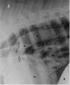
Lacerations of the axillary region occur frequently in horses. Typical complications caused by entrapment of air in the wound during locomotion are subcutaneous emphysema, with consecutive pneumomediastinum and pneumothorax. In this case report, the clinical, radiographic and laboratory diagnosis and management of these complications after an axillary laceration that finally resulted in pneumoperitoneum are described. A 1-year-old Hannoveranian was presented with a pre-existing axillary laceration of unknown duration and subcutaneous emphysema in the surrounding tissue. Due to extensive tissue loss, attempts to adequately close the wound surgically and by packing with sterile dressing material were unsuccessful. Despite stall confinement and tying of the horse, subcutaneous emphysema was progressive and pneumomediastinum as well as pneumothorax was developed. These complications were monitored radiographically. On day 5 after admission, signs of air accumulation were detected on radiographs craniodorsally in the peritoneum and a pneumoperitoneum was diagnosed. Repeated thoracentesis with a teat cannula to gradually evacuate the thoracic cavity was used in combination with nasal oxygen insufflation to treat global respiratory insufficiency. Subcutaneous emphysema and all other complications resolved progressively and the horse was discharged from the hospital 21 days after admission when the axillary wound was adequately filled with granulation tissue. The wound healed fully 1 month later and the horse did not develop long-term complications within the following year. To the authors´ knowledge, the development of pneumoperitoneum including its radiographic monitoring following an axillary laceration has not been described in horses previously.
Keywords: axillary laceration; horse; pneumomediastinum; pneumoperitoneum; pneumothorax.
© 2022 The Authors. Veterinary Medicine and Science published by John Wiley & Sons Ltd.
Conflict of interest statement
The authors declare no conflict of interest.
Figures


References
-
- Barber, S. (2016). In: Theoret, C. & Schumacher, J. (eds.), Equine wound management (pp. 294–296). John Wiley & Sons.
-
- Garcia, P. , Pizanis, A. , Massmann, A. , Reischmann, B. , Burkhardt, M. , Tosoundidis, G. , Rensing, H. , & Pohlmann, T. (2009). Bilateral pneumothoraces, pneummediastinum, pneumoperitoneum, pneumoretroperitoneum, and subcutaneous emphysema after thoracoscopic anterior fracture stabilization. Spine, 34(10), 371–375. 10.1097/BRS.0b013e3181995c87 - DOI - PubMed
-
- Greci, V. , Baio, A. , Bibbiani, L. , Borgonovo, S. , olivero, D. , Rocchi, P.M. , & Raiano, V. (2015). Pneumopericardium, Pneumomediastinum, pneumothorax and pneumoretroperitoneum complicating pulmonary metastatic carcinoma in a Cat. Journal of Small Animal Practice 56(11), 679–683. 10.1111/jsap.12366 - DOI - PubMed
Publication types
MeSH terms
LinkOut - more resources
Full Text Sources
Medical

