Review
. 2022 Jan 6:62:55-84.
doi: 10.1146/annurev-pharmtox-052220-010446.

Central Nervous System Control of Glucose Homeostasis: A Therapeutic Target for Type 2 Diabetes?

Affiliations
Review

Central Nervous System Control of Glucose Homeostasis: A Therapeutic Target for Type 2 Diabetes?

Zaman Mirzadeh et al. Annu Rev Pharmacol Toxicol. .

Abstract

Historically, pancreatic islet beta cells have been viewed as principal regulators of glycemia, with type 2 diabetes (T2D) resulting when insulin secretion fails to compensate for peripheral tissue insulin resistance. However, glycemia is also regulated by insulin-independent mechanisms that are dysregulated in T2D. Based on evidence supporting its role both in adaptive coupling of insulin secretion to changes in insulin sensitivity and in the regulation of insulin-independent glucose disposal, the central nervous system (CNS) has emerged as a fundamental player in glucose homeostasis. Here, we review and expand upon an integrative model wherein the CNS, together with the islet, establishes and maintains the defended level of glycemia. We discuss the implications of this model for understanding both normal glucose homeostasis and T2D pathogenesis and highlight centrally targeted therapeutic approaches with the potential to restore normoglycemia to patients with T2D.

Keywords: autonomic nervous system; central nervous system; diabetes; glucose effectiveness; glucose homeostasis; insulin-independent glucose disposal; therapeutics.


Figures

Figure 1
Figure 1
Mechanisms governing the biologically defended level of glycemia (BDLG). The BDLG is determined by the balance between rates of glucose appearance into and disappearance from the circulation, and imbalance in these rates contributes to the elevated BDLG in type 2 diabetes (T2D). In health, acute deviations from the BDLG are counteracted by both insulin-dependent and insulin-independent mechanisms that restore blood glucose levels into the normal range. Responses to rising blood glucose levels (dashed arrows) include increased glucose-stimulated insulin secretion (GSIS) by the pancreatic beta cell, which, together with the ability of glucose to independently facilitate its own disposal [termed glucose effectiveness (GE)], increases glucose uptake by peripheral tissues and inhibits hepatic glucose production (HGP). Conversely, a fall in blood glucose levels triggers adaptive neuroendocrine and autonomic counterregulatory responses (CRRs, solid arrows) that collectively increase glucose appearance into the circulation and decrease its removal. These responses include increased secretion of glucagon, cortisol, and epinephrine (which stimulate HGP), while insulin secretion is inhibited to prevent a further fall in blood glucose. In T2D, the lower boundary of the BDLG is increased (as reflected by a higher glycemic threshold for inducing CRRs), and the same is true of the upper boundary of the BDLG, as evidenced by diminished rates of glucose disappearance (owing to reduced GSIS, insulin resistance, and reduced GE) and failure to suppress HGP. The net outcome is a persistently elevated BDLG in T2D. Figure adapted from images created with BioRender.com.
Figure 2
Figure 2
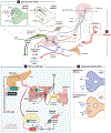
Anatomy of glycemic control: central and peripheral mechanisms. (①) After a meal, nutrients digested by the GI tract are delivered into the blood via the hepatic portal vein and circulated throughout the body. Postprandial elevations in blood glucose activate GSIS. Effects of postprandial GSIS (purple circles) include suppression of HGP (directly via activation of hepatic insulin receptors and indirectly via inhibition of glucagon release from pancreatic alpha cells) and stimulation of glucose uptake by insulin-sensitive tissues, including skeletal muscle and adipose tissue. Nutrient ingestion also stimulates endocrine cells lining the GI tract to release the incretin hormones, GLP1 and GIP. Incretins augment insulin secretion (yellow circle) and, via binding their cognate receptors expressed by sensory afferents innervating the GI tract, convey information to the brain about the size and composition of ingested nutrients (for reviews, see 26, 47, 48). (②) Spinal afferents (dark green) have their cell bodies within the dorsal root ganglia, which express molecules such as the ion channel TRPV1 (activated by noxious heat) that processes nociceptive signals conveyed to the brain via the spinothalamic tract. Spinal afferents also can exert effects by releasing inflammatory peptides such as SP and CGRP. Vagal afferents (light green) also express SP/CGRP and can sense the local microenvironment through receptors that include the serotonin receptor (5HTR3) in the pancreas and incretin receptors in the GI tract. Cell bodies of vagal sensory neurons are contained within the nodose ganglion and convey sensory information to the CNS via projections to the NTS. In addition to surveilling peripheral tissue function, the CNS can influence peripheral glucose effector function via neuroendocrine systems, including the HPA axis (③) and the ANS. In response to stressful stimuli, activation of the hypothalamic-pituitary-adrenal axis causes the adrenal cortex to secrete cortisol, which stimulates glucose production by the liver. (④) The SNS and PNS branches of the ANS innervate tissues throughout the body to influence glycemia. Stimulation of the SNS (blue circles in ①) suppresses GSIS and increases glucagon release from the islets while stimulating increased HGP from the liver via releasing the neurotransmitter NE, which binds to and activates adrenergic receptors on pancreatic islet cells and hepatocytes. Stimulation of the PNS (orange circles in ①) increases both insulin and glucagon secretion via binding of the neurotransmitter ACh to muscarinic cholinergic receptors. In the postprandial state, the responses to PNS activation promote glucose uptake and storage. Abbreviations: ACh, acetylcholine; ACTH, adrenocorticotropic hormone; ANS, autonomic nervous system; CGRP, calcitonin gene-related peptide; CNS, central nervous system; GI, gastrointestinal; GIP, glucose-stimulated insulinotropic polypeptide; GLP1, glucagon-like peptide 1; GSIS, glucose-stimulated insulin secretion; HGP, hepatic glucose production; HPA, hypothalamic-pituitary-adrenal; NE, norepinephrine; NPY, neuropeptide Y; NTS, nucleus of the solitary tract; PACAP, pituitary adenylate cyclase-activating polypeptide; PNS, parasympathetic nervous system; SNS, sympathetic nervous system; SP, substance P; TRPV1, transient receptor potential vanilloid 1. Figure adapted from images created with BioRender.com.
Figure 3
Figure 3
Hypothalamic neurocircuits in glycemic control. The hypothalamus is a forebrain structure situated ventral to the thalamus and dorsal to the pituitary gland and median eminence. It is an important component of the hypophyseal portal system. The mediobasal hypothalamus exhibits reciprocal connectivity with hindbrain structures, including the nucleus of the solitary tract (NTS), as well as with neighboring hypothalamic nuclei (arrows indicate connectivity in hypothalamus inset). The median eminence is a circumventricular organ with fenestrated capillaries that increase vascular permeability (indicated by dashed red lines in circular insets) and that receives dense innervation from hypothalamic neurons [including from parvocellular neurons of the paraventricular nucleus (PVN)], which release hypophysiotropic hormones (e.g., corticotropin-releasing factor) into the pituitary portal system, thereby directing pituitary secretions. Heightened vascular permeability of the median eminence exposes neurons in the adjacent arcuate nucleus (ARC) to higher levels of circulating nutrients and hormones than seen by brain regions located behind the blood-brain barrier (bold red line), such as the ventromedial nucleus (VMN), PVN, and lateral hypothalamic area (LHA). The ARC is also adjacent to the third ventricle (3V), permitting access to factors circulating in the cerebrospinal fluid, and receives afferent neuronal input from multiple brain regions conveying information about energetic demands, environmental cues of time and food availability, and cue-reward associations. These dynamic and varied inputs are integrated by pro-opiomelanocortin (POMC)- and agouti-related peptide (AgRP)-expressing neurons in the ARC that are regulated in a reciprocal manner and drive opposing effects on glycemia and energy balance, in part, by engaging melanocortin receptor signaling in downstream neurons. Hypothalamic network activity is also shaped by interactions with neighboring glia, including astrocytes and microglia. In addition to providing structural support for neurons and their projections, astrocytes and microglia play critical roles in neurovascular coupling, neurotransmitter uptake, synaptic pruning, immune surveillance, and inflammatory signaling (for a detailed review, see 116). In the setting of overnutrition, these cell types engage in a hypothalamic-specific reactive gliosis—characterized by cellular morphological changes, including enlarged, extended processes and heightened release of proinflammatory cytokines—that contributes to the metabolic consequences of diet-induced obesity. Figure adapted from images created with BioRender.com.
Figure 4
Figure 4
Future prospects for central nervous system (CNS)-targeted type 2 diabetes (T2D) remission/disease modulation. Device-based, CNS-targeted approaches to T2D disease modulation include (❶) vagal nerve stimulation targeting fascicles innervating liver, portal vein, and pancreatic islets, (❷) spinal cord stimulation with electrodes placed epidurally to target preganglionic sympathetic nerves in the intermediolateral cell column (blue cell bodies in inset) innervating the celiac ganglion (~T5–T9 levels), and (❸) intrathecal catheter-based targeted drug delivery (e.g., leptin, fibroblast growth factor analogs) with infusion via an implanted subcutaneous reservoir or pump. Figure adapted from images created with BioRender.com.

References

    1. Grayson BE, Seeley RJ, Sandoval DA. 2013. Wired on sugar: the role of the CNS in the regulation of glucose homeostasis. Nature 14(1):24–37 - PMC - PubMed
    1. Schwartz MW, Seeley RJ, Tschöp MH, Woods SC, Morton GJ, et al. 2013. Cooperation between brain and islet in glucose homeostasis and diabetes. Nature 503(7474):59–66 - PMC - PubMed
    1. Turner RC, Holman RR, Matthews D, Hockaday TDR, Peto J. 1979. Insulin deficiency and insulin resistance interaction in diabetes: estimation of their relative contribution by feedback analysis from basal plasma insulin and glucose concentrations. Metabolis 28(11):1086–96 - PubMed
    1. Edelman SV, Polonsky WH. 2017. Type 2 diabetes in the real world: the elusive nature of glycemic control. Diabetes Care 40(11):1425–32 - PubMed
    1. Kahn SE, Cooper ME, Prato SD. 2014. Pathophysiology and treatment of type 2 diabetes: perspectives on the past, present, and future. Lancet 383(9922):1068–83 - PMC - PubMed

Publication types