. 2022 Jan 7;375(6576):91-96.
doi: 10.1126/science.abm0594. Epub 2022 Jan 6.

CAR T cells produced in vivo to treat cardiac injury

Affiliations

CAR T cells produced in vivo to treat cardiac injury

Joel G Rurik et al. Science. .

Abstract

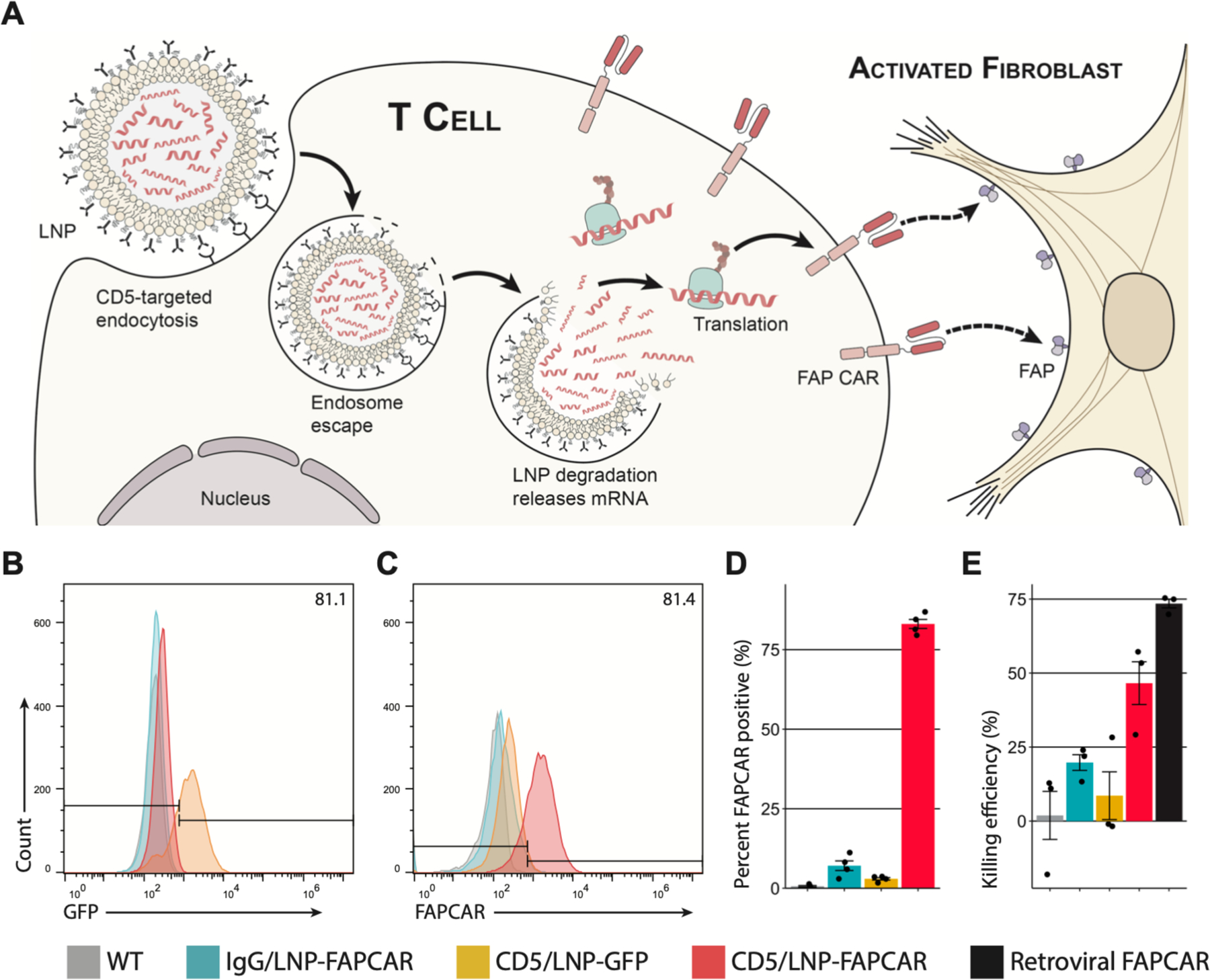
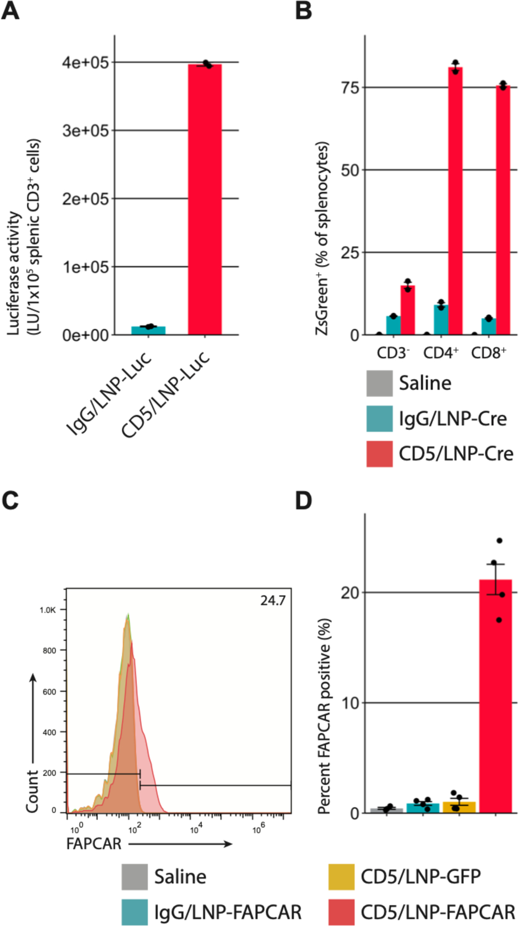
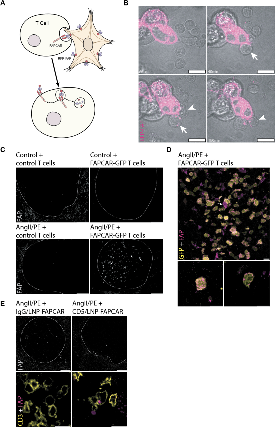
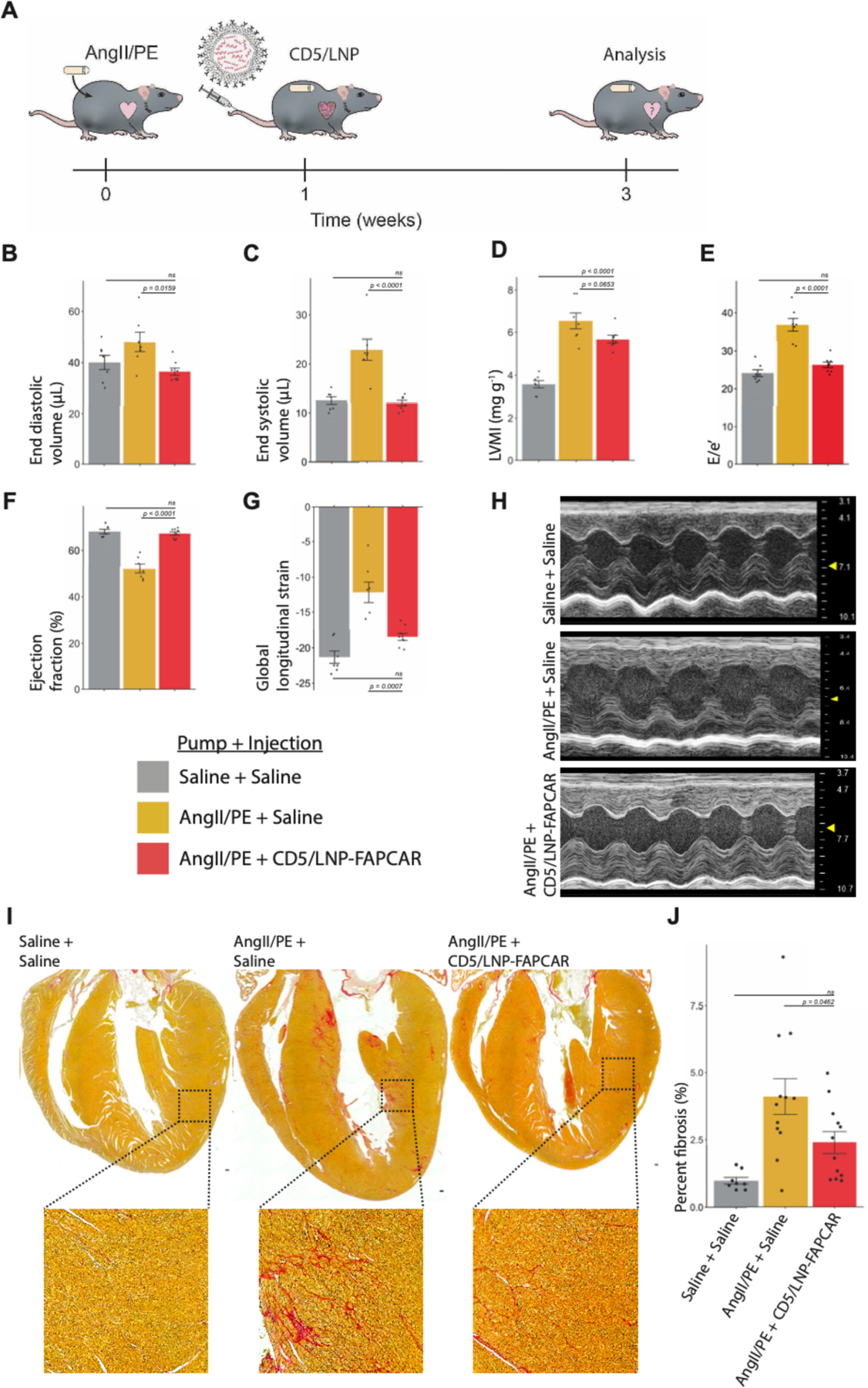
Fibrosis affects millions of people with cardiac disease. We developed a therapeutic approach to generate transient antifibrotic chimeric antigen receptor (CAR) T cells in vivo by delivering modified messenger RNA (mRNA) in T cell–targeted lipid nanoparticles (LNPs). The efficacy of these in vivo–reprogrammed CAR T cells was evaluated by injecting CD5-targeted LNPs into a mouse model of heart failure. Efficient delivery of modified mRNA encoding the CAR to T lymphocytes was observed, which produced transient, effective CAR T cells in vivo. Antifibrotic CAR T cells exhibited trogocytosis and retained the target antigen as they accumulated in the spleen. Treatment with modified mRNA-targeted LNPs reduced fibrosis and restored cardiac function after injury. In vivo generation of CAR T cells may hold promise as a therapeutic platform to treat various diseases.


Conflict of interest statement

Competing Interests: S.M.A., E.P., C.H.J., H.A., D.W., H.P., and J.A.E. are scientific founders and hold equity in Capstan Therapeutics. Y.K.T. and B.L.M. are employees and hold equity in Acuitas Therapeutics. S.M.A. is on the scientific advisory boards of Verismo and Bioardis. C.H.J. is a scientific founder and has equity in Tmunity Therapeutics and DeCART Therapeutics and reports grants from Tmunity Therapeutics and is on the scientific advisory boards of BluesphereBio, Cabaletta, Carisma, Cellares, Celldex, ImmuneSensor, Poseida, Verismo, Viracta Therapeutics, WIRB Copernicus Group and Ziopharm Oncology. D.W. receives research support from BioNTech. S.M.A., E.P., and C.H.J. are inventors (University of Pennsylvania, Wistar Institute) on a patent for a FAP CAR (US Utility Patent 9,365,641 issued 14 June 2016, WIPO Patent Application PCT/US2013/062717). S.M.A., E.P., H.A., and J.A.E. are inventors (University of Pennsylvania) on a patent for the use of CAR T therapy in heart disease (US Provisional Patent Application 62/563,323 filed 26 September 2017, WIPO Patent Application PCT/US2018/052605). J.G.R., I.T., H.A., D.W., H.P., and J.A.E. are inventors (University of Pennsylvania) on a patent for the use of CD5/LNP-FAPCAR as an anti-fibrotic therapy (US Provisional Patent Application 63/090,998 filed 13 September 2020, WIPO Patent Application PCT/ US21/54764 filed 13 October 2021). I.T., D.W., and H.P. are inventors (University of Pennsylvania) on a patent for the in vivo targeting of T cells for mRNA therapeutics (US Provisional Patent Application 63/090,985 filed 13 October 2020, WIPO Patent Application PCT/US21/54769 filed 13 October 2021). I.T., D.W., and H.P. are inventors (University of Pennsylvania) on a patent for the in vivo targeting of CD4+ T cells for mRNA therapeutics (US Provisional Patent Application 63/091,010 filed 13 October 2020, WIPO Patent Application PCT/US21/54775). In accordance with the University of Pennsylvania policies and procedures and our ethical obligations as researchers, D.W. is named on additional patents that describe the use of nucleoside-modified mRNA and targeted lipid nanoparticles as platforms to deliver therapeutic proteins and vaccines. C.H.J. is named on additional patents that describe the creation and therapeutic use of chimeric antigen receptors. These interests have been fully disclosed to the University of Pennsylvania, and approved plans are in place for managing any potential conflicts arising from licensing these patents.

Figures

Fig. 1.
Fig. 1.. CD5-targeted lipid nanoparticles produce functional, mRNA-based FAPCAR T cells in vitro.
(A) Schematic outlining the molecular process to create transient FAPCAR T cells using CD5-targeted LNP. Representative flow cytometry analysis of (B) GFP and (C) FAPCAR expression in murine T cells 48 hours after incubation with either IgG/LNP-FAPCAR, CD5/LNP-GFP, or CD5/LNP-FAPCAR. (D) Quantification of murine T cells (percent) staining positive for FAPCAR from biologically independent replicates (n = 4). (E) FAPCAR T cells were mixed with FAP-expressing target HEK293T cells overnight and assayed for killing efficiency in biologically independent replicates (n = 3). Data are mean +/− s.e.m.
Fig. 2.
Fig. 2.. CD5-targeted lipid nanoparticles produce mRNA-based FAPCAR T cells in vivo.
(A) Luciferase activity in CD3+ splenocytes 24 hours after intravenous injection of 8µg of control IgG/LNP-Luc or CD5/LNP-Luc. Bar graphs represent two biologically independent replicates. (B) Ai6 mice (Rosa26CAG-LSL-ZsGreen) were injected with 30µg of non-targeted/LNP-Cre (NT), IgG/LNP-Cre, or CD5/LNP-Cre. After 24 hours ZsGreen expression was observed in (81.1%) CD4+ and (75.6%) CD8+, but not in many (15.0%) CD3splenocytes. Bar graphs represent two biologically independent replicates. (C) T cells were isolated from the spleens of AngII/PE injured mice, 48 hours after injection of 10µg of LNP. Representative flow cytometry analysis shows FAPCAR expression in animals injected with CD5/LNP-FAPCAR, but not in control saline, IgG/LNP-FAPCAR, or CD5/LNP-GFP animals. (D) Quantification of murine T cells staining positive for FAPCAR in C. n = 4 biologically independent mice in two separate cohorts. Data are mean +/− s.e.m.
Fig. 3.
Fig. 3.. FAPCAR T cells trogocytose FAP from activated cardiac fibroblasts and return FAP to the spleen only in AngII/PE injured, FAPCAR T-treated animals.
(A) Schematic representation of FAPCAR-expressing T cells trogocytosing FAP from activated fibroblasts. (B) Confocal time-lapse micrographs of two FAPCAR T cells first forming an immunological synapse at 40min (arrow), and 85min (arrowhead) then trogocytosing RFP-FAP (magenta) from HEK293T cells (punctae seen at 85min, arrow, and 150min, arrowhead within FAPCAR T cells). Scale bars: 10μm. (C) Widefield images of FAP-stained spleens (white pulp regions highlighted by the dashed line) of an uninjured animal 24 hours after adoptive transfer of 107 MigR1-control T cells, an uninjured animal 24 hours after adoptive transfer of 107 FAPCAR-GFP T cells, an AngII/PE-injured (7 days) animal 48 hours after adoptive transfer of 107 MigR1-control T cells, and an AngII/PE-injured (7 days) animal 48 hours after adoptive transfer of 107 FAPCAR-GFP T cells. Scale bars: 100μm. (D) Confocal micrograph of FAP (magenta) and FAPCAR-GFP (yellow) in a white pulp region of the spleen of an AngII/PE-injured (7 days) animal 48 hours after adoptive transfer of 107 FAPCAR-GFP T cells. Max-Z projection (lower left subpanel) and a single Z slice (lower right subpanel) of a representative FAP+/FAPCAR+ T cell. Scale bars: 10μm. (E) Confocal micrographs of a white pulp region (dashed outline) of FAP-stained spleens from AngII/PE-injured (7 days) animals injected with 10μg of IgG/LNP-FAPCAR or CD5/LNP-FAPCAR for 48 hours. FAP (grey and magenta) and CD3 (yellow) overlap specifically in CD5/LNP-FAPCAR-treated condition. Scale bars: 100μm (top row; greyscale) or 10μm (bottom row; merged pseudo-colored).
Fig. 4.
Fig. 4.. In vivo generation of transient FAPCAR T cells improves cardiac function after injury.
Wild-type adult C57BL/6 mice were continually dosed with saline or AngII/PE via implanted 28-day osmotic minipump. After one week of cardiac pressure-overload injury, CD5-targeted LNP were injected. Mice were analyzed after an additional two weeks. (A) Schematic representation of experimental timeline. Echocardiograph measurements show improvements in left ventricle (LV) volumes, diastolic and systolic function following a single injection of 10μg of CD5/LNP-FAPCAR. Measurements of (B) end diastolic and (C) end systolic volumes (μL). (D) M-mode estimate of weight-normalized LV mass (mg/g). (E) Diastolic function (E/e’, an estimate of LV filling pressure) (F) ejection fraction (%) and (G) global longitudinal strain. (H) Representative m-mode echocardiography images. Echocardiograph data represent n = 7, 7, 8 biologically independent mice per condition, spread over three cohorts. (I) Picrosirius red (PSR) staining highlights collagen (pink) in coronal cardiac sections of mock uninjured animals (3 weeks after saline pump implant + saline injection at week 1), injured control animals (AngII/PE + saline), isotype non-targeted LNP control (AngII/PE + IgG/LNP-FAPCAR) and treated animals (AngII/PE + CD5/LNP-FAPCAR). Inset shows magnification of left ventricular myocardium. Scale bar: 100μm. (J) Quantification of percent fibrosis of the ventricles seen in (I). Histology data represent n = 8, 11, 12, biologically independent mice per condition, spread over five cohorts. Data are mean +/− s.e.m. Displayed p-values are from Tukey’s post-hoc test following one-way ANOVA p<0.05.

Comment in

References

    1. Khalil H, Kanisicak O, Prasad V, Correll RN, Fu X, Schips T, Vagnozzi RJ, Liu R, Huynh T, Lee SJ, Karch J, Molkentin JD, Fibroblast-specific TGF-β-Smad2/3 signaling underlies cardiac fibrosis. J Clin Invest 127, 3770–3783 (2017). - PMC - PubMed
    1. Schafer S, Viswanathan S, Widjaja AA, Lim W-W, Moreno-Moral A, DeLaughter DM, Ng B, Patone G, Chow K, Khin E, Tan J, Chothani SP, Ye L, Rackham OJL, Ko NSJ, Sahib NE, Pua CJ, Zhen NTG, Xie C, Wang M, Maatz H, Lim S, Saar K, Blachut S, Petretto E, Schmidt S, Putoczki T, Guimarães-Camboa N, Wakimoto H, van Heesch S, Sigmundsson K, Lim SL, Soon JL, Chao VTT, Chua YL, Tan TE, Evans SM, Loh YJ, Jamal MH, Ong KK, Chua KC, Ong B, Chakaramakkil MJ, Seidman JG, Seidman CE, Hubner N, Sin KYK, Cook SA, IL-11 is a crucial determinant of cardiovascular fibrosis. Nature 552, 110–115 (2017). - PMC - PubMed
    1. Moore-Morris T, Guimarães-Camboa N, Banerjee I, Zambon AC, Kisseleva T, Velayoudon A, Stallcup WB, Gu Y, Dalton ND, Cedenilla M, Gomez-Amaro R, Zhou B, Brenner DA, Peterson KL, Chen J, Evans SM, Resident fibroblast lineages mediate pressure overload–induced cardiac fibrosis. J Clin Invest 124, 2921–2934 (2014). - PMC - PubMed
    1. Yokota T, McCourt J, Ma F, Ren S, Li S, Kim TH, Kurmangaliyev YZ, Nasiri R, Ahadian S, Nguyen T, Tan XHM, Zhou Y, Wu R, Rodriguez A, Cohn W, Wang Y, Whitelegge J, Ryazantsev S, Khademhosseini A, Teitell MA, Chiou PY, Birk DE, Rowat AC, Crosbie RH, Pellegrini M, Seldin M, Lusis AJ, Deb A, Type V Collagen in Scar Tissue Regulates the Size of Scar after Heart Injury. Cell 182, 545–562.e23 (2020). - PMC - PubMed
    1. Rurik JG, Aghajanian H, Epstein JA, Immune Cells and Immunotherapy for Cardiac Injury and Repair. Circ Res 128, 1766–1779 (2021). - PMC - PubMed

Publication types

MeSH terms