. 2022 Feb 8;7(3):e151893.
doi: 10.1172/jci.insight.151893.

The Purkinje-myocardial junction is the anatomic origin of ventricular arrhythmia in CPVT

Affiliations

The Purkinje-myocardial junction is the anatomic origin of ventricular arrhythmia in CPVT

Daniel J Blackwell et al. JCI Insight. .

Abstract

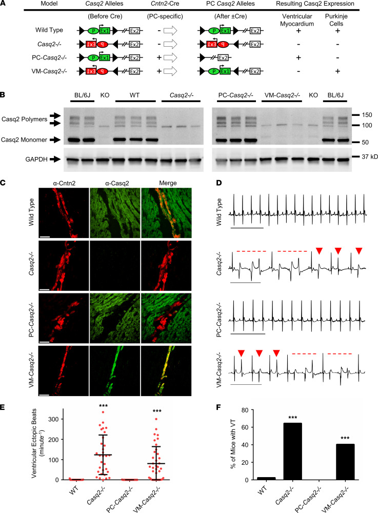
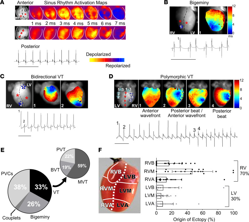
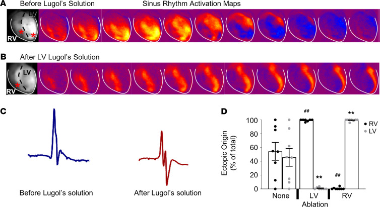
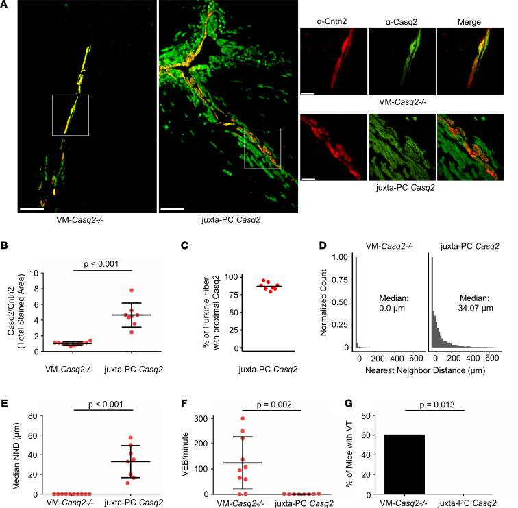
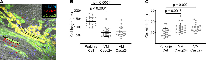
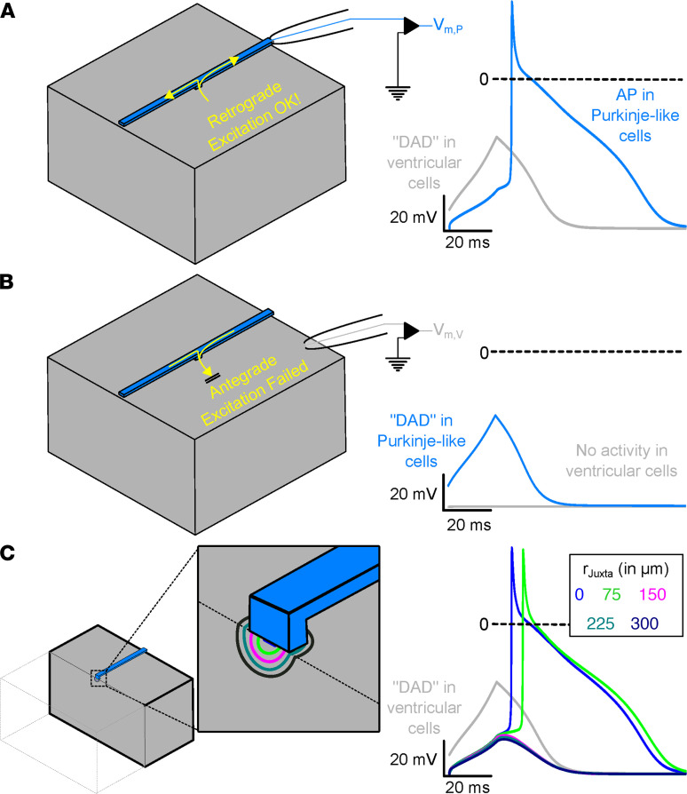
Catecholaminergic polymorphic ventricular tachycardia (CPVT) is an arrhythmia syndrome caused by gene mutations that render RYR2 Ca release channels hyperactive, provoking spontaneous Ca release and delayed afterdepolarizations (DADs). What remains unknown is the cellular source of ventricular arrhythmia triggered by DADs: Purkinje cells in the conduction system or ventricular cardiomyocytes in the working myocardium. To answer this question, we used a genetic approach in mice to knock out cardiac calsequestrin either in Purkinje cells or in ventricular cardiomyocytes. Total loss of calsequestrin in the heart causes a severe CPVT phenotype in mice and humans. We found that loss of calsequestrin only in ventricular myocytes produced a full-blown CPVT phenotype, whereas mice with loss of calsequestrin only in Purkinje cells were comparable to WT mice. Subendocardial chemical ablation or restoration of calsequestrin expression in subendocardial cardiomyocytes neighboring Purkinje cells was sufficient to protect against catecholamine-induced arrhythmias. In silico modeling demonstrated that DADs in ventricular myocardium can trigger full action potentials in the Purkinje fiber, but not vice versa. Hence, ectopic beats in CPVT are likely generated at the Purkinje-myocardial junction via a heretofore unrecognized tissue mechanism, whereby DADs in the ventricular myocardium trigger full action potentials in adjacent Purkinje cells.

Keywords: Arrhythmias; Calcium signaling; Cardiology; Genetic diseases.


Figures

Figure 1
Figure 1. Generation and characterization of cardiac Casq2 tissue-specific mouse models.
(A) Each mouse model is shown with its representative Casq2 alleles before and after Cre expression in the Purkinje cells (PCs) and resulting Casq2 protein expression in the VM and PCs. The Casq2 floxed allele contains the promoter (P) and exon 1 (Ex1) in either the forward or reverse gene orientation flanked by loxP sites (triangles). Cre expression (if present) flips the orientation of the promoter and exon 1, resulting in 4 models: WT (global Casq2+/+ expression); Casq2–/– (global Casq2–/–); PC-Casq2–/– (Casq2 knocked out only in PCs); and VM-Casq2–/– (Casq2 knocked out only in the VM). Cntn2 is only expressed in PCs. (B) Western blots from Casq2 tissue-specific mouse hearts. C57BL/6J (BL/6J) was included as a positive control. Casq2 null (KO) is an independent Casq2 germline deletion model (7) and was included as a negative control (the same Bl/6J and KO samples were loaded on both gels). GAPDH was used as a loading control. (C) Representative immunostaining for Cntn2 and Casq2 from sectioned mouse hearts. Scale bar: 50 μm. (D) Representative ECG traces from each mouse model after i.p. administration of 3 mg/kg ISO plus 60 mg/kg caffeine. Arrows denote premature ventricular contractions, and dashed lines denote episodes of VT. Scale bar: 500 ms. Quantification of VEBs (E) and VT (F) incidence in 8–38-week-old mice. WT sample, n = 39; Casq2KO, n = 31; PC-Casq2–/–, n = 16; VM-Casq2–/–, n = 28. (E) Data reported with mean ± SD. ***P < 0.001 versus WT or PC-Casq2–/– by Kruskal-Wallis test followed by Dunn’s multiple comparisons post hoc test (E) or the Fisher exact test (F).
Figure 2
Figure 2. Ex vivo optical mapping and continuous ECG recording in Langendorff-perfused Casq2 null mouse hearts.
(A) ECGs were continuously recorded while optical voltage maps were acquired from anterior and posterior epicardial surfaces during sinus rhythm. Sinus rhythm epicardial breakthroughs are denoted by red stars in the left-most panel. (B) Example temporal activation maps and associated ECG traces of bigeminy; (C) bidirectional VT; and (D) polymorphic VT after perfusion of a 100 nM ISO bolus. Ectopic foci are denoted by blue stars and indicated numerically on the accompanying ECG traces. (E) Classification of arrhythmia episodes from 8 hearts captured by ECG and optical mapping (n = 246 total episodes). BVT, bidirectional VT; MVT, monomorphic VT; PVC, premature ventricular contraction; PVT, polymorphic VT. ECG scale bars: 500 ms. (F) Quantification of the site of epicardial breakthroughs (n = 21 for each group) during voltage mapping from the same recordings as in E. Data reported as mean ± SD. R/LVB, right/left ventricular base; R/LVM, mid right/left ventricle; R/LVA, right/left ventricle apex.
Figure 3
Figure 3. Endocardial ablation reduces arrhythmia burden in Casq2 null hearts.
Lugol’s solution was injected either into the LV or RV to ablate the endocardial wall. Optical mapping before (A) and after (B) injection of Lugol’s solution into the LV. Breakthroughs are denoted in the left-most panels by red stars. Activation of the LV epicardium was maintained even after endocardial ablation. (C) Representative QRS waveform morphology before and after injection with Lugol’s solution. (D) Origin of VEBs after injection of vehicle or Lugol’s solution into the LV or RV. Arrhythmias were stimulated by perfusion with 100 nM ISO (n = 8 hearts/group). Data are reported as mean ± SD. ##P < 0.01 by 2-sided Student’s t test when comparing ectopic origin percentage from the RV with that of no ablation; **P < 0.01 by 2-sided Student’s t test when comparing ectopic origin percentage from the LV with that of no ablation.
Figure 4
Figure 4. Casq2 expression in subendocardial ventricular myocytes juxtaposed to Purkinje cells reduces PVC burden and prevents arrhythmia.
(A) Immunostaining for Cntn2 (a Purkinje cell marker) and Casq2 in selected hearts from VM-Casq2–/– mice. Scale bar: 200 μm. A subset of mice expressed Casq2, in addition to the Purkinje cells, also in ventricular myocytes next to Purkinje cells, denoted as “juxta-PC Casq2” (see top right image in A). Other mice co-expressed Casq2 only in Cntn2-positive cells (see lower right-side image in A). Scale bar in right-side images: 50 μm. (B) Ratio of Casq2 to Cntn2-positive immunostaining in hearts categorized as VM-Casq2–/– or juxta-PC Casq2 by a reviewer blinded to the genotype. (C) Percentage of Cntn2-labeled fibers having contiguous Casq2 staining in ventricular myocytes juxtaposed to the fiber. (D) NND distributions for Casq2-positive immunostaining relative to Cntn2-positive immunostaining. Data are displayed in 15 μm bins (individual distributions are shown in Supplemental Figure 2). (E) Median NND for each heart. (F) VEB and (G) VT incidence (>2 consecutive VEBs); n = 10 and 8 hearts/group, respectively. (B, D, and E) Data are reported with mean ± SD and compared using a 2-sided Mann-Whitney test. (F) Data were compared using the Fisher exact test.
Figure 5
Figure 5. Juxta-Purkinje ventricular myocytes expressing cardiac Casq2 are morphometrically like ventricular myocytes lacking Casq2.
(A) Representative image showing Purkinje cells stained by Cntn2 (resulting in yellow) alongside ventricular myocytes expressing Casq2 (green) or lacking Casq2 (gray). Cell boundaries are drawn for 1 cell of each type and selected cells used for analysis are marked with *. Scale bar: 50 μm. (B) Cell length. (C) Cell width. Data collected from 6 fields of view for a total of 20 cells/group. Data are reported with mean ± SD and compared using 1-way ANOVA with Tukey’s multiple comparisons test.
Figure 6
Figure 6. Subthreshold DAD-like activity in ventricular cells of the Purkinje–myocardial junction cause retrograde excitation of PFs.
(A) Representation of the tissue block (left) used in the computational model. Recording electrode is illustrated for the ventricular (gray) and Purkinje (blue) tissue subtypes. The membrane voltage recording (at right) shows a ventricular DAD (gray) triggering an AP in the PF (blue). (B) Reciprocal experiment demonstrating that Purkinje DADs fail to generate ventricular APs. (C) Schematic representation of the computational model. Clipping plain and zoomed-in inset show the boundaries of the hemispherical juxta-cell region with characteristic rJuxta. Membrane voltage traces (right), showing DAD-like activity in ventricular tissue (gray; identical regardless of rJuxta value) and the response in a coupled PF for various rJuxta values. Evolution of membrane voltage over time in this model for all rJuxta values can be found in Supplemental Video 1.

References

    1. Leenhardt A, et al. Catecholaminergic polymorphic ventricular tachycardia in children. A 7-year follow-up of 21 patients. Circulation. 1995;91(5):1512–1519. doi: 10.1161/01.CIR.91.5.1512. - DOI - PubMed
    1. Priori SG, et al. Clinical and molecular characterization of patients with catecholaminergic polymorphic ventricular tachycardia. Circulation. 2002;106(1):69–74. doi: 10.1161/01.CIR.0000020013.73106.D8. - DOI - PubMed
    1. Swan H, et al. Arrhythmic disorder mapped to chromosome 1q42-q43 causes malignant polymorphic ventricular tachycardia in structurally normal hearts. J Am Coll Cardiol. 1999;34(7):2035–2042. doi: 10.1016/S0735-1097(99)00461-1. - DOI - PubMed
    1. Lahat H, et al. A missense mutation in a highly conserved region of CASQ2 is associated with autosomal recessive catecholamine-induced polymorphic ventricular tachycardia in Bedouin families from Israel. Am J Hum Genet. 2001;69(6):1378–1384. doi: 10.1086/324565. - DOI - PMC - PubMed
    1. Postma AV, et al. Absence of calsequestrin 2 causes severe forms of catecholaminergic polymorphic ventricular tachycardia. Circ Res. 2002;91(8):e21–e26. - PubMed

Publication types