. 2022 Feb 8;55(2):341-354.e7.
doi: 10.1016/j.immuni.2021.12.003. Epub 2022 Jan 5.

Analysis of antibodies from HCV elite neutralizers identifies genetic determinants of broad neutralization

Affiliations

Analysis of antibodies from HCV elite neutralizers identifies genetic determinants of broad neutralization

Timm Weber et al. Immunity. .

Abstract

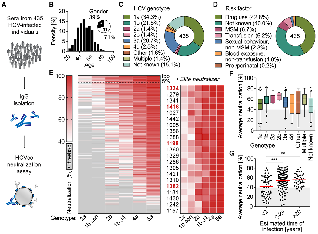
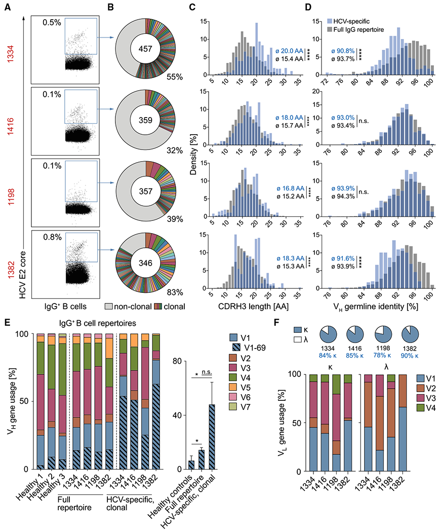
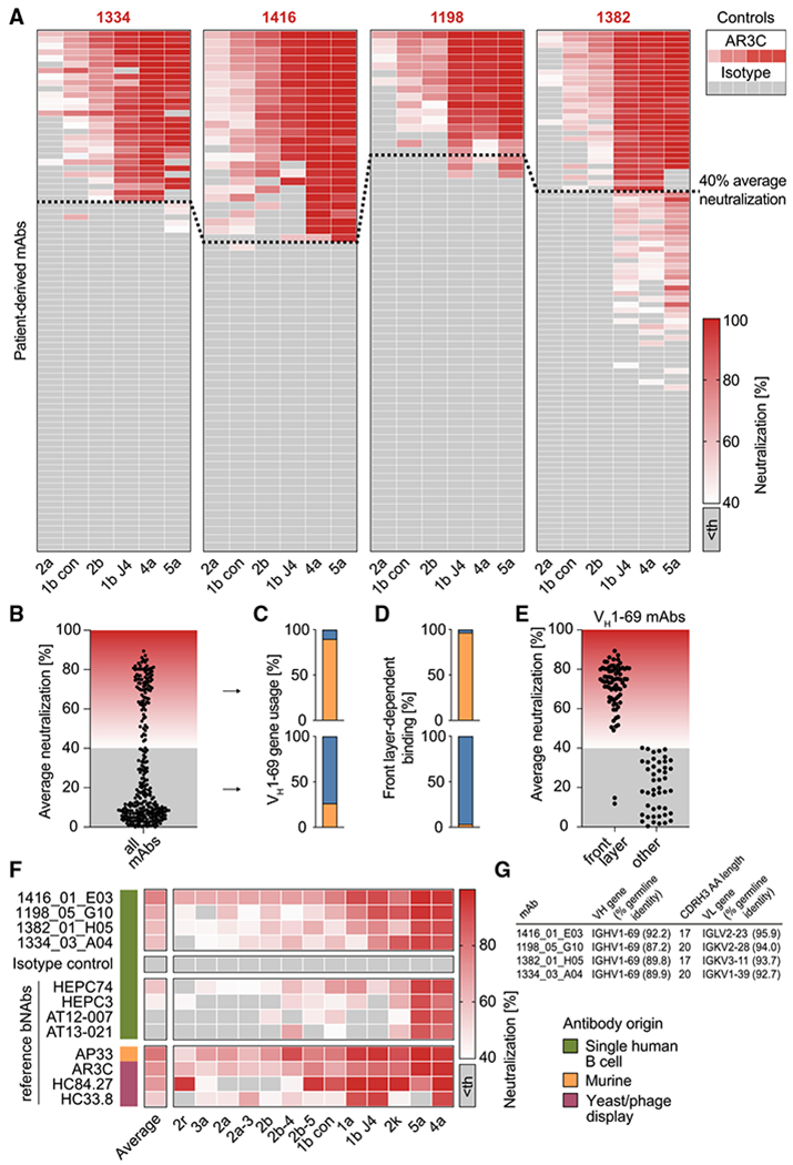
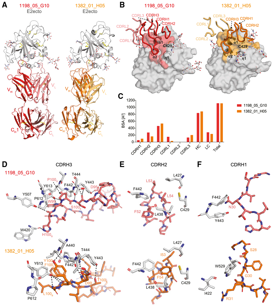
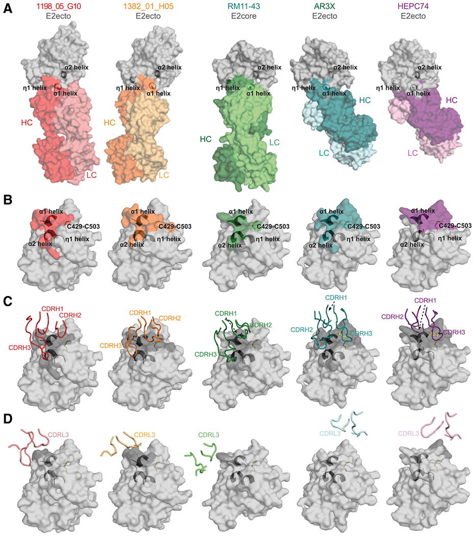
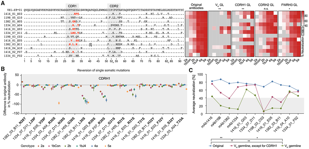
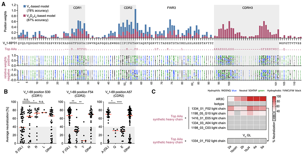
The high genetic diversity of hepatitis C virus (HCV) complicates effective vaccine development. We screened a cohort of 435 HCV-infected individuals and found that 2%-5% demonstrated outstanding HCV-neutralizing activity. From four of these patients, we isolated 310 HCV antibodies, including neutralizing antibodies with exceptional breadth and potency. High neutralizing activity was enabled by the use of the VH1-69 heavy-chain gene segment, somatic mutations within CDRH1, and CDRH2 hydrophobicity. Structural and mutational analyses revealed an important role for mutations replacing the serines at positions 30 and 31, as well as the presence of neutral and hydrophobic residues at the tip of the CDRH3. Based on these characteristics, we computationally created a de novo antibody with a fully synthetic VH1-69 heavy chain that efficiently neutralized multiple HCV genotypes. Our findings provide a deep understanding of the generation of broadly HCV-neutralizing antibodies that can guide the design of effective vaccine candidates.

Keywords: HCV; V(H)1-69; elite neutralizer; hepatitis C virus; machine learning; monoclonal antibody; neutralizing antibody; single B cell analysis; somatic mutation.


Conflict of interest statement

Declaration of interests Reported antibodies are in the process of being patented.

Figures

Figure 1.
Figure 1.. HCVcc screening identified 5% of HCV-infected individuals with outstanding “neutralizing activity”
(A) pIgG from 435 HCV-infected individuals was screened for neutralizing activity in an HCVcc assay. (B) Age and gender distribution of HCV-infected individuals. (C) HCV genotypes that individuals were infected with. (D) Self-reported most likely risk factor for HCV infection. (E) Left panel: screening of pIgG for neutralizing activity against authentic HCV virions. Huh 7.5 cells were infected with 6 different HCVcc strains (columns) carrying a Renilla luciferase reporter gene, in the presence of 300 μg/mL pIgG from HCV-infected individuals (rows). HCV neutralization was determined by reduction of luciferase activity in Huh 7.5 cells relative to cells infected with virus alone. Sorted by row means (average neutralization). Medians of triplicate measurements. Screening was performed once. Right panel: enlarged heatmap showing IDs of individuals with average neutralization in the top 5% range and neutralization of all HCVcc strains. Red IDs: individuals who later donated peripheral blood mononuclear cells. (F) Influence of the HCV genotype on average neutralization of pIgG samples. Lines, boxes, and whiskers indicate medians, 25%–75%, and 5%–95% percentiles, respectively. (G) Influence of estimated time of HCV infection on average neutralization. Data are plotted for 209 individuals. No data on time of infection were available for 226 individuals. Lines indicate means.**p ≤ 0.01, ***p ≤ 0.001; two-tailed Mann-Whitney test with Bonferroni correction. See also Figure S1.
Figure 2.
Figure 2.. HCV elite neutralizer B cell repertoires are polyclonal and VH1-69 biased
(A) Exemplary dot plots of IgG+ B cell analysis (see also Figure S2A). Numbers in red are IDs of HCV-infected individuals (see also Table S1). Numbers in plots indicate percent frequencies of E2 core-reactive B cells within IgG+ B cells (see also Table S2). Sorting was performed once. (B) Clonal relationship of E2 core-reactive B cells. Clones are shown in different colors. Numbers of productive heavy-chain sequences are depicted in the chart center. Clone sizes are proportional to the total number of productive heavy-chains per clone. (C–E) Frequencies of CDR3 length (C), V gene germline identity (D), as well as V gene segments (E) of productive heavy-chain sequences obtained from E2 core-reactive B cells compared with NGS reference data from full IgG+ B cell repertoires of the same individuals. Reference data from three randomly selected healthy donors in (E) are also included in a previous publication (Kreer et al., 2020b). Right panel of (E): quantification of VH1-69 heavy-chain frequencies. Means ± SDs are plotted. Not significant (n.s.), p > 0.05, *p ≤ 0.05, **p ≤ 0.01, ****p ≤ 0.0001; two-tailed Mann-Whitney test with Bonferroni correction (C and D) or two-tailed t test with Bonferroni correction (E). (F) Characteristics of amplified light chains. See also Figure S2 and Tables S1 and S2.
Figure 3.
Figure 3.. B cells of HCV elite neutralizers encode highly broad and potent bNAbs
(A) 310 antibodies isolated from HCV elite neutralizers were screened at a concentration of 50 μg/mL for neutralizing activity against HCV as in Figure 1E. Medians of triplicate measurements. Screening was performed once. Antibodies with an average neutralization above 40% (indicated by a dashed line) were defined as potent neutralizers. Th, threshold. (B) Distribution of average neutralization for all patient-derived antibodies from (A). (C) Percentage of antibodies utilizing the VH gene segment 1-69 among potent (upper panel) or low/non-neutralizers (lower panel). (D) Percentage of antibodies binding the E2 protein in a front-layer-dependent manner within all antibodies binding the E2 protein. Determined by ELISA with either wildtype or front layer knockout E2 protein of strain 1a157. (E) Average neutralization as in (B) for VH1-69 antibodies that bind E2 in either a front layer-dependent or a front layer-independent way. (F) Four top hits from the neutralization screen in (A) along with a set of reference HCV-specific antibodies were re-tested for neutralizing activity against an extended panel of 13 HCVcc strains, including those used for the first screen. Medians of triplicate measurements, representative of 3 independent experiments. (G) Genetic properties of the 4 isolated HCV bNAbs assayed in (F). Germline identity is indicated on nucleotide level. See also Figure S3 and Table S3.
Figure 4.
Figure 4.. Crystal structures of 1198_05_G10-E2ecto and 1382_01_H05-E2ecto complexes reveal a distinct binding mode of human HCV bNAbs
(A) Crystal structures of the 1198_05_G10-E2ecto and 1382_01_H05-E2ecto complexes. E2ecto is shown as a cartoon representation with N-glycans highlighted as sticks and disulfide bonds shown as yellow sticks. Fabs are shown as cartoons with 1198_05_G10-HC colored in red; 1198_05_G10-LC—light red, 1382_01_H05-HC—orange, and 1382_01_H05-LC—yellow. The structures were superimposed on E2 proteins. (B) Comparison of 1198_05_G10 and 1382_01_H05 CDR loop positions and corresponding E2 epitopes. Epitopes on the E2 front-layer surface were defined as residues in E2 containing an atom within 4 Å of the bound Fab. The positions of three α1-helices and Cys429 residue are indicated. (C) Comparison of buried surface areas (BSAs). (D) Interactions of CDRH3 loops with E2ecto. Potential hydrogen bonds are shown as black dashed lines and residues at the interface are indicated. Hydrogen bonds for the 3.2 Å 1382_01 _H05-E2ecto structure should be considered tentative. (E) Interactions of Fab CDRH2 loops with E2ecto. Residues at the interface are indicated. (F) Interactions of Fab CDRH1 loops with E2ecto. Residues at the interface are indicated. See also Tables S4 and S5.
Figure 5.
Figure 5.. 1198_05_G10 and 1382_01_H05 share a similar binding mode with vaccine-induced bNAbs
(A) Surface representations of 1198_05_G10-E2, 1382_01_H05-E2, and other bNAb-E2 crystal structures. The location of α1-, α2-, and η1-helices in E2 are indicated by black cartoon representations. The structures were superimposed on E2 proteins. (B) Comparison of 1198_05_G10, 1382_01_H05, RM11-43, AR3X, and HEPC74 epitopes. Epitopes on the E2 front layer (surface representation) were defined as residues in E2 containing an atom within 4 Å of the bound Fab. The location of α1-, α2-, and η1-helices in E2 are indicated by black cartoon representations and the C429–C503 disulfide bond is indicated by yellow sticks. (C) CDRH loops mapped onto the E2 surface. HC interacting residues are colored in gray on the E2 surface. (D) CDRL loops mapped onto the E2 surface. LC interacting residues are colored in gray on the E2 surface. See also Tables S4 and S5.
Figure 6.
Figure 6.. VH1-69 CDRH1 somatic mutations are critical for broad HCV neutralization
(A) Left panel: alignment of amino acid sequences of 12 HCV antibodies to VH1-69*01 (K73 is encoded by the VH1-69*06 germline allele and therefore is not necessarily a somatic mutation). Right panel: effect of partial germline-reversion on HCVcc neutralization at an antibody concentration of 50 μg/mL. Medians of triplicate measurements. Th, threshold. (B) Effect of single VH1-69 amino acid germline-reversions on HCVcc neutralization. The reverted mutations are also highlighted in (A) in red. Means ± SD of triplicate measurements. (C) HCVcc average neutralization (6-virus panel) by antibodies with VH gene segments reverted to germline, except for the CDRH1. Medians of triplicate measurements. Representative of 3 (A) or 2 (B and C) independent experiments. Not significant (n.s.), *p ≤ 0.05, **p ≤ 0.01; Wilcoxon matched-pairs signed rank test with Bonferroni correction. Average neutralization values across all 6 tested HCVcc strains for each mutated antibody were compared pairwise with average neutralization values of corresponding original antibodies (A) or among each other (C). GL, germline. See also Figure S4.
Figure 7.
Figure 7.. Potent HCV-neutralizing activity requires a pattern of genetic features and can be predicted
(A) Top panel: total weights of VH1-69-amino-acid positions contributing to the classification as an efficient neutralizer in VH-based or VHDHJH-based machine learning models. The VH1-69*01 germline amino acids are shown in black and the most impactful amino acids for the VHDHJH-based model (top AAs) are shown in violet. Bottom panel: logo plot with letter heights representing relative weights of amino acids at each position for positive or negative classification of the encoded antibodies as efficient neutralizers. Letter widths represent absolute weights. Insertions are omitted for clarity. (B) Average neutralization values of antibodies with indicated amino acid sequence features in the HCVcc screen shown in Figure 3A. Lines indicate medians. (C) HCVcc neutralization by antibodies with a synthetic heavy-chain amino acid sequence as depicted in (A) in violet. Medians of triplicate measurements. Representative of 2 independent experiments. Th, threshold. Not significant (n.s.), p > 0.05, *p ≤ 0.05, **p ≤ 0.01, ****p ≤ 0.0001; two-tailed Mann-Whitney test with Bonferroni correction. GL, germline.

Comment in

References

    1. Adams PD, Afonine PV, Bunkóczi G, Chen VB, Davis IW, Echols N, Headd JJ, Hung L-W, Kapral GJ, Grosse-Kunstleve RW, et al. (2010). PHENIX: a comprehensive Python-based system for macromolecular structure solution. Acta Crystallogr. D Biol. Crystallogr 66, 213–221. - PMC - PubMed
    1. Bailey JR, Barnes E, and Cox AL (2019). Approaches, progress, and challenges to hepatitis C vaccine development. Gastroenterology 156, 418–430. - PMC - PubMed
    1. Bailey JR, Flyak AI, Cohen VJ, Li H, Wasilewski LN, Snider AE, Wang S, Learn GH, Kose N, Loerinc L, et al. (2017). Broadly neutralizing antibodies with few somatic mutations and hepatitis C virus clearance. JCI Insight 2, e92872. - PMC - PubMed
    1. Bankwitz D, Bahai A, Labuhn M, Doepke M, Ginkel C, Khera T, Todt D, Ströh LJ, Dold L, Klein F, et al. (2021). Hepatitis C reference viruses highlight potent antibody responses and diverse viral functional interactions with neutralising antibodies. Gut 70, 1734–1745. - PMC - PubMed
    1. Battye TGG, Kontogiannis L, Johnson O, Powell HR, and Leslie AGW (2011). iMOSFLM: a new graphical interface for diffraction-image processing with MOSFLM. Acta Crystallogr. D Biol. Crystallogr 67, 271–281. - PMC - PubMed

Publication types

MeSH terms

Substances