. 2022 Jan 6;20(1):18.
doi: 10.1186/s12967-021-03218-1.

Identification of COPA as a potential prognostic biomarker and pharmacological intervention target of cervical cancer by quantitative proteomics and experimental verification

Affiliations

Identification of COPA as a potential prognostic biomarker and pharmacological intervention target of cervical cancer by quantitative proteomics and experimental verification

Huiqiong Bao et al. J Transl Med. .

Abstract

Background: Cervical cancer is the most fatal gynecological carcinoma in the world. It is urgent to explore novel prognostic biomarkers and intervention targets for cervical cancer.

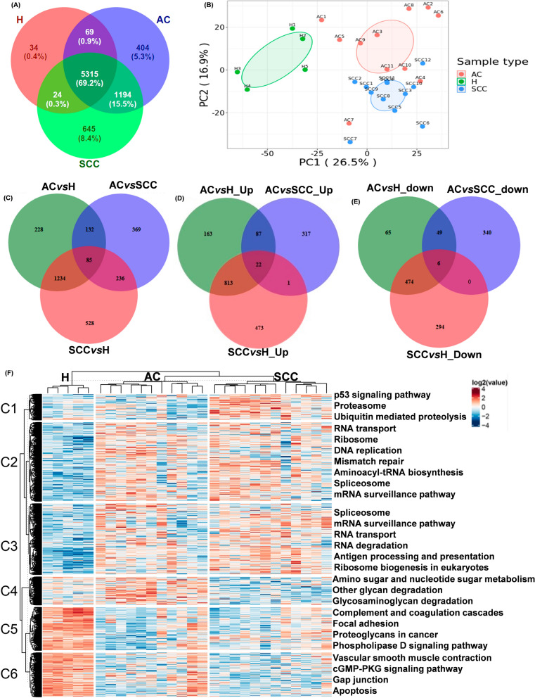
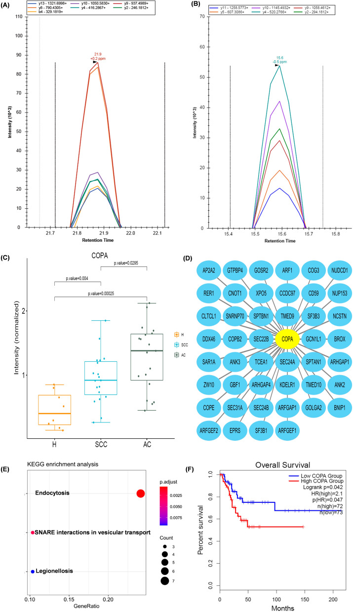
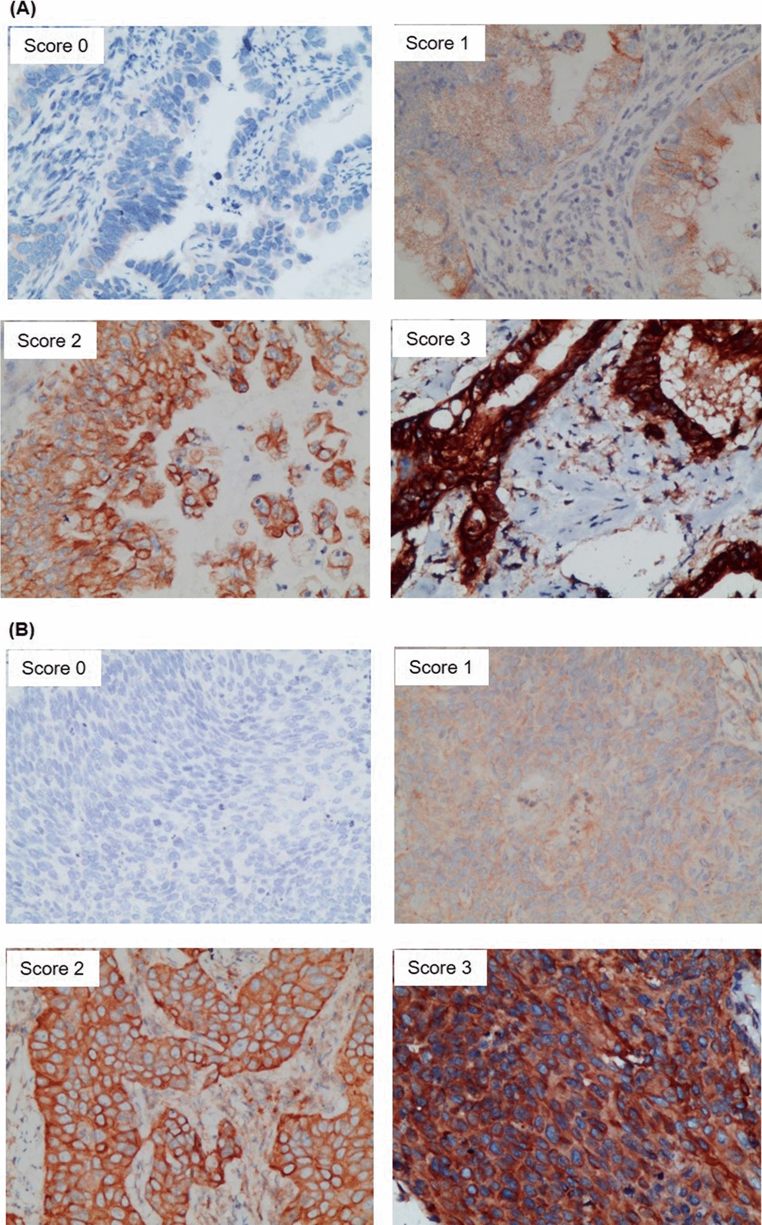
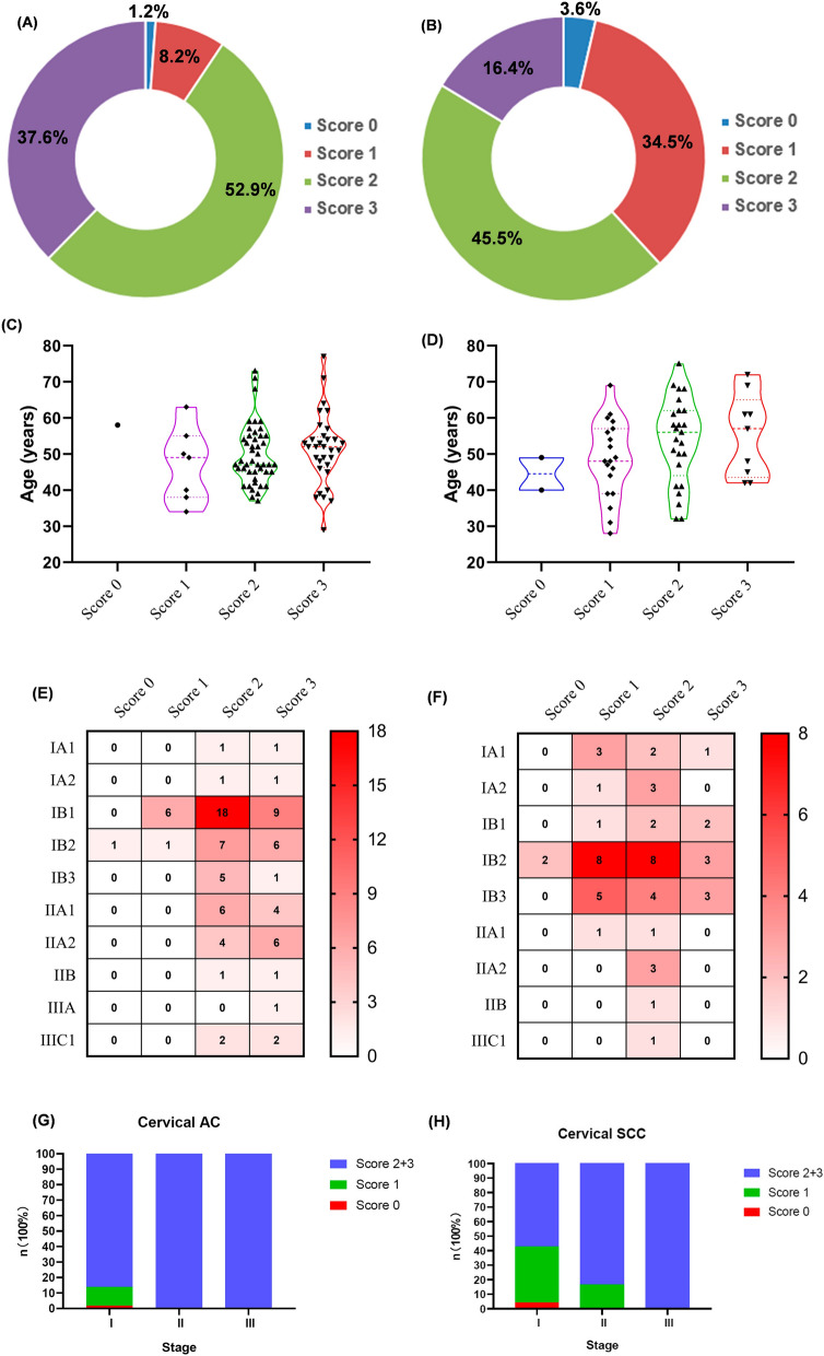
Methods: Through integrated quantitative proteomic strategy, we investigated the protein expression profiles of cervical cancer; 28 fresh frozen tissue samples (11 adenocarcinoma (AC), 12 squamous cell carcinoma (SCC) and 5 normal cervixes (HC)) were included in discover cohort; 45 fresh frozen tissue samples (19 AC, 18 SCC and 8 HC) were included in verification cohort; 140 paraffin-embedded tissues samples of cervical cancer (85 AC and 55 SCC) were used for immunohistochemical evaluation (IHC) of coatomer protein subunit alpha (COPA) as a prognostic biomarker for cervical cancer; how deficiency of COPA affects cell viability and tumorigenic ability of cervical cancer cells (SiHa cells and HeLa cells) were evaluated by cell counting kit-8 and clone formation in vitro.

Results: We identified COPA is a potential prognostic biomarker for cervical cancer in quantitative proteomics analysis. By retrospective IHC analysis, we additionally verified the proteomics results and demonstrated moderate or strong IHC staining for COPA is an unfavourable independent prognostic factor for cervical cancer. We also identified COPA is a potential pharmacological intervention target of cervical cancer by a series of in vitro experiments.

Conclusion: This study is the first to demonstrate that COPA may contribute to progression of cervical cancer. It can serve as a potential prognostic biomarker and promising intervention target for cervical cancer.

Keywords: Cervical cancer; Coatomer protein subunit alpha; Immunohistochemistry; Proteomics; Tumor mechanism.


Conflict of interest statement

The authors declare no conflict of interest.

Figures

Fig. 1
Fig. 1
Methodological workflow of the present study
Fig. 2
Fig. 2
Quantitative proteomic study. A Venn diagram demonstrating the number of quantified proteins from the three groups. H, healthy control; AC, adenocarcinoma; SCC, squamous cell carcinoma. B Principal component analysis for the quantified proteins from cervical AC, cervical SCC, and healthy control groups. C Venn diagram of the DEPs among the three comparable groups. D The venn diagram showing the up-regulated DEPs among the three comparable groups. E Venn diagram showing the down-regulated DEPs in the three comparable groups. F The cluster heatmap for the significantly different proteins (p < 0.05) between the groups calculated by the ANOVA method, the right side of the heatmap indicating the KEGG pathway analysis
Fig. 3
Fig. 3
Targeted proteomics study, bioinformatics analysis of COPA. PRM verification of COPA using unique peptides: A GITGVDLFGTTDAVVK and B CPLSGACYSPEFK. C Box-plot exhibiting protein levels of COPA in the three comparable groups. D PPI network analysis of all DEPs from cervical cancer (both AC and SCC) vs. healthy controls showed the first neighbour relationships (undirected) to COPA. E KEGG pathway analysis of the COPA and its first neighbour relationships. F Kaplan–Meier analyses for COPA (log-rank tests) based on TCGA database using the GEPIA2 software
Fig. 4
Fig. 4
IHC staining for COPA in cervical cancer. Representative photomicrographs of IHC staining for COPA (score 0–3) in tissue samples of A cervical AC and B cervical SCC. AC, adenocarcinoma; COPA, coatomer protein subunit alpha; IHC, immunohistochemistry; SCC, squamous cell carcinoma
Fig. 5
Fig. 5
Distribution characteristics of IHC staining for COPA in cervical cancer. A Relative proportions of COPA staining in cervical AC scored as negative (score 0), weak (score 1), moderate (score 2), and strong (score 3) staining. B Relative proportions of COPA staining in cervical SCC scored as negative (score 0), weak (score 1), moderate (score 2), and strong (score 3) staining. C Violin plot displays the correlation of COPA staining in cervical AC (score 0, 1, 2, 3) and age. D Violin plot displays the correlation of COPA staining in cervical SCC (score 0, 1, 2, 3) and age. E Heatmap displays the distribution of COPA staining in cervical AC (score 0, 1, 2, 3) of each stage. F Stacked column chart shows the percentage of COPA staining in cervical AC (score 0, 1, 2, 3) of each stage. G Heatmap displays the distribution of COPA staining in cervical SCC (score 0, 1, 2, 3) of each stage. H Stacked column chart shows the percentage of COPA staining in cervical SCC (score 0, 1, 2, 3) of each stage
Fig. 6
Fig. 6
Correlation between COPA staining and histopathological risk factors of cervical cancer. A Correlation between COPA staining in cervical AC (score 0, 1, 2, 3) and histopathological risk factors of cervical cancer. B Correlation between COPA staining in cervical SCC (score 0, 1, 2, 3) and histopathological risk factors of cervical cancer
Fig. 7
Fig. 7
Survival analysis of COPA in clinical samples. A Kaplan–Meier survival analysis for cervical cancer patients with different IHC staining for COPA (score 0-1vs score 2–3). B ROC curve analysis for cervical cancer patients with moderate (score 2) or strong (score 3) COPA staining
Fig. 8
Fig. 8
Depletion COPA inhibited the aggressive behaviors of cervical cancer cells in vitro. The effect of COPA knockdown was examined by western blotting analysis in A SiHa cells and B HeLa cells. β-actin was used as loading control. CCK8 assays showed COPA depletion inhibited cell viability of C SiHa cells and D HeLa cells. Quantification of foci formation were induced by the indicated clones of E SiHa cells and F HeLa cells. CCK8, cell counting kit-8. *p < 0.05

References

    1. Ahmad A, Ansari IA. A Comprehensive Review on Crosstalk of Human Papilloma Virus oncoproteins and developmental/self-renewal pathways during the pathogenesis of uterine cervical cancer. Curr Mol Med. 2020;89:234. - PubMed
    1. Cohen JD, Li L, Wang Y, Thoburn C, Afsari B, Danilova L, Douville C, Javed AA, Wong F, Mattox A. Detection and localization of surgically resectable cancers with a multi-analyte blood test. Science. 2018;359:926–930. doi: 10.1126/science.aar3247. - DOI - PMC - PubMed
    1. Lu X, Wang Z, Zhang S, Konijnenberg AP, Ouyang Y, Zhao C, Cai Y. Microscopic phase reconstruction of cervical exfoliated cell under partially coherent illumination. J Biophotonics. 2021;14:e202000401. - PubMed
    1. Zorzi M, Mistro A, Farruggio A, Bartolomeis L, Frayle-Salamanca H, Baboci L, Bertazzo A, Cocco P, Fedato C, Gennaro M, et al. Use of a high-risk human papillomavirus DNA test as the primary test in a cervical cancer screening programme: a population-based cohort study. BJOG. 2013;120:1260–1267. doi: 10.1111/1471-0528.12272. - DOI - PubMed
    1. Han R, Shi Q, Wu S, Yin D, Peng M, Dong D, Zheng Y, Guo Y, Zhang R, Hu F. Dissemination of Carbapenemases (KPC, NDM, OXA-48, IMP, and VIM) Among Carbapenem-Resistant Enterobacteriaceae Isolated From Adult and Children Patients in China. Front Cell Infect Microbiol. 2020;10:314. doi: 10.3389/fcimb.2020.00314. - DOI - PMC - PubMed

Publication types