. 2022 Jan 6;19(1):11.
doi: 10.1186/s12974-021-02324-7.

Argonaute-2 protects the neurovascular unit from damage caused by systemic inflammation

Affiliations

Argonaute-2 protects the neurovascular unit from damage caused by systemic inflammation

Marta Machado-Pereira et al. J Neuroinflammation. .

Abstract

Background: The brain vasculature plays a pivotal role in the inflammatory process by modulating the interaction between blood cells and the neurovascular unit. Argonaute-2 (Ago2) has been suggested as essential for endothelial survival but its role in the brain vasculature or in the endothelial-glial crosstalk has not been addressed. Thus, our aim was to clarify the significance of Ago2 in the inflammatory responses elicited by these cell types.

Methods: Mouse primary cultures of brain endothelial cells, astrocytes and microglia were used to evaluate cellular responses to the modulation of Ago2. Exposure of microglia to endothelial cell-conditioned media was used to assess the potential for in vivo studies. Adult mice were injected intraperitoneally with lipopolysaccharide (LPS) (2 mg/kg) followed by three daily intraperitoneal injections of Ago2 (0.4 nM) to assess markers of endothelial disruption, glial reactivity and neuronal function.

Results: Herein, we demonstrated that LPS activation disturbed the integrity of adherens junctions and downregulated Ago2 in primary brain endothelial cells. Exogenous treatment recovered intracellular Ago2 above control levels and recuperated vascular endothelial-cadherin expression, while downregulating LPS-induced nitric oxide release. Primary astrocytes did not show a significant change in Ago2 levels or response to the modulation of the Ago2 system, although endogenous Ago2 was shown to be critical in the maintenance of tumor necrosis factor-α basal levels. LPS-activated primary microglia overexpressed Ago2, and Ago2 silencing contained the inflammatory response to some extent, preventing interleukin-6 and nitric oxide release. Moreover, the secretome of Ago2-modulated brain endothelial cells had a protective effect over microglia. The intraperitoneal injection of LPS impaired blood-brain barrier and neuronal function, while triggering inflammation, and the subsequent systemic administration of Ago2 reduced or normalized endothelial, glial and neuronal markers of LPS damage. This outcome likely resulted from the direct action of Ago2 over the brain endothelium, which reestablished glial and neuronal function.

Conclusions: Ago2 could be regarded as a putative therapeutic agent, or target, in the recuperation of the neurovascular unit in inflammatory conditions.

Keywords: Argonaute-2; Brain endothelial cells; Glia; Lipopolysaccharide; Neuroprotection; Secretome.


Conflict of interest statement

The authors declare that they have no competing or conflicting interests.

Figures

Fig. 1
Fig. 1
LPS-induced Ago2 downregulation correlates with loss of endothelial function. Brain endothelial cells stimulated with lipopolysaccharide (LPS, 100 ng/ml), for 24 h, exhibited endogenous Ago2 levels significantly lower than untreated cells (control, CTR). This effect was similarly obtained with Ago2 silencing (0.05 µM). Ago2 treatment per se did not change intracellular levels (a). Ago2 silencing compromised cell survival (b) and induced cytotoxicity (c). LPS only caused loss of protein content (d). Ago2 treatment had no effect on any of these parameters associated to cell survival (bd). While siAgo2 had no effect on NRP1 expression, the receptor was downregulated by LPS and upregulated by Ago2 treatment alone. Ago2 and LPS co-administration maintained NRP1 expression (e). Ago2 co-treatment (0.4 nM) restored its intracellular levels in LPS-activated endothelial cells. The same result was produced by autophagy inhibition with bafilomycin A1 (bafA1; 100 nM), measured by western blotting (f). LPS-activated cells released nitric oxide (NO) and Ago2 treatment significantly reverted LPS-induced increase of NO levels, measured by Griess assay (g). LPS-activated cells showed a decrease in VE-cadherin expression and Ago2 treatment maintained the levels of this intercellular junction protein, measured by western blotting (h). LPS-activated brain endothelial cells released pro-inflammatory factors, such as tumor necrosis factor-alpha (TNF-α) and interleukin-6 (IL-6), the replenishment of Ago2 intracellular levels failed to normalize the levels of these cytokines (i and j, respectively), measured by ELISA. Data are expressed as the mean ± SEM of the indicated number of repeats and as a percentage relative to untreated controls (*p < 0.05, **p < 0.01, ***p < 0.001, ****p < 0.0001 compared to untreated controls; #p < 0.05, ##p < 0.01, ###p < 0.001, ####p < 0.0001 compared to LPS-activated cells; one-way ANOVA for all figures; in i, Student’s t test was used for the comparison between CTR and LPS). Ago2 argonaute-2, BafA1 bafilomycin A1, IL-6 interleukin-6, LPS lipopolysaccharide, NO nitric oxide, NRP1 neuropilin-1, TNF-α tumor necrosis factor-alpha, VE-cadherin vascular endothelial-cadherin
Fig. 2
Fig. 2
Ago2 is not involved in LPS-induced astrocyte activation. Direct exposure to lipopolysaccharide (LPS, 100 ng/ml) did not change Ago2 levels (a) or caused cytotoxicity in primary astrocytes (b). LPS-activated primary astrocytes overexpressed GFAP (c) and GDNF (f), measured by western blot, and released TNF-α (e), measured by ELISA. Ago2 silencing decreased Ago2 levels (a) and lowered the basal release of TNF-α (e). No effect was observed on NO (d). Data are expressed as the mean ± SEM of the indicated number of repeats and as a percentage relative to untreated controls (*p < 0.05, **p < 0.01, ****p < 0.0001 compared to untreated controls; one-way ANOVA for all figures; in e, Student’s t test was used for the comparison between CTR and siAgo2). Ago2 argonaute-2, LPS lipopolysaccharide, NO nitric oxide, TNF-α tumor necrosis factor-alpha, GDNF glial-derived neurotrophic factor
Fig. 3
Fig. 3
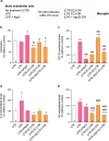
Ago2 silencing reduces microglial inflammatory responses. LPS (LPS, 100 ng/ml) increased Ago2 intracellular levels in primary microglia, while the transfection with 0.05 µM of Ago2 siRNA reduced the intracellular levels of Ago2, even after LPS stimulation, measured by western blotting (a). Ago2 silencing was cytotoxic only in conjunction with LPS (b). Ago2-silenced microglial revealed lower levels of NO (c) and IL-6 (e), but not TNF-α (d), in an inflammatory environment, measured by ELISA. TRAF6 expression was reduced after LPS challenge but Ago2 silencing counteracted this effect, measured by western blotting (f). Data are expressed as the mean ± SEM of the indicated number of repeats and as a percentage relative to untreated controls (*p < 0.05, **p < 0.01; ***p < 0.001, compared to untreated controls; #p < 0.05, ##p < 0.01, ###p < 0.001, compared to LPS-activated cells; one-way ANOVA for all figures; in d, Student’s t test was used for the comparison between CTR and LPS). Ago2 argonaute-2, IL-6 interleukin-6, LPS lipopolysaccharide, NO nitric oxide, TNF-α tumor necrosis factor-alpha, TRAF6 TNF receptor-associated factor 6
Fig. 4
Fig. 4
Ago2-restored endothelium normalizes microglia response. Schematic representation of the experimental setup (a). Endothelial cell-conditioned media (EC-CM) from cells exposed to LPS and treated with Ago2 (0.4 nM) ((LPS + Ago2 EC)-CM) reproduced the protective effect of EC-CM collected from healthy endothelial cells ((CTR EC)-CM), measured by LDH activity assay (b). Microglial cells exposed to (LPS + Ago2 EC)-CM presented CD11b levels similar to untreated cells (CTR) and cells exposed to (CTR EC)-CM, measured by western blotting, while (LPS EC)-CM had the same effect as the LPS treatment (c). EC-CM had no significant effect over IL-1β release (d), but (CTR EC)-CM and (LPS + Ago2 EC)-CM decreased NO below control levels (e). Data are expressed as the mean ± SEM of the indicated number of repeats and as a percentage relative to untreated controls (*p < 0.05, **p < 0.01, ***p < 0.001, ****p < 0.0001, compared to untreated controls; #p < 0.05, ##p < 0.01, ###p < 0.001, ####p < 0.0001, compared to LPS-activated cells; §p < 0.05, §§§p < 0.001, §§§§p < 0.0001, compared to (CTR EC)-CM; $p < 0.05, $$$p < 0.001, $$$$p < 0.0001, compared to (LPS EC)-CM; one-way ANOVA; in d, Student’s t test was used for the comparison between CTR and LPS)). Ago2 argonaute-2, CD11b cluster of differentiation molecule 11b, EC-CM endothelial cell-conditioned media, IL-1β interleukin-1 beta, LPS lipopolysaccharide, NO nitric oxide
Fig. 5
Fig. 5
Systemic administration of Ago2 restores endothelial barrier function and normalizes glial activation in the cortex. Mice injected intraperitoneally with lipopolysaccharide (LPS, 2 mg/kg) showed the activation of p38 signaling pathway, i.e., increased p38 phosphorylation (Pp38) (b) and produced higher levels of eNOS (c), iNOS (d), NOX2 (e), Iba-1 (g), GFAP (h) and S100B (i), and lower levels of VE-cadherin (a) and p47phox (f). LPS-injected mice treated with Ago2 (0.4 nmol/l) had marker levels similar to sham controls (animals injected with phosphate-buffered saline, CTR), with the exception of the Pp38 and iNOS markers. The same pattern of reduction below control levels was obtained with Ago2 injection in sham controls. Ago2 cortical levels did not change significantly in any of the experimental conditions (j). The levels of NRP1 were only raised in LPS-injected mice treated with Ago2 (k). The table summarizes data from these experiments (l). Schematic representation of the experimental setup used herein (and in this figure) (m). Data are expressed as the mean ± SEM of the indicated number of repeats and as a percentage relative to sham controls (*p < 0.05, **p < 0.01, ***p < 0.001, ****p < 0.0001 compared to sham controls; #p < 0.05, ##p < 0.01, ####p < 0.0001 compared to LPS-injected animals; one-way ANOVA; in a, c and f, Student’s t test was used for the comparison between CTR and LPS). Ago2 argonaute-2, eNOS endothelial nitric oxide synthase, GFAP glial fibrillary acidic protein, Iba-1 ionized calcium-binding adaptor molecule-1, iNOS inducible nitric oxide synthase, LPS lipopolysaccharide, NG2 oligodendrocyte precursor cells, NO nitric oxide, NOX2 NADPH oxidase 2, NRP1 neuroplilin-1, p47phox neutrophil cytosol factor 1, Pp38 phosphorylated p38 mitogen-activated protein kinases signaling pathway, S100B S100 calcium-binding protein B, VE-cadherin vascular endothelial-cadherin
Fig. 6
Fig. 6
Systemic administration of Ago2 normalizes inflammatory activation and induces neuroprotection in the hippocampus. LPS-injected mice (LPS, 2 mg/kg) produced higher levels of Iba-1 (a) and S100B (b), and lower levels of CREB (c), MAP2 (d) and PSD-95 (e). Mice treated with Ago2 (0.4 nmol/L) had marker levels similar to sham controls (animals injected with phosphate-buffered saline, CTR), with the exception of the Iba-1 marker. Similar to the mouse cortex, the Ago2 hippocampus levels did not change significantly in the experimental conditions, with the exception of the Ago2-injected mice, in which a decrease of Ago2 levels occurred (f). NRP1 levels did not change significantly in any of the experimental conditions (g). The table summarizes data from these experiments (h). Data are expressed as the mean ± SEM of the indicated number of repeats and as a percentage relative to sham controls (*p < 0.05, **p < 0.01, ***p < 0.001, ****p < 0.0001 compared to sham controls; #p < 0.05, ###p < 0.001, ####p < 0.0001 compared to LPS-injected animals; one-way ANOVA; in c to e, Student’s t test was used for the comparison between CTR and LPS). Ago2 argonaute-2, CREB cAMP response element-binding protein, Iba-1 ionized calcium-binding adaptor molecule-1, LPS lipopolysaccharide, MAP2 microtubule-associated protein, NRP1 neuropilin-1, PSD-95 postsynaptic density protein-95, S100B S100 calcium-binding protein B
Fig. 7
Fig. 7
Proposed model for Ago2 regulation of the endothelial and glial crosstalk. LPS activation of the p38 signaling pathway promotes VE-cadherin downregulation and the transcription of mRNA related to pro-inflammatory mediators (e.g., cytokines). The translation of these transcripts is facilitated by low Ago2 levels and low RISC activity. These events trigger the activation of microglia and astrocytes associated with blood vessels, and cause neuronal damage. The exogenous application and internalization of Ago2, via NRP1, recuperates RISC activity, which is conducive to eNOS degradation and low NO, reducing glial activation and protecting neuronal cells. Ago2 argonaute-2, IL-6 interleukin-6, LPS lipopolysaccharide, mRNA messenger ribonucleic acid, NO nitric oxide, NRP1 neuropilin-1, p38 p38 mitogen-activated protein kinases signaling pathway, RISC RNA-induced silencing complex, TNF-α tumor necrosis factor alpha, TLR4 toll-like receptor 4, VE-cadherin vascular endothelial-cadherin

Similar articles

Cited by

References

    1. Daneman R, Prat A. The blood-brain barrier. Cold Spring Harb Perspect Biol. 2015;7(1):a020412. - PMC - PubMed
    1. Varatharaj A, Galea I. The blood-brain barrier in systemic inflammation. Brain Behav Immun. 2017;60:1–12. - PubMed
    1. Machado-Pereira M, Santos T, Bernardino L, Ferreira R. Vascular inter-regulation of inflammation: molecular and cellular targets for CNS therapy. J Neurochem. 2016;140(5):692–702. - PubMed
    1. Dauphinee SM, Karsan A. Lipopolysaccharide signaling in endothelial cells. Lab Investig. 2006;86(1):9–22. - PubMed
    1. Akira S, Takeda K. Toll-like receptor signalling. Nat Rev Immunol. 2004;4:499–511. - PubMed

LinkOut - more resources