. 2022 Jan 6;19(1):8.
doi: 10.1186/s12974-021-02357-y.

Nafamostat reduces systemic inflammation in TLR7-mediated virus-like illness

Affiliations

Nafamostat reduces systemic inflammation in TLR7-mediated virus-like illness

Abi G Yates et al. J Neuroinflammation. .

Abstract

Background: The serine protease inhibitor nafamostat has been proposed as a treatment for COVID-19, by inhibiting TMPRSS2-mediated viral cell entry. Nafamostat has been shown to have other, immunomodulatory effects, which may be beneficial for treatment, however animal models of ssRNA virus infection are lacking. In this study, we examined the potential of the dual TLR7/8 agonist R848 to mimic the host response to an ssRNA virus infection and the associated behavioural response. In addition, we evaluated the anti-inflammatory effects of nafamostat in this model.

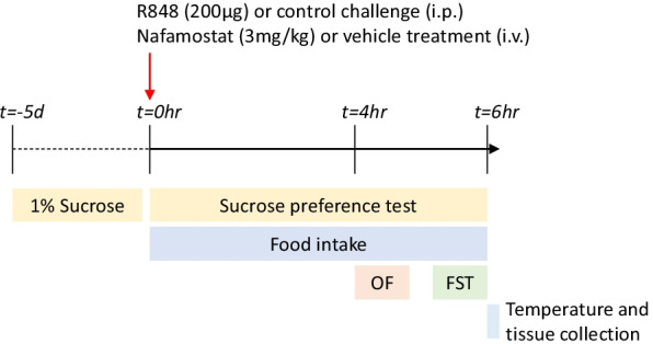
Methods: CD-1 mice received an intraperitoneal injection of R848 (200 μg, prepared in DMSO, diluted 1:10 in saline) or diluted DMSO alone, and an intravenous injection of either nafamostat (100 μL, 3 mg/kg in 5% dextrose) or 5% dextrose alone. Sickness behaviour was determined by temperature, food intake, sucrose preference test, open field and forced swim test. Blood and fresh liver, lung and brain were collected 6 h post-challenge to measure markers of peripheral and central inflammation by blood analysis, immunohistochemistry and qPCR.

Results: R848 induced a robust inflammatory response, as evidenced by increased expression of TNF, IFN-γ, CXCL1 and CXCL10 in the liver, lung and brain, as well as a sickness behaviour phenotype. Exogenous administration of nafamostat suppressed the hepatic inflammatory response, significantly reducing TNF and IFN-γ expression, but had no effect on lung or brain cytokine production. R848 administration depleted circulating leukocytes, which was restored by nafamostat treatment.

Conclusions: Our data indicate that R848 administration provides a useful model of ssRNA virus infection, which induces inflammation in the periphery and CNS, and virus infection-like illness. In turn, we show that nafamostat has a systemic anti-inflammatory effect in the presence of the TLR7/8 agonist. Therefore, the results indicate that nafamostat has anti-inflammatory actions, beyond its ability to inhibit TMPRSS2, that might potentiate its anti-viral actions in pathologies such as COVID-19.

Keywords: COVID-19; Inflammation; Nafamostat; Sickness behaviour; Viral infection.


Conflict of interest statement

The authors declare that they have no competing interests.

Figures

Fig. 1
Fig. 1
Schematic of behavioural tests performed. In summary, animals were exposed to 1% sucrose solution during the 5 days of acclimatisation. On the day of the experiment, male, CD-1 mice received an intraperitoneal injection of R848 (200 μg, dissolved in DMSO and sterile saline) or control solution (DMSO diluted in sterile saline), together with an intravenous injection of nafamostat (3 mg/kg) or vehicle (sterile saline). For the duration of the experiment, animals underwent the sucrose preference test (SPT), and food intake was examined. 4-h post-challenge, animals completed the open field (OF) paradigm. 6 h post-challenge, animals completed the forced swim test (FST), after which their temperature was determined and animals were culled. d = days, hr = hours
Fig. 2
Fig. 2
R848 induces a sickness behaviour phenotype, which is not ameliorated by nafamostat. Male, CD-1 mice received an intraperitoneal injection of R848 (200 μg, dissolved in DMSO and sterile saline) or control solution (DMSO diluted in sterile saline), together with an intravenous injection of nafamostat (3 mg/kg) or vehicle (sterile saline), and sickness behaviour was assessed 4–6 h later. Animal temperature (A) and food intake (B) were determined to assess any fever and anorexia, respectively. Anhedonia was measured with the SPT; total sucrose water intake (C) and preference for the sucrose water (D) were evaluated. OF was used to assess exploratory behaviour and grid crossings/minute (E), number of rears (F) and time spent in the centre (G) were calculated. Latency to immobility (H) and total immobility (I) in the FST were measured. Naïve animals were included to establish baseline (dotted line). Data presented as mean ± SEM, n = 4–10/group, and analysed by two-way ANOVA, *p < 0.05, **p < 0.01, ***p < 0.001, ****p < 0.0001 main effect, #p < 0.05, ##p < 0.01, ###p < 0.001, ####p < 0.0001 control vs. R848 in Sidak’s post hoc test
Fig. 3
Fig. 3
R848 causes a depletion in circulating leukocytes, which is partially restored by nafamostat. Male, CD-1 mice received an intraperitoneal injection of R848 (200 μg, dissolved in DMSO and sterile saline) or control solution (DMSO diluted in sterile saline), together with an intravenous injection of nafamostat (3 mg/kg) or vehicle (sterile saline). Blood was collected 6-h post-challenge and total concentration of total white blood cells (A), as well as the concentrations of lymphocytes (B), monocytes (C), neutrophils (D), basophils (E) and eosinophils (F), specifically, were measured. Naïve animals were included to establish baseline (dotted line). Data presented as mean ± SEM, n = 4–10/group, and analysed by two-way ANOVA, **p < 0.01 main effect, #p < 0.05, ##p < 0.01 control vs. R848 with Sidak’s post-hoc test, &p < 0.05 vehicle vs. nafamostat with Sidak’s post-hoc test
Fig. 4
Fig. 4
R848 induces neutrophil infiltration of peripheral and central tissues, but depletion of monocytes and B lymphocytes. Male, CD-1 mice received an intraperitoneal injection of R848 (200 μg, dissolved in DMSO and sterile saline) or control solution (DMSO diluted in sterile saline), together with an intravenous injection of nafamostat (3 mg/kg) or vehicle (sterile saline). Fresh liver was collected 6-h post-challenge and a subset of each group was cut at 12 μm thickness (n = 5/group). Immunostaining for neutrophils (MBS+), macrophages (Iba-1+) and B lymphocytes (B220+) was performed to determine leukocyte density. Liver neutrophils (A) and macrophages (B), lung neutrophils (C), macrophages (D) and B lymphocytes (E), and brain neutrophils (F) and microglia (G) were quantified, blinded, and presented as cells per mm2. Naïve animals were included to establish baseline (dotted line). Data presented as mean ± SEM and analysed by two-way ANOVA, ****p < 0.0001, **p < 0.01 main effect, #p < 0.05, ##p < 0.01, ###p < 0.001, ####p < 0.0001 control vs. R848 with Sidak’s post hoc test
Fig. 5
Fig. 5
R848 induces pro-inflammatory gene expression in the liver, which is ameliorated with nafamostat treatment. Male, CD-1 mice received an intraperitoneal injection of R848 (200 μg, dissolved in DMSO and sterile saline) or control solution (DMSO diluted in sterile saline), together with an intravenous injection of nafamostat (3 mg/kg) or vehicle (sterile saline). Fresh liver was collected 6-h post-challenge and relative expression of TNF (A), IFN-γ (B), CXCL1 (C), CXCL10 (D), SAA-2 (E) and CRP (F) were determined by qPCR. Naïve animals were included to establish baseline (dotted line). Data presented as mean ± SEM, n = 4–10/group, and analysed by two-way ANOVA, ****p < 0.0001 main effect, ##p < 0.01, ####p < 0.0001 control vs. R848 with Sidak’s post hoc test, &p < 0.05, &&p < 0.01 vehicle vs. nafamostat with Sidak’s post hoc test
Fig. 6
Fig. 6
R848 induces pro-inflammatory gene expression in the lung, which is unaffected by treatment with nafamostat. Male, CD-1 mice received an intraperitoneal injection of R848 (200 μg, dissolved in DMSO and sterile saline) or control solution (DMSO diluted in sterile saline), together with an intravenous injection of nafamostat (3 mg/kg) or vehicle (sterile saline). Fresh lung was collected 6-h post-challenge and relative expression of TNF (A), IFN-γ (B), CXCL1 (C) and CXCL10 (D) was determined by qPCR. Naïve animals were included to establish baseline (dotted line). Data presented as mean ± SEM, n = 4–10/group, and analysed by two-way ANOVA, ****p < 0.0001 main effect, #p < 0.05, ##p < 0.01, ###p < 0.001 control vs. R848 with Sidak’s post hoc test
Fig. 7
Fig. 7
R848 causes pro-inflammatory gene expression in the brain which is not attenuated by nafamostat. Male, CD-1 mice received an intraperitoneal injection of R848 (200 μg, dissolved in DMSO and sterile saline) or control solution (DMSO diluted in sterile saline), together with an intravenous injection of nafamostat (3 mg/kg) or vehicle (sterile saline). Fresh brain was collected 6-h post-challenge. Relative expression of TNF (A), IFN-γ (B), CXCL1 (C) and CXCL10 (D) in the prefrontal cortex were determined by qPCR. Naïve animals were included to establish baseline (dotted line). Data presented as mean ± SEM, n = 4–10/group, and analysed by two-way ANOVA, ***p < 0.001, ****p < 0.0001 main effect, #p < 0.05, ##p < 0.01, ####p < 0.0001 control vs. R848 with Sidak’s post hoc test

References

    1. Hoffmann M, Kleine-Weber H, Schroeder S, Kruger N, Herrler T, Erichsen S, Schiergens TS, Herrler G, Wu NH, Nitsche A, et al. SARS-CoV-2 cell entry depends on ACE2 and TMPRSS2 and is blocked by a clinically proven protease inhibitor. Cell. 2020;181:271–280.e278. - PMC - PubMed
    1. Yamamoto M, Kiso M, Sakai-Tagawa Y, Iwatsuki-Horimoto K, Imai M, Takeda M, Kinoshita N, Ohmagari N, Gohda J, Semba K, et al. The anticoagulant nafamostat potently inhibits SARS-CoV-2 S protein-mediated fusion in a cell fusion assay system and viral infection in vitro in a cell-type-dependent manner. Viruses. 2020;12(6):629. - PMC - PubMed
    1. Yamamoto M, Matsuyama S, Li X, Takeda M, Kawaguchi Y, Inoue JI, Matsuda Z. Identification of nafamostat as a potent inhibitor of middle east respiratory syndrome coronavirus s protein-mediated membrane fusion using the split-protein-based cell-cell fusion assay. Antimicrob Agents Chemother. 2016;60:6532–6539. - PMC - PubMed
    1. Takahashi W, Yoneda T, Koba H, Ueda T, Tsuji N, Ogawa H, Asakura H. Potential mechanisms of nafamostat therapy for severe COVID-19 pneumonia with disseminated intravascular coagulation. Int J Infect Dis. 2021;102:529–531. - PMC - PubMed
    1. Liu Y, Li C, Wang J, Fang Y, Sun H, Tao X, Zhou XF, Liao H. Nafamostat mesilate improves neurological outcome and axonal regeneration after stroke in rats. Mol Neurobiol. 2017;54:4217–4231. - PubMed

MeSH terms