. 2022 Jan 6;17(1):1.
doi: 10.1186/s13024-021-00511-x.

Constitutively active SARM1 variants that induce neuropathy are enriched in ALS patients

Affiliations

Constitutively active SARM1 variants that induce neuropathy are enriched in ALS patients

A Joseph Bloom et al. Mol Neurodegener. .

Abstract

Background: In response to injury, neurons activate a program of organized axon self-destruction initiated by the NAD+ hydrolase, SARM1. In healthy neurons SARM1 is autoinhibited, but single amino acid changes can abolish autoinhibition leading to constitutively active SARM1 enzymes that promote degeneration when expressed in cultured neurons.

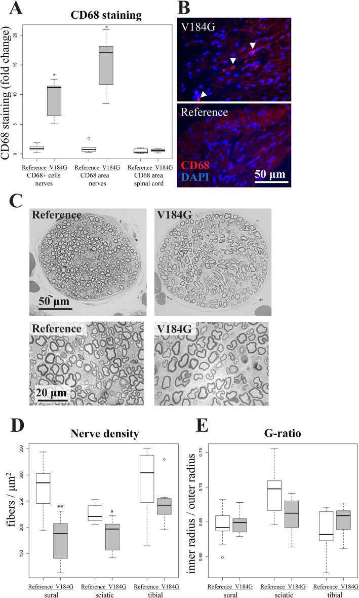
Methods: To investigate whether naturally occurring human variants might disrupt SARM1 autoinhibition and potentially contribute to risk for neurodegenerative disease, we assayed the enzymatic activity of all 42 rare SARM1 alleles identified among 8507 amyotrophic lateral sclerosis (ALS) patients and 9671 controls. We then intrathecally injected mice with virus expressing SARM1 constructs to test the capacity of an ALS-associated constitutively active SARM1 variant to promote neurodegeneration in vivo.

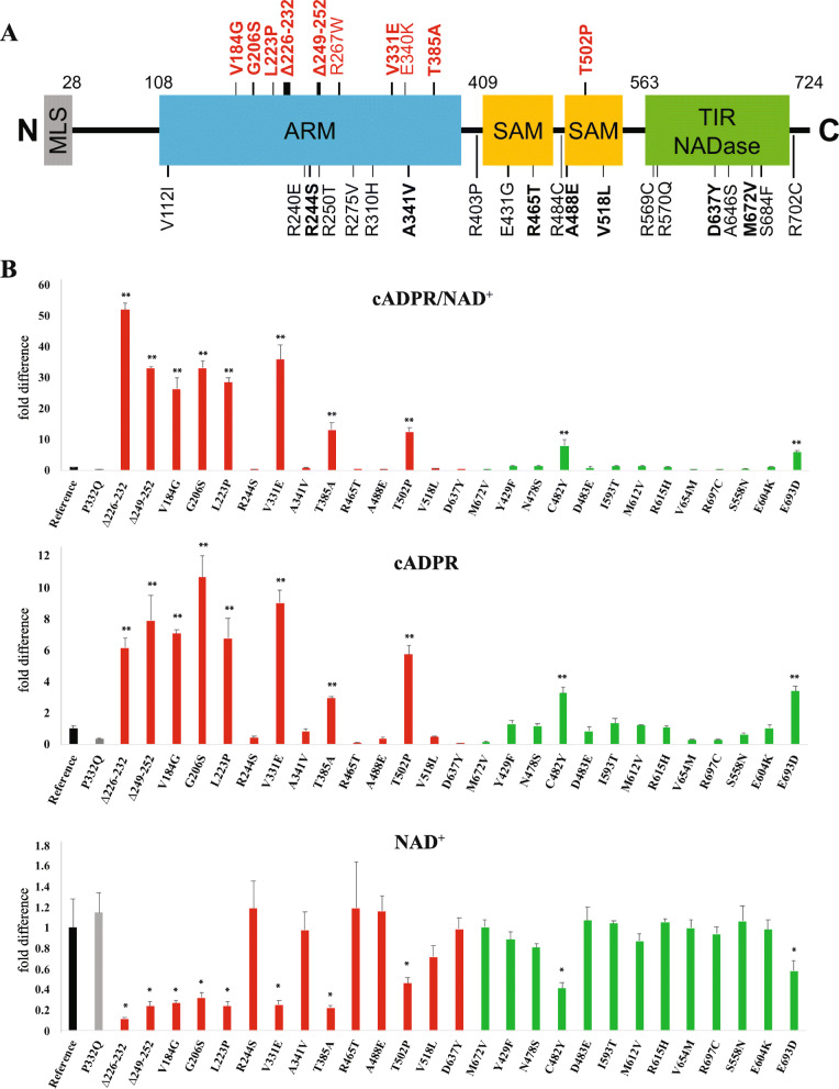
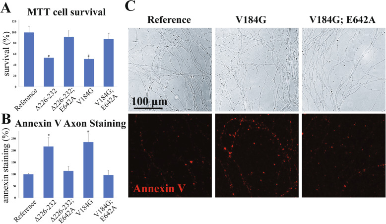
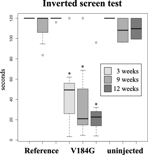
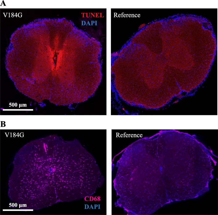
Results: Twelve out of 42 SARM1 missense variants or small in-frame deletions assayed exhibit constitutive NADase activity, including more than half of those that are unique to the ALS patients or that occur in multiple patients. There is a > 5-fold enrichment of constitutively active variants among patients compared to controls. Expression of constitutively active ALS-associated SARM1 alleles in cultured dorsal root ganglion (DRG) neurons is pro-degenerative and cytotoxic. Intrathecal injection of an AAV expressing the common SARM1 reference allele is innocuous to mice, but a construct harboring SARM1V184G, the constitutively active variant found most frequently among the ALS patients, causes axon loss, motor dysfunction, and sustained neuroinflammation.

Conclusions: These results implicate rare hypermorphic SARM1 alleles as candidate genetic risk factors for ALS and other neurodegenerative conditions.

Keywords: ALS; Axon; Human genetics; NAD; Neurodegeneration; Neuropathy; SARM1.


Conflict of interest statement

A.D. and J.M. are co-founders, scientific advisory board members and shareholders of Disarm Therapeutics, a wholly owned subsidiary of Eli Lilly. A.J.B. and Y.S. are consultants to Disarm Therapeutics. The authors have no other competing conflicts or financial interests.

Figures

Fig. 1
Fig. 1
Identification of dysregulated SARM1 variants found in ALS patients. (A) Schematic representation of the domain structure of SARM1 marked with every rare variant found in ALS patients. Constitutively-active variants are indicated above in red. Bold variants were prioritized because they were identified in multiple ALS patients or were unique to ALS patients. Δ indicates an in-frame deletion. MLS, mitochondrial localization signal; ARM, HEAT/Armadillo motif; SAM, sterile alpha motif; TIR, Toll/interleukin-1 receptor homology domain. (B) The ratio of cADPR/NAD+, cADPR, and NAD+ levels from cultured Sarm1−/− DRG neurons infected with human SARM1 constructs carrying every rare variants identified in multiple ALS patients or unique to ALS patients (red), every rare variant found in controls (green), and the most common SARM1 variant, P332Q (gray), relative to the common reference human genome allele of SARM1 (black). *p < 0.05; **p < 0.0005 difference from reference allele. We demonstrate that strongly constitutively active variants predominate among those SARM1 variants identified in multiple ALS patients or unique to ALS patients, while the only two significantly active variants discovered in controls are comparatively weak
Fig. 2
Fig. 2
Neurodegeneration in cultured neurons transfected with ALS-associated SARM1 variants. (A) Neuron death as measured by the MTT assay and (B) axon degeneration as measured by Annexin V staining in Sarm1−/− DRG neurons infected with lentivirus expressing SARM1 variant constructs as well as double mutant constructs including E642A, a point mutation that disrupts SARM1 NAD+ hydrolase activity, relative to the common SARM1 reference allele. All data are expressed as the percent of surviving cells or area of Annexin stained axons relative to neurons infected with the reference allele. (C) Representative bright-field and Annexin V-stained images of axons from Sarm1−/− DRG cultures infected with variant and SARM1 reference allele constructs. *p < 0.005 difference from reference allele. These results demonstrate that constitutive SARM1 activity causes cell death and axon degeneration while expression of control SARM1 does not
Fig. 3
Fig. 3
Motor dysfunction in mice injected intrathecally with a SARM1V184G AAV construct. Average time suspended from an inverted screen (maximum 120 s) for C57BL/6 mice injected with a human SARM1 reference allele (n = 8) or SARM1V184G (n = 7) AAV compared to uninjected controls (n = 3) 3, 9 and 12 weeks post-injection. *p < 0.005 difference from both the reference allele and uninjected controls. These results demonstrate that infection with SARM1V184G causes persistent motor dysfunction while the control SARM1 construct does not
Fig. 4
Fig. 4
Rapid cell death and neuroinflammation in mice injected intrathecally with a SARM1V184G AAV construct. This figure demonstrates anatomical findings from the small subset of mice with rapid onset pathology. (A) Representative images of spinal cord sections stained with DAPI and the apoptosis marker TUNEL from mice 2 days after injection with a SARM1V184G or SARM1 human reference allele construct. (B) Representative images of spinal cord stained with DAPI and the macrophage marker anti-CD68 from mice 2 days after injection with a SARM1V184G or reference allele construct. These images demonstrate that mice that become paralyzed shortly after injection with SARM1V184G exhibit cell death and activated macrophages in their spinal cords while mice injected with the control SARM1 construct do not
Fig. 5
Fig. 5
Persistent neuroinflammation and axon loss in mice injected intrathecally with a SARM1V184G AAV construct. This figure demonstrates anatomical findings from the larger subset of mice with slower onset pathology. (A) The normalized average number of cells stained by the macrophage marker anti-CD68 in nerve, and the average percent area of total anti-CD68 staining in nerve and in spinal cord sections, from C57BL/6 mice injected with a SARM1V184G (n = 7) AAV construct relative to those injected with a human SARM1 reference allele construct (n = 8) 12 weeks post-injection; *p < 10− 4 difference from reference allele. (B) Representative images of sural nerve stained with DAPI and anti-CD68 from mice 12 weeks after injection with a SARM1V184G or reference allele construct. White arrows indicate CD68-positive activated macrophages. (C) Representative images of toluidine blue stained sural nerve sections. (D) Average fibers per cross-sectional μm2 in sural, sciatic and tibial nerves from mice 12 weeks after injection with a SARM1V184G (n = 7 mice) or reference allele construct (n = 8); *p < 0.05, **p < 0.001. (E) Average g-ratio (ratio between the inner and outer myelin sheath) for axons in the sural, sciatic and tibial nerves from mice 12 weeks after injection with a SARM1V184G or reference allele construct. These analyses demonstrate that mice injected with SARM1V184G exhibit axon loss and persistent neuroinflammation in their nerves 12 weeks after infection, compared to mice injected with a control SARM1 construct

References

    1. Al-Chalabi A. Don’t keep it in the family. Nature. 2017;550(7676):S112. doi: 10.1038/550S112a. - DOI - PubMed
    1. Gelfman S, Dugger S, de Araujo Martins Moreno C, et al. A new approach for rare variation collapsing on functional protein domains implicates specific genic regions in ALS. Genome Res. 2019. 10.1101/gr.243592.118. - PMC - PubMed
    1. Farhan SMK, Howrigan DP, Abbott LE, et al. Exome sequencing in amyotrophic lateral sclerosis implicates a novel gene, DNAJC7, encoding a heat-shock protein. Nat Neurosci. 2019;22(12):1966–1974. doi: 10.1038/s41593-019-0530-0. - DOI - PMC - PubMed
    1. Cirulli ET, Lasseigne BN, Petrovski S, et al. Exome sequencing in amyotrophic lateral sclerosis identifies risk genes and pathways. Science. 2015;(80). 10.1126/science.aaa3650. - PMC - PubMed
    1. Lattante S, Doronzio PN, Marangi G, Conte A, Bisogni G, Bernardo D, Russo T, Lamberti D, Patrizi S, Apollo FP, Lunetta C, Scarlino S, Pozzi L, Zollino M, Riva N, Sabatelli M. Coexistence of variants in TBK1 and in other ALS-related genes elucidates an oligogenic model of pathogenesis in sporadic ALS. Neurobiol Aging. 2019;84:239.e9–239.e14. doi: 10.1016/j.neurobiolaging.2019.03.010. - DOI - PubMed

Publication types

MeSH terms