Review
. 2022 Jul:89:101039.
doi: 10.1016/j.preteyeres.2021.101039. Epub 2022 Jan 4.

The impact of sensory neuropathy and inflammation on epithelial wound healing in diabetic corneas

Affiliations
Review

The impact of sensory neuropathy and inflammation on epithelial wound healing in diabetic corneas

Fu-Shin X Yu et al. Prog Retin Eye Res. 2022 Jul.

Abstract

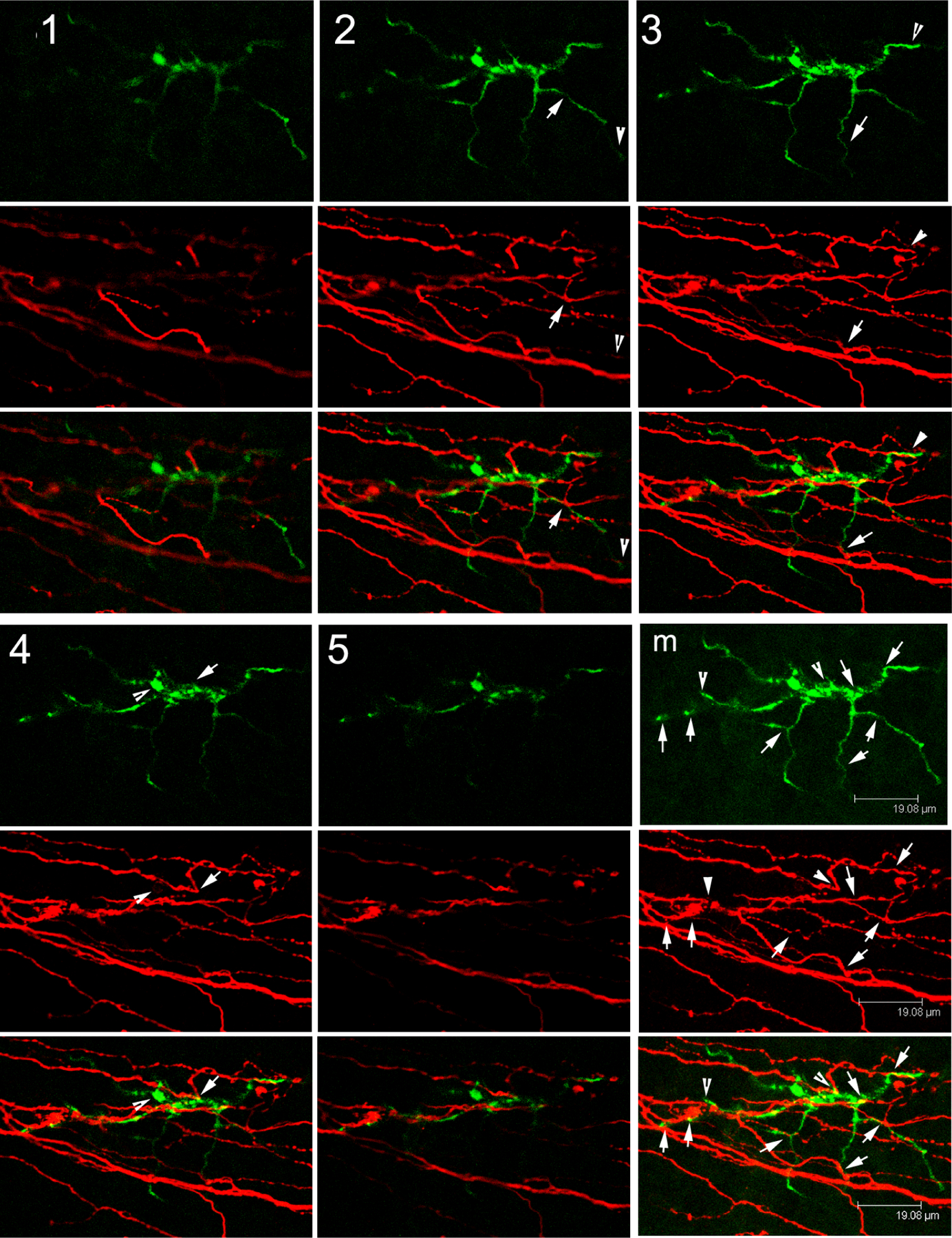
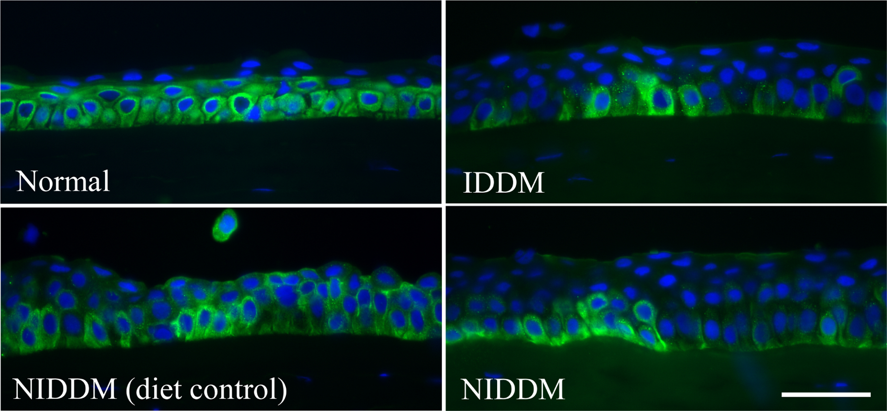
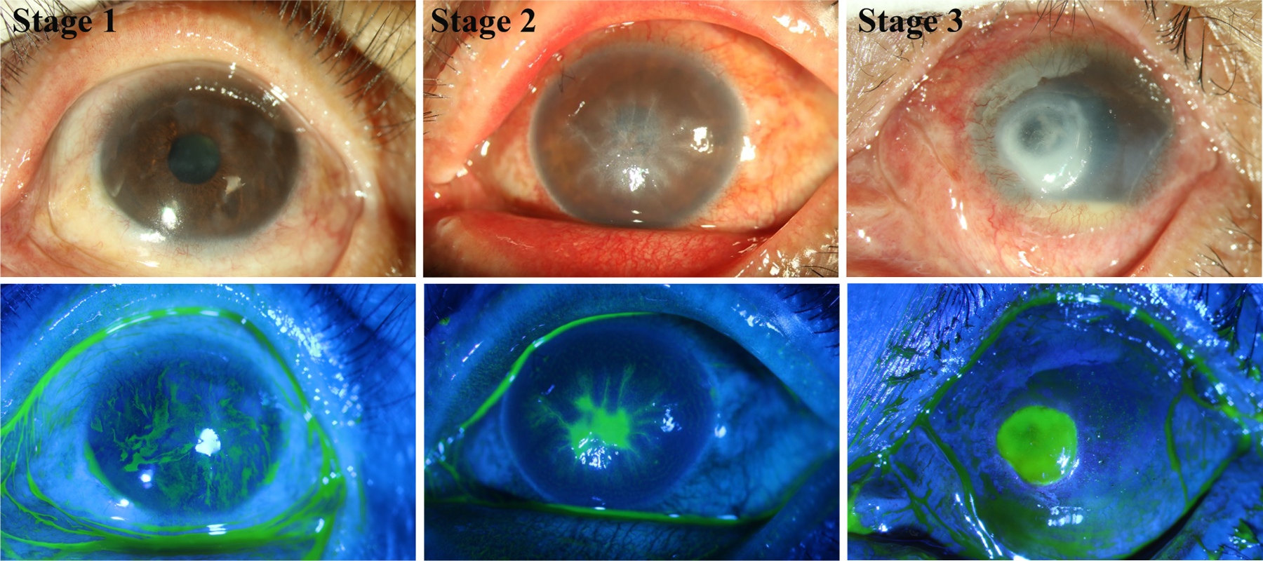
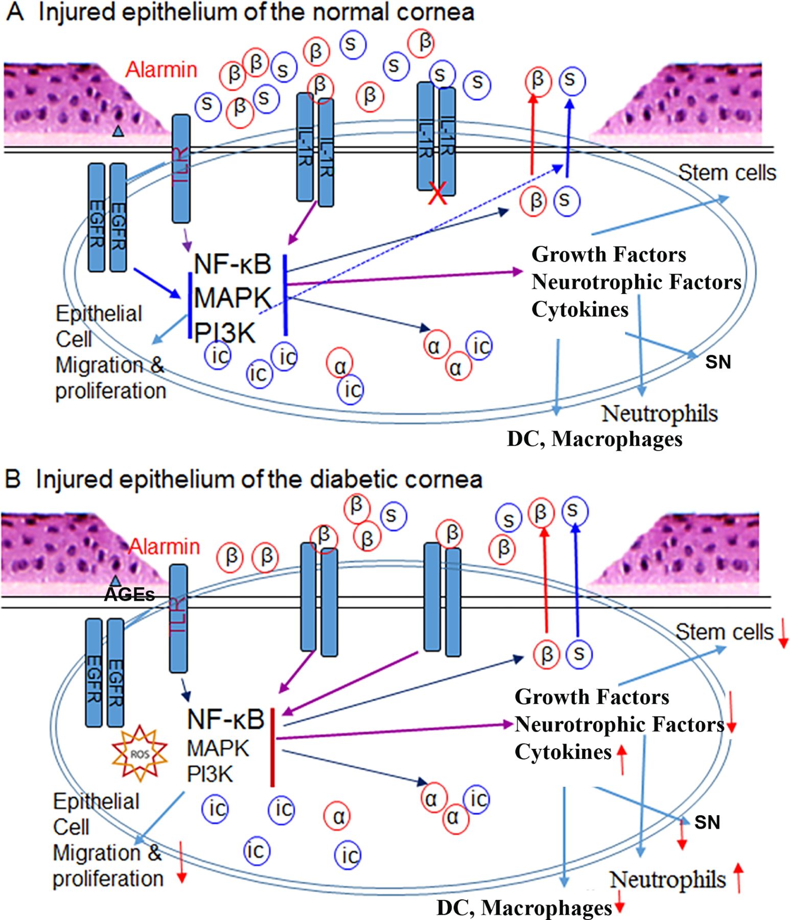
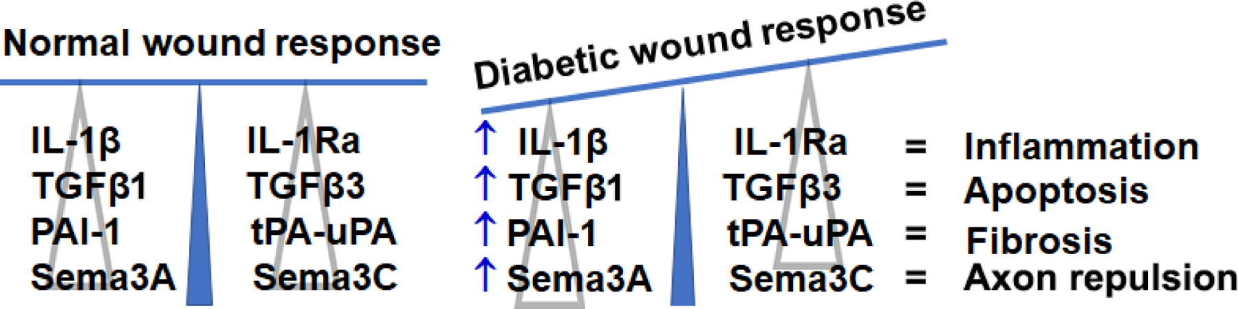
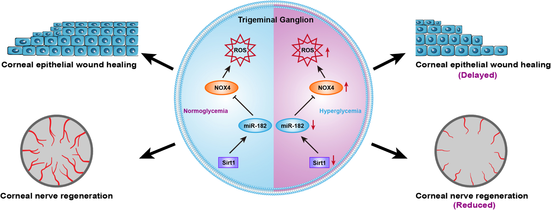
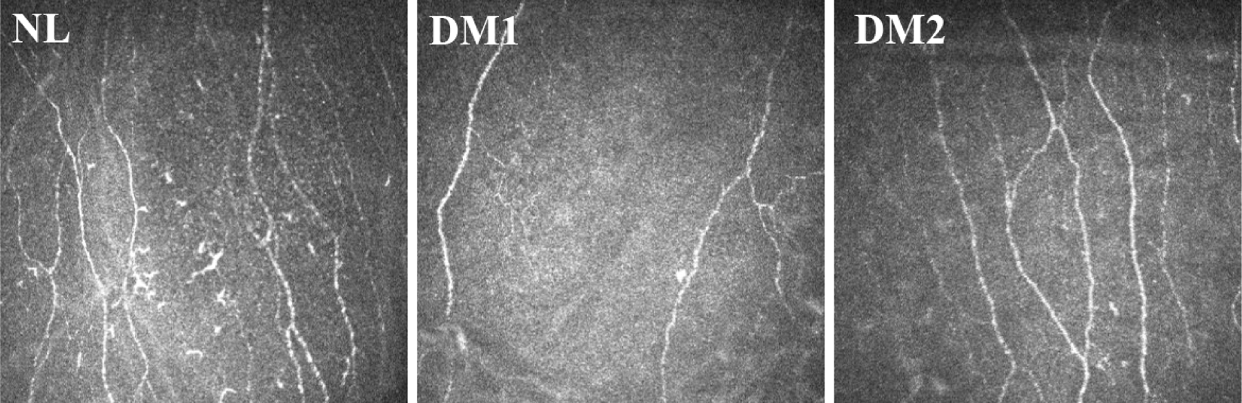
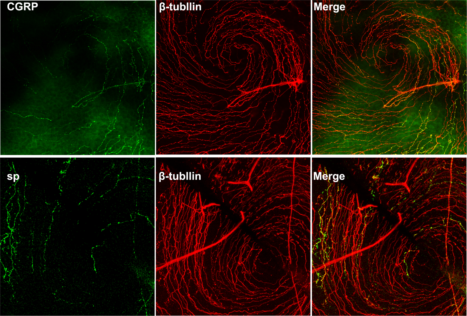
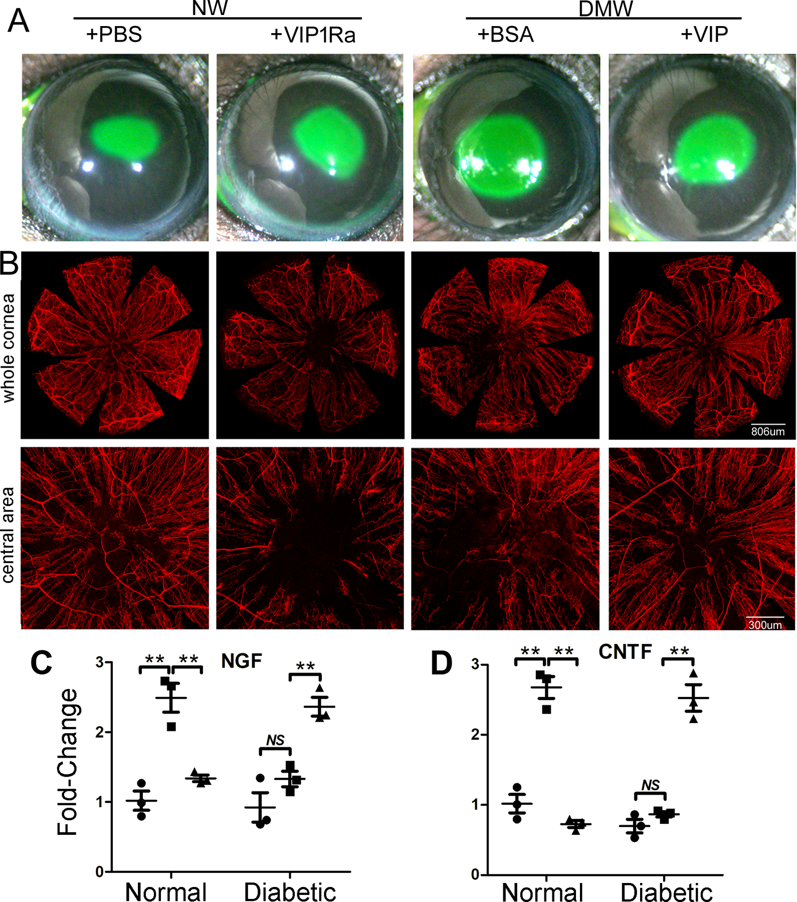
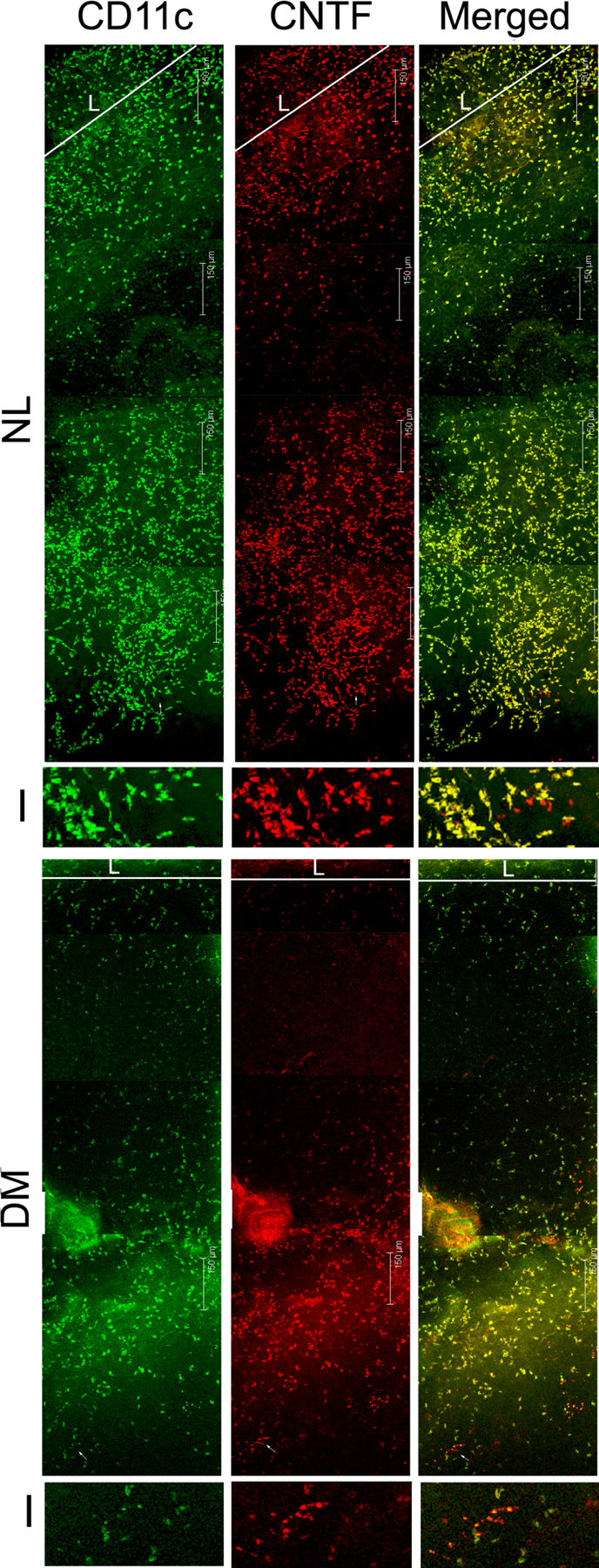
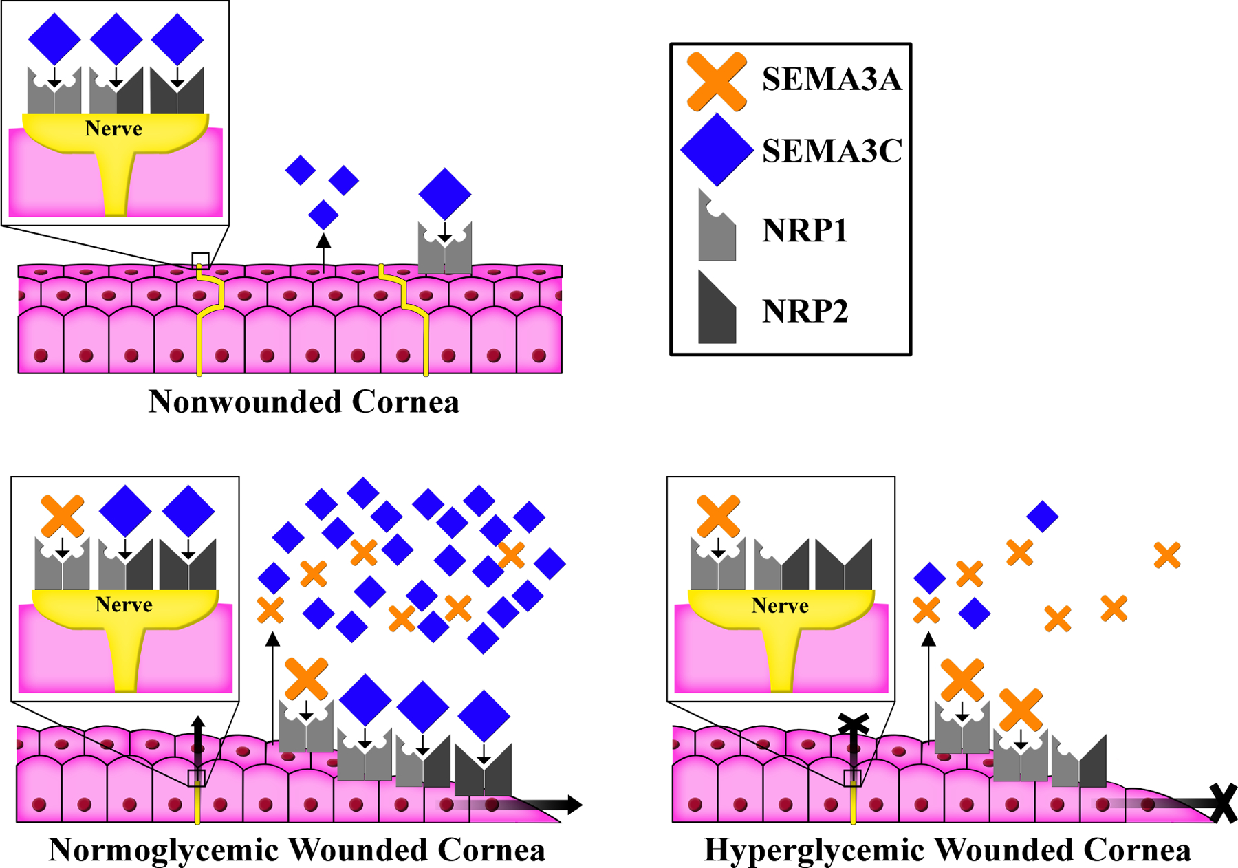
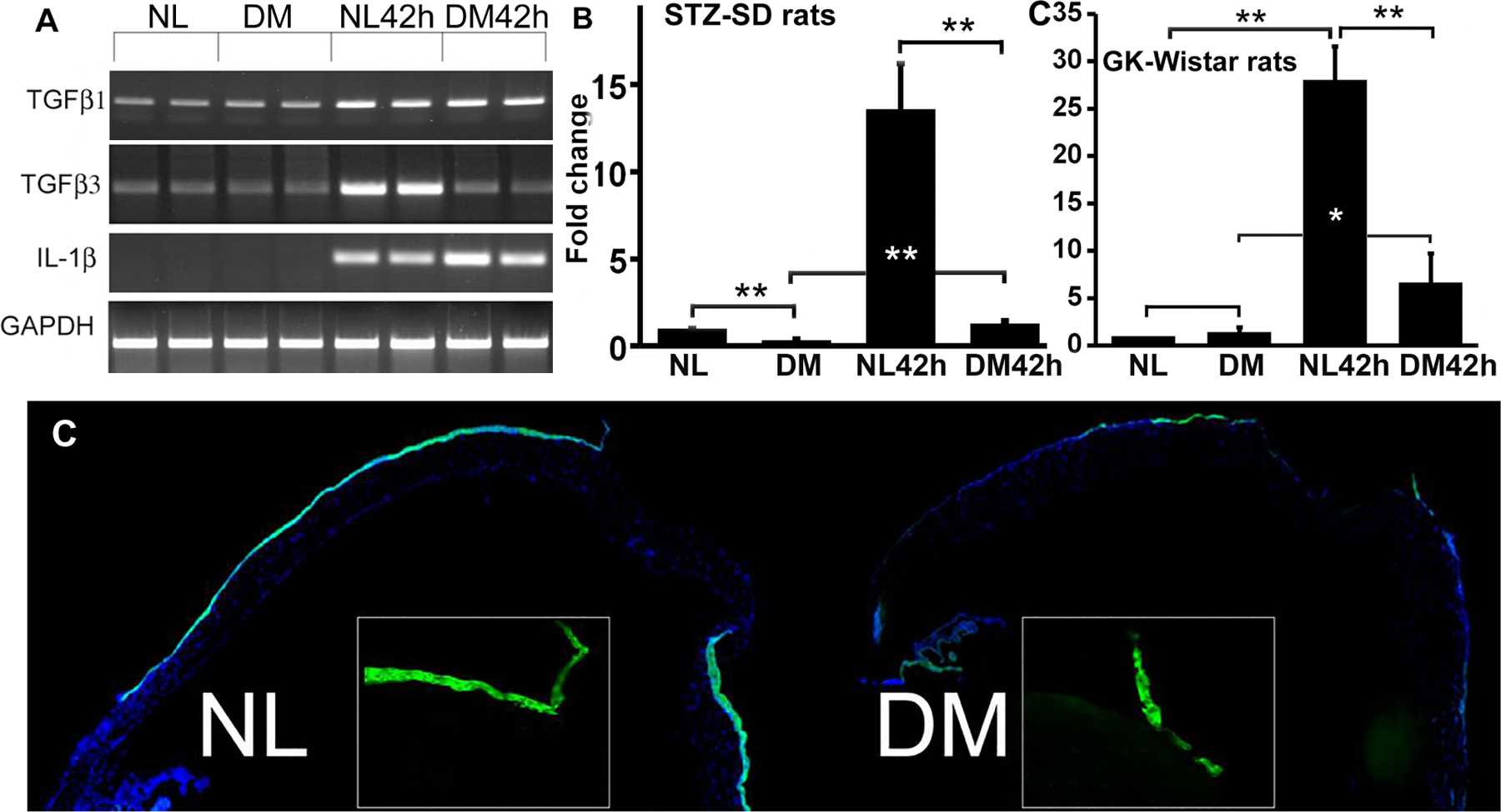
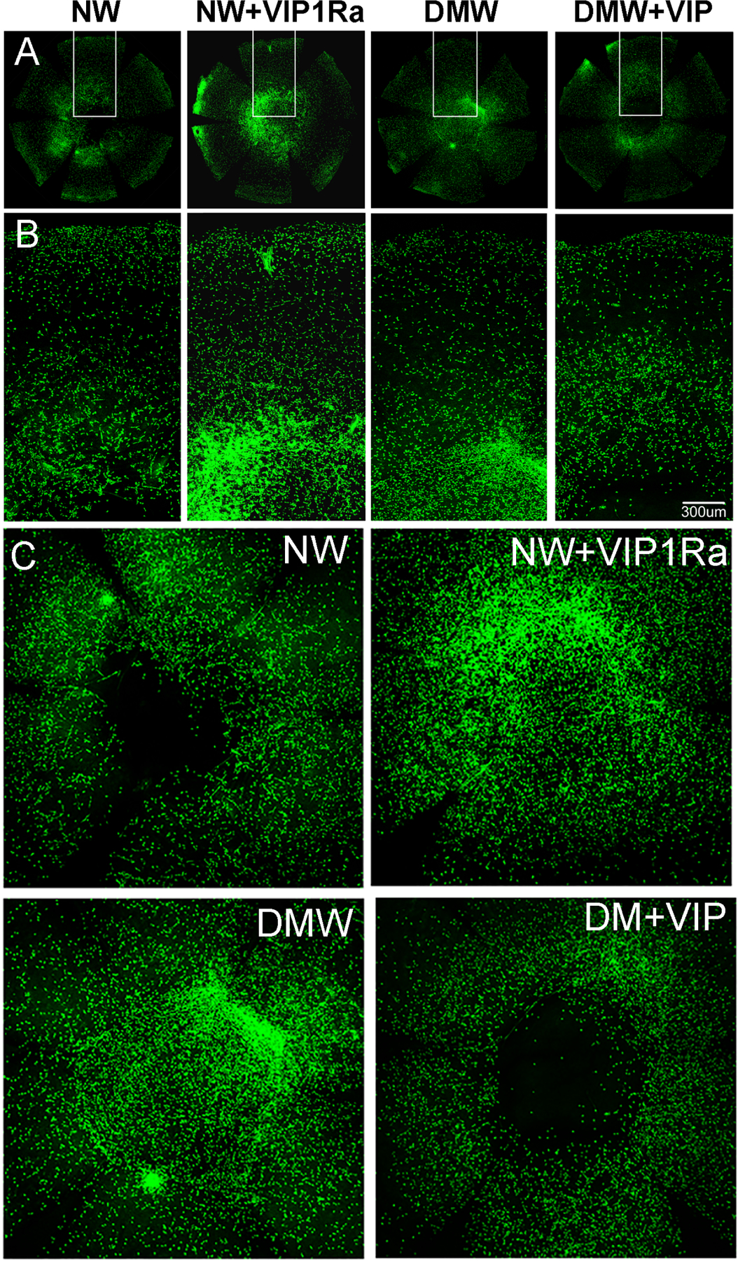
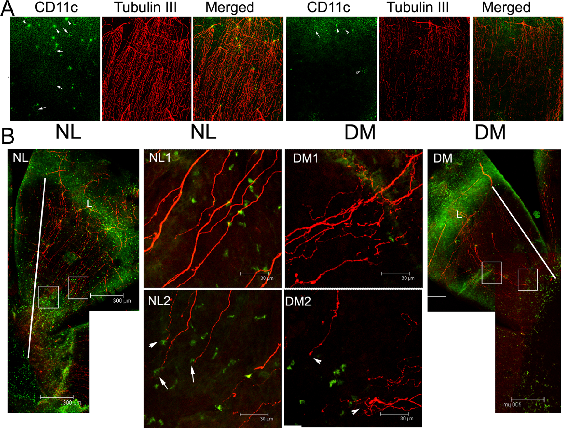
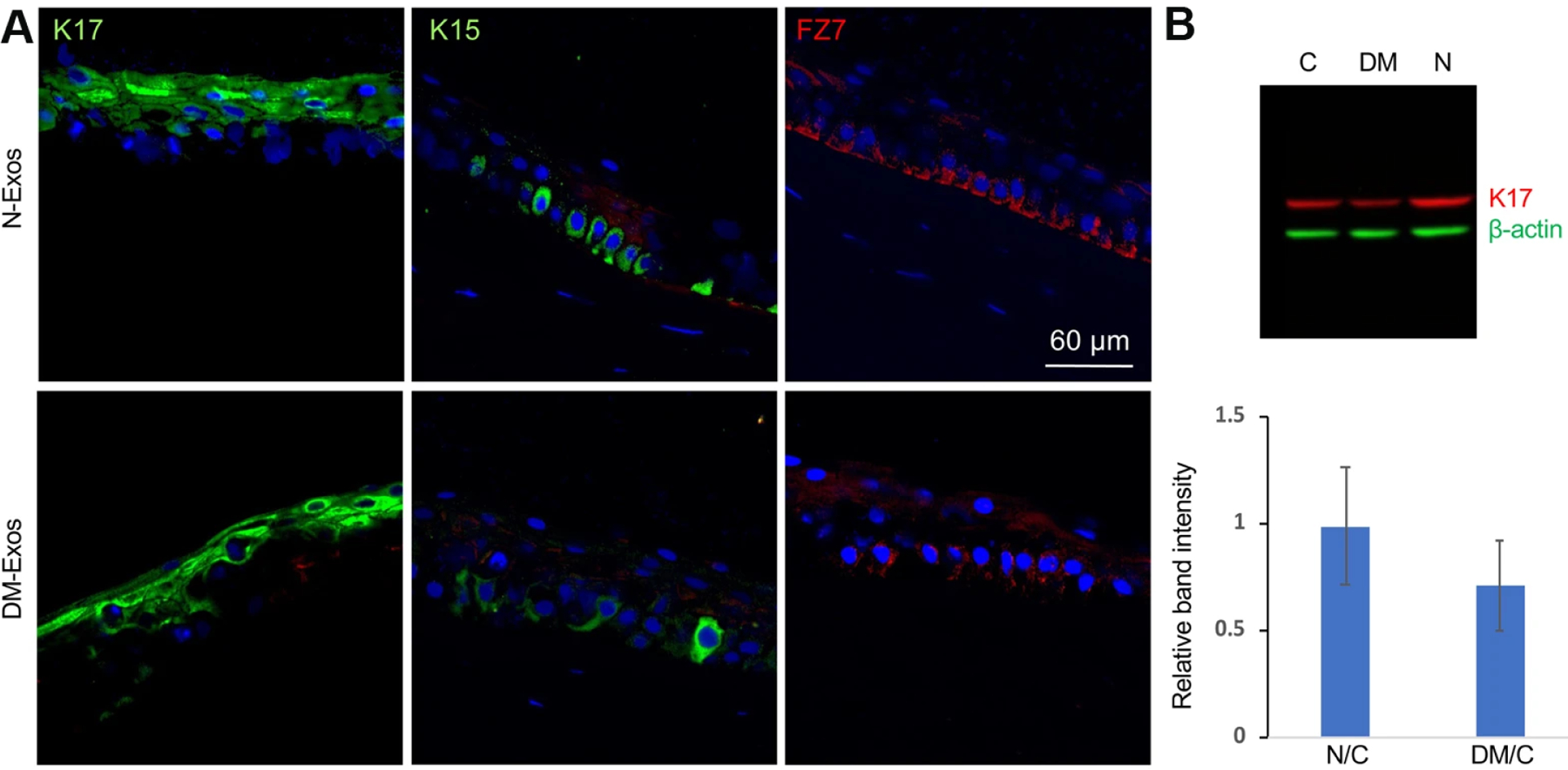
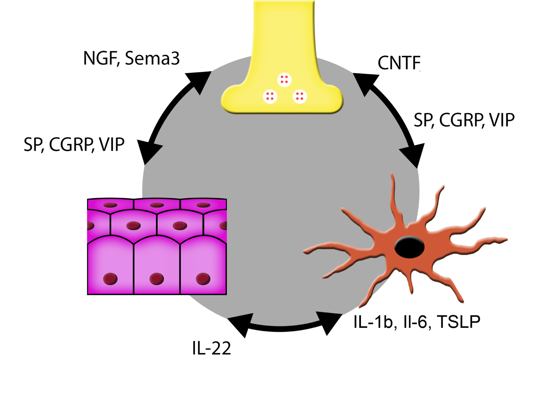
Diabetic peripheral neuropathy (DPN) is the most common complication of diabetes, with several underlying pathophysiological mechanisms, some of which are still uncertain. The cornea is an avascular tissue and sensitive to hyperglycemia, resulting in several diabetic corneal complications including delayed epithelial wound healing, recurrent erosions, neuropathy, loss of sensitivity, and tear film changes. The manifestation of DPN in the cornea is referred to as diabetic neurotrophic keratopathy (DNK). Recent studies have revealed that disturbed epithelial-neural-immune cell interactions are a major cause of DNK. The epithelium is supplied by a dense network of sensory nerve endings and dendritic cell processes, and it secretes growth/neurotrophic factors and cytokines to nourish these neighboring cells. In turn, sensory nerve endings release neuropeptides to suppress inflammation and promote epithelial wound healing, while resident immune cells provide neurotrophic and growth factors to support neuronal and epithelial cells, respectively. Diabetes greatly perturbs these interdependencies, resulting in suppressed epithelial proliferation, sensory neuropathy, and a decreased density of dendritic cells. Clinically, this results in a markedly delayed wound healing and impaired sensory nerve regeneration in response to insult and injury. Current treatments for DPN and DNK largely focus on managing the severe complications of the disease. Cell-based therapies hold promise for providing more effective treatment for diabetic keratopathy and corneal ulcers.

Keywords: Corneal wound healing; Diabetic keratopathy; Diabetic peripheral nerve degeneration.


Conflict of interest statement

Author Statement

The authors declare that there is no duality of interest associated with this manuscript.

Figures

Figure 1.
Figure 1.. Intimate contacts between an intraepithelial DC and sensory nerve endings at sub-basal space between basal side of epithelium and its basement membrane.
Confocal images of a mouse cornea stained with CD11c (green for DC) and β-tubulin 3 (red for neuron). Intimate contacts of DC body and processes with sensory nerve endings which are likely derived from the same epithelium insertion site of a sensory nerve fiber. This Figure wis originally published in (Gao et al., 2016a).
Figure 2.
Figure 2.. Altered pAkt staining pattern in diabetic human corneal epithelium.
Human corneal frozen sections from patients with type 1 diabetes (IDDM) and non–insulin-dependent type 2 diabetes (NIDDM), with normal subjects and diet-controlled type 2 diabetic patients as the control subjects, were stained by immunofluorescence with antibody against pAkt. Photos show merged images of immunoreactivity of pAkt and nuclear staining of DAPI. Scale bar 50 μm. This Figure wis originally published in (Gao et al., 2016a).
Figure 3.
Figure 3.. Representative images of diabetic keratopathy progression.
Slit lamp images with or without sodium fluorescein staining showing different severity of DNK. A cornea at first stage with scattered superficial stromal scarring and neovascularization in sclerotic scatter illumination photograph (top) and adjacent irregular hyperplastic epithelium and superficial punctate staining in cobalt blue scatter illumination photograph (bottom). A cornea at second stage with diffuse stromal edema and Descemet folds in sclerotic scatter illumination photograph (top) and lamellar staining indicating epithelial lamellar defect in cobalt blue scatter illumination photograph (bottom). A cornea at third stage with stromal infiltrate, neovascularization and hypopyon in sclerotic scatter illumination photograph (top) and stromal deep ulcerations in cobalt blue scatter illumination photograph (bottom), indicating excessive inflammation. The images were taken at the Qingdao Eye Hospital of Shandong First Medical University clinic.
Figure 4.
Figure 4.. Inflammatory responses of the cornea in response to epithelial injury.
(A) In a normal cornea, injury causes the release of alarmins such as IL-1α (a) and ATP, resulting in activation of intracellular signaling pathways which trigger the expression and secretion of growth factors, neurotrophic factors, and cytokines, including IL-1β and soluble IL-1Ra (sIL-1Ra). The balanced expression of both IL-1β and its antagonist sIL-1Ra ensures the controlled inflammation and infiltration of neutrophils, dendritic cells (DCs), and macrophages. Growth factors stimulate limbal stem cell proliferation to replenish lost epithelial cells. (B) In diabetic corneas, hyperglycemia causes accumulation of Advanced Glycation End-Products (AGEs) extracellularly and generation of reactive oxygen species (ROS) intracellularly. AGEs and ROS inhibits epithelial proliferation and migration, resulting in delayed wound healing. Hyperglycemia promotes IL-1β secretion and neutrophil infiltration but suppresses sIL-1Ra expression and DC and macrophage infiltration, resulting in an imbalance favoring excessive inflammation and increased cell death, further delaying epithelial wound closure and sensory nerve regeneration (not shown in this diagram). This figure is adapted from supplemental Figure 1 of (Yan et al., 2016).
Figure 5.
Figure 5.. Epithelium response to wounding in normal and diabetic corneas.
In normal corneas, wounding induces the balanced expressions of IL-1β/1Ra, transforming growth factor (TGF) β1/β3, PAI/tPA-uPA, and Semaphorin (Sema) 3A/3C. In diabetic corneas, wounding sufficiently induces the expressions of IL-1β, TGFβ1, PAI, and Sema3A, but not IL-1Ra, TGFβ3, uPA-tPA, and Sema3C, resulting in increased inflammation, apoptosis, fibrosis, and repulsion of regenerating sensory nerves, hence delaying corneal epithelial wound closure and sensory nerve regeneration seen in diabetic patients. This diagram has not been published and was made by FS Yu.
Figure 6.
Figure 6.. The downregulation of Sirt1/miR-182 in the trigeminal ganglion of diabetic type 2 db/db mice.
In diabetic trigeminal ganglion, the normal expression of sirt1 was impaired, resulting in inhibiting expression of its downstream miRNA miR-182, causing excess expression of the target gene NOX4 of miR-182, thereby induing an overexpression of ROS. These changes eventually lead to the delay of corneal epithelial wound healing and corneal nerve regeneration. This diagram has not been published and was made by B Zhang.
Figure 7.
Figure 7.. In vivo corneal confocal microscopy of sensory fibers in human normal and T1DM and T2DM corneas.
In vivo confocal microscopy images of the cornea in a healthy control subject (NL), an age-matched patient with type 1 (T1DM), and an age-matched patient with type 2 (T2DM) diabetes. The images were taken at the Shandong Eye Institute clinic. The images were taken at the Qingdao Eye Hospital of Shandong First Medical University clinic.
Figure 8.
Figure 8.. Co-staining of CGRP and SP with β-tubulin-3.
B6 mouse corneas were staining with CGRP/Tubulin3 or SP/Tubulin-3 and the center of the corneas were photographed and the images of sensory nerve ending and nerve expressing CGRP or SP were merged. Note only small portion of sensory nerve endings express CGRP or SP. Unpublished results (Gao and Yu).
Figure 9.
Figure 9.. VIP accelerates diabetic wound healing and nerve regeneration in healing corneas through VIPR1.
(A) NL corneas were pretreated with VIPR1 antagonist or PBS and diabetic corneas with recombinant VIP or BSA as the control 4h prior to epithelial debridement. At 0h, the corneas were wounded by epithelium-debridement (2 mm diameter). At 22 hpw, the remaining wounds were stained with fluorescein and photographed. The wound sizes were calculated and presented as percent of healed area over the size of original wounds. (B) Another set of mice were allowed to heal for 3 days and the corneas were processed for WMCM with beta-tubulin III staining for nerve fibers and endings. The images of whole corneas (upper panels) and high-magnification images of central area (bottom panels) were shown. The Figure shows that VIP regulates epithelial wound healing and nerve regeneration in the corneas, suggesting a therapeutic potential for these molecules in treating diabetic keratopathy. (C) The expression of NGF in wounded, with unwounded (●) as the control, NL or DM corneas treated with (▲) or without VIP antagonist (■). (D) The expression of CNTF in wounded, with unwounded (●) as the control, NL or DM corneas treated with (▲) or without VIP antagonist (■). VIP antagonist suppresses and exogenous VIP promotes wound-induced NGF and CNTF expression in NL and DM corneas, respectively. This figure was originally published in (Zhang et al., 2020a).
Figure 10.
Figure 10.. CNTF expression and co-localization with CD11c-positive cells in the normal and diabetic corneas with or without epithelium debridement.
Whole mount confocal microscopy showing DC and CNTF co-localization in healing corneas of normal and diabetic mice. I: 3.8x magnification of the images showing CD11c-negative, CNTF-positive cells; L: limbal region. Note the shorter distance between limbal region of leading edge and significantly less numbers of CD11c and CNTF positive cells in DM versus NL corneas. This figure was originally published in (Gao et al., 2016c).
Figure 11.
Figure 11.. Proposed Mechanism of Sema3-Neuropilin Signaling in the Diabetic and Nondiabetic Wounded Cornea.
In the nonwounded cornea, low amounts of Semaphorin (Sema) 3C signal through neuropilin (NRP) 1 complexes of the epithelium and NRP1 and/or NRP2 complexes on sensory nerves. Corneal wounding induces expression of both SEMA3A and its preferred receptor, NRP1, as well as additional SEMA3C and its preferred receptor, NRP2. SEMA3C-NRP2 signaling is crucial for both epithelial wound healing and sensory nerve regeneration. In the diabetic cornea, reduced expression of both SEMA3C and NRP2 results in a relative excess of SEMA3A-NRP1 signaling (resulting in axon repulsion), compared to SEMA3C-NRP2 signaling. This imbalance leads to delayed epithelial wound healing and nerve regeneration. This original figure was made by P SY Lee.
Figure 12.
Figure 12.. mRNA Expression of TGFβ isoforms detected by RT-PCR and verification of TGFβ3 expression by real-time PCR.
The RNA samples were obtained as described in Fig. 1. The scraped CECs from NL and STZ-DM rat and mouse corneas for creating a wound were marked as UW and from the wound bed at 42 hpw (rats) or 24 hpw (mice) and were subjected to RT-PCR with GAPDH as the internal control and IL-1β as a positive control. The data were presented as fold changes over non-DM, homeostatic CECs (1). For each condition three samples were collected from three rats/mice. Four independent experiments were performed: two with SD and STZ-SD, one with Wistar and GK rats, and one with B6 and STZ-B6 mice.*P < 0.05; **P < 0.01. Immunohistochemistry of TGF-β3 distribution in healing and UW corneas. The corneas of SD and STZ-SD were wounded and O.C.T. snap frozen at 42 hpw, followed by sectioning and immunostaining with antibodies against TGF-β3; DAPI was used to stain nuclei. Low magnification (×5) images of the entire cornea were taken and images stitched to present the whole from limbus to wound center. Inserts are high magnification (×20) images of the leading edge. This figure was originally published in (Bettahi et al., 2014a).
Figure 13.
Figure 13.. VIP dampens neutrophil infiltration in NL and diabetic healing corneas.
NL (NW), pretreated with VIP1Ra (NW1VIP1Ra), and diabetic (DMW), pretreated with recombinant VIP (DMW1VIP), mouse corneas were wounded by epithelium debridement (2-mm diameter). Healing corneas collected at 22 hpw from NL and diabetic mice were subjected to WMCM using Ly6G-FITC antibody for mouse neutrophil staining. The images of the whole cornea were captured (A), the marked areas of whole corneas were amplified (B), and central areas were shown (C). (D): Cell numbers in the whole corneas (A) were calculated with ImageJ and presented (mean 6 SD; n 5 3). *P>0.05 (one-way ANOVA). This figure was originally published in (Zhang et al., 2020a).
Figure 14.
Figure 14.. DCs and sensory nerve fibers/endings in healing normal and diabetic mouse corneas.
(A) Unwounded corneas were double-stained for β-tubulin III (nerve marker) and CD11c (dendritic cell marker) and the limbal region was photographed. Normal (NL, left) corneas showed dendritic cells (DCs) located where sensory nerves of the basal plexus enter the superficial epithelium. In the diabetic (DM, right) cornea, the number of DCs and nerves are diminished, and this anatomical relationship appears to be lost. Arrows, dendriform DCs; arrowheads, round-shaped DCs. (B) The NL and DM corneas were wounded using epithelial debridement. The whole cornea from the limbus (L) to the leading edge was photographed (10x, lateral panels). Higher magnification images (40x) at the middle (NL1, DM1) and the leading edge (NL2, DM2) of healing migratory sheets are also shown. DCs appear at each end of the healing nerve fibers in the NL cornea, but this relationship is lost in the DM cornea. Arrow, co-localization of nerve endings with a DC in NL cornea; Arrowhead, free nerve ending in the DM cornea. The results are representative of two independent experiments (N=3). Diabetes delays epithelial wound closure, and disturbs the interactions of DC and regenerative sensory nerves. This figure was originally published in (Gao et al., 2016c).
Figure 15.
Figure 15.. Effect of N- or DM-Exos on LESC marker expression in normal organ-cultured corneas and primary LEC.
(A) Normal Exo treatment in normal organ-cultured corneas led to increased expression of putative LESC markers, K15 and FZ7, and no significant change in K17 protein level compared to fellow corneas treated with DM-Exos (immunofluorescent staining of limbal corneal sections). The same exposure time was used for each set of compared stained sections, and the assessment was done by more than one observer. (B) Western analysis shows that N-Exos treatment increased, whereas DM-Exo treatment decreased K17 protein expression level in primary LEC compared to control treated cells, which did not reach significance. Antibody to β-actin was used as equal loading control and for semi-quantitation. This figure was originally published in (Leszczynska et al., 2018)
Figure 16.
Figure 16.. The Functional “Epineuroimmune Unit”.
Corneal epithelium, sensory nerves, and immune cells are anatomically and functionally interdependent in maintaining the corneal barrier. Epithelial-derived cytokines and neurotrophic factors/axonal guidance factors allow regulation of immune cells and nerves, respectively. CNTF produced by dendritic cells promotes epithelial healing and nerve regeneration. Neuropeptides derived from sensory nerves allow regulation of both epithelium and immune cells. Details regarding interactions are described in the text. CGRP, Calcitonin Gene-Related Peptide; CNTF, Ciliary Neurotrophic Factor; IL, Interleukin; MANF, Mesencephalic Astrocyte-Derived Neurotrophic Factor; NGF, Nerve Growth Factor; Sema3, class 3 semaphorins; SP, Substance P; TGF, Transforming Growth Factor α; TSLP, Thymic Stromal Lymphopoietin; VIP, Vasoactive Intestinal Peptide. This original figure was made by P SY Lee.

References

    1. Abdelaziz MH, Abdelwahab SF, Wan J, Cai W, Huixuan W, Jianjun C, Kumar KD, Vasudevan A, Sadek A, Su Z, Wang S, Xu H, 2020. Alternatively activated macrophages; a double-edged sword in allergic asthma. J Transl Med 18, 58. - PMC - PubMed
    1. Abdelkader H, Patel DV, McGhee C, Alany RG, 2011. New therapeutic approaches in the treatment of diabetic keratopathy: a review. Clin Experiment Ophthalmol 39, 259–270. - PubMed
    1. Adeghate E, Rashed H, Rajbandari S, Singh J, 2006. Pattern of distribution of calcitonin gene-related Peptide in the dorsal root ganglion of animal models of diabetes mellitus. Ann N Y Acad Sci 1084, 296–303. - PubMed
    1. Ahmed A, Bril V, Orszag A, Paulson J, Yeung E, Ngo M, Orlov S, Perkins BA, 2012. Detection of diabetic sensorimotor polyneuropathy by corneal confocal microscopy in type 1 diabetes: a concurrent validity study. Diabetes Care 35, 821–828. - PMC - PubMed
    1. Ahuja AS, Bowden FW 3rd, Robben JL, 2020. A Novel Treatment for Neurotrophic Corneal Ulcer Using Topical Cenegermin (OXERVATE) Containing Recombinant Human Nerve Growth Factor. Cureus 12, e11724. - PMC - PubMed

Publication types