Review
. 2021 Nov;13(11):6602-6617.
doi: 10.21037/jtd-2021-23.

Ex vivo lung perfusion

Affiliations
Review

Ex vivo lung perfusion

Tatsuaki Watanabe et al. J Thorac Dis. 2021 Nov.

Abstract

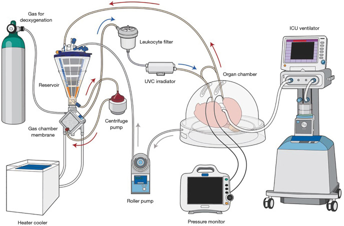
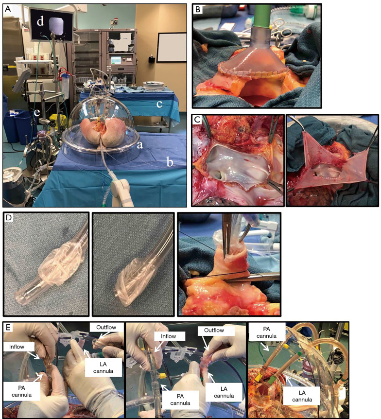
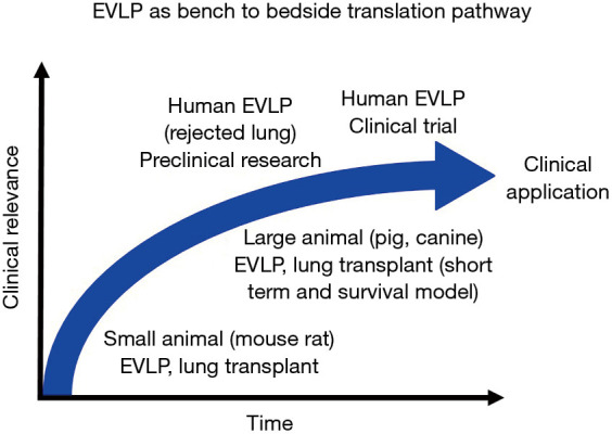
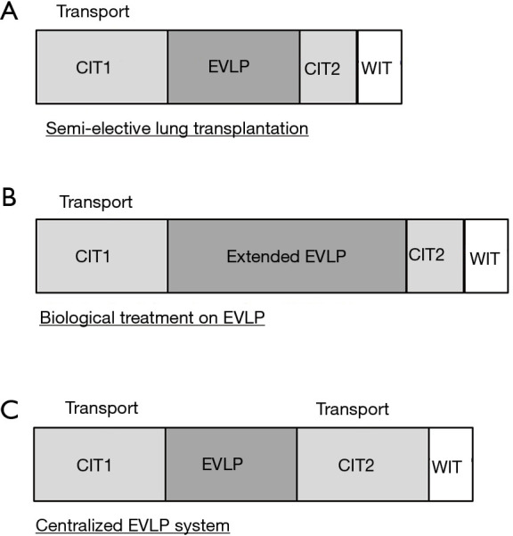
Lung transplantation is a life-saving treatment for patients with end stage lung disease. The imbalance between lung graft supply and recipients has been a serious issue and barrier to successful lung transplantation. Ex vivo lung perfusion is a strategy wherein lungs are perfused and ventilated outside of the body. This technology has emerged as a safe preservation method that also enables the reassessment and reconditioning of marginal lung grafts. Ex vivo lung perfusion has successfully expanded the donor pool and led to greater lung transplant activity worldwide. Furthermore, ex vivo lung perfusion can be used as a platform for advanced diagnostics that enable specific targeted or personalized treatments that can be developed along a bench to bedside pathway leading to safe ex vivo intervention. Recent findings have shown that ex vivo lung perfusion could significantly and safely extend the preservation period, which enables transplant programs further optimization of the logistics around transplantation surgeries, and create a new paradigm whereby donor lungs are assessed at a centralized ex vivo lung perfusion center prior to delivery to a transplant clinic in need. The introduction of ex vivo lung perfusion to clinical lung transplantation has been a major step in the evolution and practice of lung transplantation.

Keywords: Ex vivo lung perfusion; donation after cardiocirculatory death donor; extended criteria donor.


Conflict of interest statement

Conflicts of Interest: All authors have completed the ICMJE uniform disclosure form (available at https://dx.doi.org/10.21037/jtd-2021-23). The series “Lung Transplantation: Past, Present, and Future” was commissioned by the editorial office without any funding or sponsorship. MC is a co-founder of XOR Labs Toronto Inc., a company dedicated to the development of EVLP machines. The XOR EVLP machine was not used in the performance of this study. SK reports grants from XVIVO Perfusion, personal fees from United Therapeutics/Lung Bioengineering, during the conduct of the study; and SK is a co-founder of XOR Labs Toronto Inc., a company dedicated to the development of EVLP machines. The XOR EVLP machine was not used in the performance of this study. SK is the co-founder and Chief Scientific Officer of Perfusix Canada Inc. The authors have no other conflicts of interest to declare.

Figures

Figure 1
Figure 1
The schema of Toronto EVLP. Gas for deoxygenation consists of 86% N2, 8% CO2, 6% O2. *, bridge between inflow and outflow. The bridge is closed during perfusion. The bridge can be used for de-air from inflow line. Arrows indicate the direction of perfusate flow. Arrow colors indicate oxygenation of the perfusate; red indicates oxygenated perfusate and blue indicates de-oxygenated perfusate. UVC irradiator is optional. EVLP, ex vivo lung perfusion; UVC, ultraviolet C.
Figure 2
Figure 2
Toronto EVLP technique. (A) Toronto EVLP system. a: organ chamber; b: table for organ chamber; c: back table; d: monitor for bronchoscope; e: EVLP circuit. (B) LA and LA cannula connection. LA and LA cannula are sewn with 4-0 polypropylene sutures. (C) In the case of a short LA. Left: short LA. Right: LA is fixed with pericardium and 4-0 polypropylene sutures before cannulation. (D) Left and middle: the tip of endotracheal tube was removed. Right: the trachea was closed with a clamp and the endotracheal tube is fixed to the trachea with heavy silk ties. (E) Connection of PA and LA cannulas to the circuit. Left: connecting Inflow tube to PA cannula after de-air PA cannula and pulmonary vasculature. LA cannula was open at this time. Middle: connecting outflow tube and LA cannula. Clamp on outflow cannula was removed immediately after connection. Right: cannulas and circuit were connected. PA and LA cannulas were fixed to the chamber without kinking of PA and pulmonary veins. EVLP, ex vivo lung perfusion; LA, left atrium; PA, pulmonary artery.
Figure 3
Figure 3
Bench to bedside translation pathway. EVLP can be used to help translate outcomes across physiological conditions in in vitro, small animal, large animal, and human research models. Small animal EVLP can be used to test the effect of drugs in isolated lungs and develop novel therapies with a variety of biochemical assessment tools at a relatively low cost. Large aminal EVLP can be used to test the effects and safety of novel therapies that represents physiologically close conditions to a clinical scenario. EVLP can utilize clinically declined human lungs as a translational step toward clinical application of each investigated therapy. EVLP, ex vivo lung perfusion.
Figure 4
Figure 4
Schema of emergent clinical applications of EVLP. (A) EVLP as a preservation strategy for semi-elective lung transplantation. (B) EVLP as a therapeutic platform for advanced approaches relevant to organ repair and regeneration. (C) Model for high-volume specialized organ assessment and repair centers. The figure is modified from Ref. (45). EVLP, ex vivo lung perfusion.

References

    1. Valapour M, Paulson K, Smith JM, et al. OPTN/SRTR 2011 Annual Data Report: lung. Am J Transplant 2013;13 Suppl 1:149-77. 10.1111/ajt.12024 - DOI - PubMed
    1. Keeshan BC, Rossano JW, Beck N, et al. Lung transplant waitlist mortality: height as a predictor of poor outcomes. Pediatr Transplant 2015;19:294-300. 10.1111/petr.12390 - DOI - PubMed
    1. Tuttle-Newhall JE, Krishnan SM, Levy MF, et al. Organ donation and utilization in the United States: 1998-2007. Am J Transplant 2009;9:879-93. 10.1111/j.1600-6143.2009.02565.x - DOI - PubMed
    1. Yeung JC, Cypel M, Waddell TK, et al. Update on donor assessment, resuscitation, and acceptance criteria, including novel techniques--non-heart-beating donor lung retrieval and ex vivo donor lung perfusion. Thorac Surg Clin 2009;19:261-74. 10.1016/j.thorsurg.2009.02.006 - DOI - PubMed
    1. Divithotawela C, Cypel M, Martinu T, et al. Long-term Outcomes of Lung Transplant With Ex Vivo Lung Perfusion. JAMA Surg 2019;154:1143-50. 10.1001/jamasurg.2019.4079 - DOI - PMC - PubMed