Review
. 2021 Jul 1:5:24715492211023302.
doi: 10.1177/24715492211023302. eCollection 2021.

Maximizing Muscle Function in Cuff-Deficient Shoulders: A Rehabilitation Proposal for Reverse Arthroplasty

Affiliations
Review

Maximizing Muscle Function in Cuff-Deficient Shoulders: A Rehabilitation Proposal for Reverse Arthroplasty

Helen Razmjou et al. J Shoulder Elb Arthroplast. .

Abstract

Purpose: The purpose of this review is to describe the role of altered joint biomechanics in reverse shoulder arthroplasty and to propose a rehabilitation protocol for a cuff-deficient glenohumeral joint based on the current evidence.Methods and Materials: The proposed rehabilitation incorporates the principles of pertinent muscle loading while considering risk factors and surgical complications.

Results: In light of altered function of shoulder muscles in reverse arthroplasty, scapular plane abduction should be more often utilized as it better activates deltoid, teres minor, upper trapezius, and serratus anterior. Given the absence of supraspinatus and infraspinatus and reduction of external rotation moment arm of the deltoid in reverse arthroplasty, significant recovery of external rotation may not occur, although an intact teres minor may assist external rotation in the elevated position.

Conclusion: Improving the efficiency of deltoid function before and after reverse shoulder arthroplasty is a key factor in the rehabilitation of the cuff deficient shoulders. Performing exercises in scapular plane and higher abduction angles activates deltoid and other important muscles more efficiently and optimizes surgical outcomes.

Keywords: biomechanics; complications; cuff tear arthropathy; deltoid; rehabilitation.


Conflict of interest statement

Declaration of Conflicting Interests: The author(s) declared no potential conflicts of interest with respect to the research, authorship, and/or publication of this article.

Figures

Figure 1.
Figure 1.
A, AP view of the right shoulder RTSA with humeral component displaced anteroinferiorly in a 53 year old woman. B, Transcapular view of the same patient showing anteroinferior dislocation of the humeral component.
Figure 2.
Figure 2.
A, AP view of the prosthesis showing an erosive lesion at the inferior scapular neck in a 91-year female at 2 and ½ years post reverse arthroplasty. B, Axillary view of the same shoulder showing a linear lucency anteriorly along the component-cement interface at 2 and ½ years post reverse arthroplasty.
Figure 3.
Figure 3.
A, No obvious signs of stress fracture on plain radiographs in a 74 year-old male at 3 and ½ months post reverse arthroplasty. B, 1.2 mm helical CT scan images taken two weeks later showing healing fracture of the base of the acromion without significant displacement. C, Healing fracture of the base of the acromion now obvious on plain radiographs, taken two weeks after the CT scan and 4 weeks after the initial plain radiographs.
Figure 4.
Figure 4.
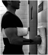
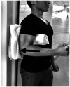
Anterior deltoid, pectoralis major and coracobrachialis. Isometric flexionin neutral position.
Figure 5.
Figure 5.
Posterior deltoid and latissimus dorsi. Isometric extension in neutral position. Hyperextension to be avoided at all times.
Figure 6.
Figure 6.
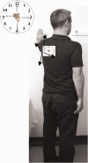
Scapular exercises (upper, middle and lower trapezius muscles, levator scapulae, rhomboids, latissimus dorsi and serrates anterior). • Face-clock exercises are done in standing, facing the wall with the hand pointed straight up and at any height that is comfortable. • Without moving hand during the exercise, the scapula is moved towards the numbers on the clock, starting with 12:00, 3:00, 6:00 and back over to 9:00. This will isolate the scapular stabilizers.
Figure 7.
Figure 7.
Anterior and middle deltoid. Isometric diagonal flexion and abduction in scapular plane.
Figure 8.
Figure 8.
Middle deltoid. Isometric abduction in scapular plane to activate middle deltoid.
Figure 9.
Figure 9.
Posterior deltoid. Isometric diagonal extension and abduction in scapular plane to activate posterior deltoid. A strap is held by the opposite hand to provide resistance.
Figure 10.
Figure 10.
Posterior deltoid and teres minor. Isometric abduction and external rotation in the scapular plane. A, Initial position: affected arm is kept at 30° of scapular plane abduction and the forearm is pressed against the opposite hand outward. B, Progression: affected arm is externally rotated while resisting against the opposite hand and keeping the elbow inward. C, The hornblower sign, commonly seen in patients with CTA should be avoided by focusing on external rotation and moving the elbow inward.
Figure 11.
Figure 11.
Anterior deltoid. Isotonic diagonal flexion and abdduction against resistance band in the scapular plane.
Figure 12.
Figure 12.
Middle deltoid. Isotonic abduction against resistance band in the scapular plane.
Figure 13.
Figure 13.
Posterior deltoid and teres minor. Advanced isotonic abduction and external rotation against resistance band in the scapular plane. While above elbow strap provides resistance in isometric abduction, the affected forearm is externally rotated against the resistance band above the wrist. Caution: limit the amount of stretch when using two bands simultaneously to avoid strain.
Figure 14.
Figure 14.
Ttrapezius, rhomboids, teres minor, anterior, middle and posterior deltoid. Bilateral isotonic arm elevation with external rotation using a resistance band. Elbows should be kept inward during the elevation. A, Initial position with squeezing shoulder blades. B, Final position.
Figure 15.
Figure 15.
Posterior deltoid and latissimus dorsi. Isotonic lats/pull downs is performed with a band placed over top of a door or secured on a wall and the resistance band held in both hands. Patient pulls down in scapular plane abduction with the elbows in a 90° angle. Hyperextension to be avoided at all times. A, Initial position. B, Final position.
Figure 16.
Figure 16.
Posterior deltoid, rhomboids and latissimus dorsi. Isotonic rowing is performed with a band placed around a door knob or secured in the front. The resistance band is held in both hands and pulled backward maintaining the scapular plane abduction with the elbows in a 90° angle. Hyperextension to be avoided at all times. A, Initial position. B, Final position.

References

    1. Lugli T.Artificial shoulder joint by Pean (1893): the facts of an exceptional intervention and the prosthetic method. Clin Orthop Relat Res. 1978;133:215–218. - PubMed
    1. Baulot E, Sirveaux F, Boileau P.Grammont’s idea: the story of Paulgrammont’s functional surgery concept and the development of the reverse principle. Clin Orthop Relat Res. 2011; 469(9):2425–2431. - PMC - PubMed
    1. Grammont P, Laffay J, Deries X.Concept study and reaslization of a new total shoulder prosthesis [French]. Rhumatologie. 1987; 39:407–418.
    1. Grammont P, Bourgon J, Pelzer P.Study of a Mechanical Model for a Shoulder Total Prosthesis: Realization of a Prototype [in French] [thèse de sciences de l’Ingénieur]. Dijon, France: Universitédijon; Lyon, France: ECAM de lyon; 1981.
    1. Ernstbrunner L, Andronic O, Grubhofer F, Camenzind RS, Wieser K, Gerber C. Long-term results of reverse total shoulder arthroplasty for rotator cuff dysfunction: a systematic review of longitudinal outcomes. J Shoulder Elbow Surg. 2019; 28(4):774–781. - PubMed

LinkOut - more resources