Case Reports
. 2021 Nov 8;5(12):ytab444.
doi: 10.1093/ehjcr/ytab444. eCollection 2021 Dec.

Cardiac magnetic resonance in the assessment of pericardial abnormalities: a case series

Affiliations
Case Reports

Cardiac magnetic resonance in the assessment of pericardial abnormalities: a case series

T Branco Mano et al. Eur Heart J Case Rep. .

Abstract

Background: Cardiac magnetic resonance (CMR) has a unique role in evaluating pericardial disease, permitting non-invasive tissue analysis, and haemodynamic assessment.

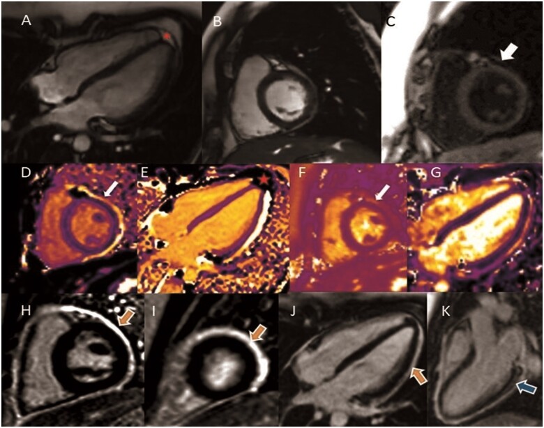
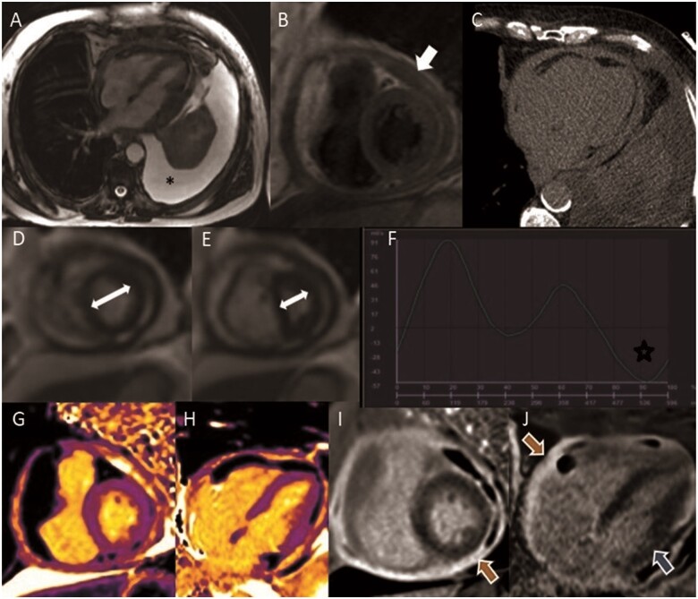
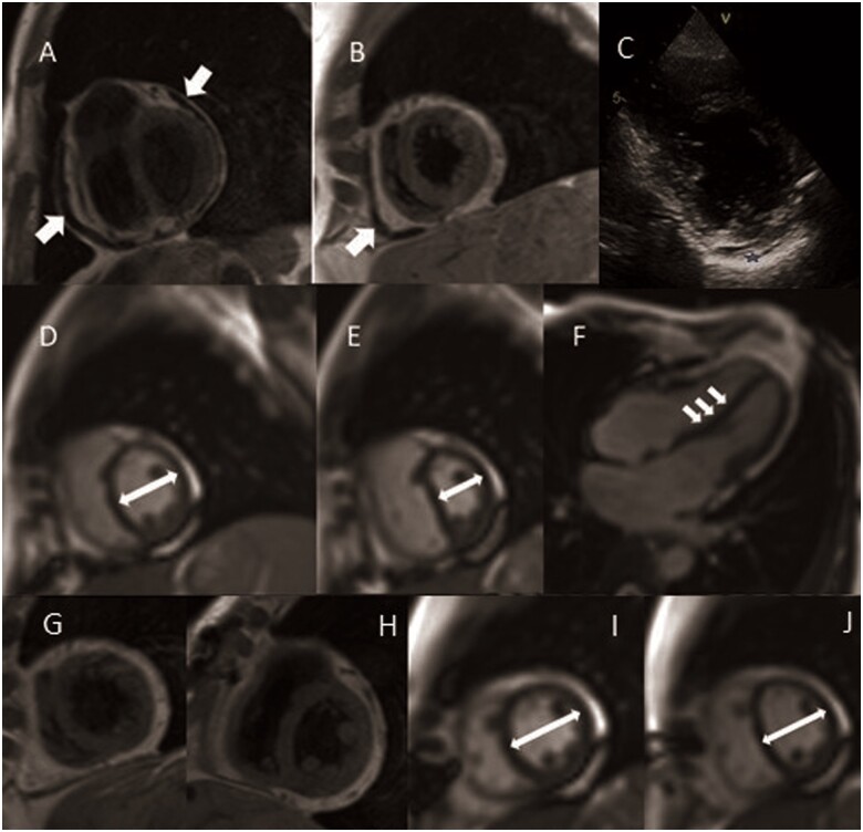
Case summary: In Case 1 of recurrent pericarditis, CMR confirmed reactivation of inflammation with late gadolinium enhancement and native T1/T2 mapping techniques, prompting therapeutic changes. In constrictive pericarditis, CMR is the only modality capable of differentiating a subacute potentially reversible form (Case 2), from a chronic, burnt out irreversible phase characterized by constrictive physiology (Case 3).

Discussion: Cardiac magnetic resonance is an effective tool to tailor individual therapy, particularly in cases of recurrent and constrictive pericarditis. Late gadolinium enhancement provides diagnostic and prognostic information, and multiparametric mapping has emerged as a promising tool with incremental diagnostic value.

Keywords: Cardiac magnetic resonance; Case series; Constrictive pericarditis; Pericardial disease; Pericarditis.


Figures

Figure 1
Figure 1
Steady-state free precession (SSFP) sequence in long-axis (A) and short-axis (B). (C) Black blood T1 weighted (double inversion recovery) showing maximum pericardium thickness of 2.5 mm (arrow). Native T1 mapping [980 ms—normal value 990.66 (17.5) ms] in short-axis (D) and long-axis (E), contributing to differentiate between fat (red star) from fluid/inflammation (bright/arrow). (F) Native T2 mapping sequence showing pericardial inflammation (arrow). (G) Increased myocardium native T2 mapping [52 ms—normal value 47.69 (4.28) ms], suggestive of concomitant myocardial oedema. (H–K) Late gadolinium enhancement (LGE) sequence in short-axis (median and apical slices) and long-axis views showing hyperenhancement in the pericardium (orange arrows) and in myocardium (subepicardial pattern in the basal infero-lateral wall—blue arrow).
Figure 2
Figure 2
Cardiac magnetic resonance study of Case 2 constrictive pericarditis. (A) Steady-state free precession sequence, noticing the large left pleural effusion (*). (B) Black blood T1 weighted Turbo Spin Echo sequences showing circumferential thickening of the pericardium (arrow). (C) Computed tomography scan excluded calcification of the pericardium. (D, E) Real-time cine imaging functional assessment of ventricular inter-dependence (D) normal septal position during expiration and (E) marked leftward septal shift seen immediately after deep inspiration (white arrows demonstrate the relative change in cavity size due to the septal shift). (F) Phase contrast study showing reverse diastolic flow in inferior vena cava (star). (G and H) Native T1 mapping with high native T1 values in the pericardium. (I and J) Late gadolinium enhancement sequence in short-axis view and four-chamber view with severe hyperenhancement in the pericardium (orange arrow) and in the myocardium (subendocardial pattern in the basal inferolateral wall—blue arrow).
Figure 3
Figure 3
Cardiac magnetic resonance study of Case 3—(A–I) Chronic pericardial constriction, previous to pericardiectomy. Black blood T1 weighted Turbo Spin Echo showing thickening of the pericardium (A, B). (C) Echocardiography, in parasternal short axis view, showing bright and thickened pericardium (blue star). (D–F) Functional assessment of ventricular inter-dependence (short axis in midcavity and long-axis view): (D) normal septal position during expiration, (E) marked leftward septal shift after deep inspiratory effort (white arrows demonstrate the relative change in cavity size due to the septal shift); (F) evidence of septal bounce in long-axis four-chamber views. (G–J) Cardiac magnetic resonance study after pericardiectomy. (G and H) Black blood T1 weighted turbo spin echo confirmed successful partial pericardiectomy. Real-time free breathing: end-expiratory (I) and end-inspiratory (J) septal position, revealing almost completely resolution of constriction signs.
None

References

    1. Chetrit M, Xu B, Kwon D, Ramchand J, Rodriguez RE, Tan CD. et al. Imaging-guided therapies for pericardial diseases. JACC Cardiovasc Imaging 2020;13:1422–1437. - PubMed
    1. Zurick A, Bolen M, Kwon D, Tan CD, Popovic ZB, Rajeswaran J. et al. Pericardial delayed hyperenhancement with CMR imaging in patients with constrictive pericarditis undergoing surgical pericardiectomy - a case series with histopathological correlation. J Am Coll 2011;4:1180–1191. - PubMed
    1. Vidalakis E, Kolentinis M, Gawor M, Vasquez M, Nagel E.. CMR in pericardial diseases - an update. Curr Cardiovasc Imaging Rep 2020;13:14.
    1. Cosyns B, Plein S, Nihoyanopoulos P, Smiseth O, Achenbach S, Andrade MJ, et al. ; European Society of Cardiology Working Group (ESC WG) on Myocardial and Pericardial diseases. European Association of Cardiovascular Imaging (EACVI) position paper: multimodality imaging in pericardial disease. Eur Heart J Cardiovasc Imaging 2015;16:12–31. - PubMed
    1. Hassan O, Kwon D.. Update on MRI techniques for evaluation of pericardial disease. Curr Cardiol Rep 2020;22:147. - PubMed

Publication types

LinkOut - more resources