. 2021 Dec 22;4(1):fcab301.
doi: 10.1093/braincomms/fcab301. eCollection 2022 Feb.

Phenotyping of multiple sclerosis lesions according to innate immune cell activation using 18 kDa translocator protein-PET

Affiliations

Phenotyping of multiple sclerosis lesions according to innate immune cell activation using 18 kDa translocator protein-PET

Marjo Nylund et al. Brain Commun. .

Abstract

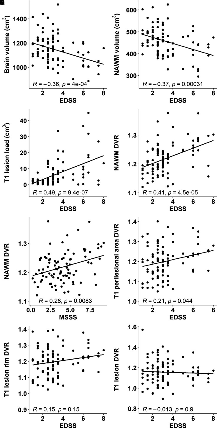
Chronic active lesions are promotors of neurodegeneration and disease progression in multiple sclerosis. They harbour a dense rim of activated innate immune cells at the lesion edge, which promotes lesion growth and thereby induces damage. Conventional MRI is of limited help in identifying the chronic active lesions, so alternative imaging modalities are needed. Objectives were to develop a PET-based automated analysis method for phenotyping of chronic lesions based on lesion-associated innate immune cell activation and to comprehensively evaluate the prevalence of these lesions in the various clinical subtypes of multiple sclerosis, and their association with disability. In this work, we use 18 kDa translocator protein-PET imaging for phenotyping chronic multiple sclerosis lesions at a large scale. For this, we identified 1510 white matter T1-hypointense lesions from 91 multiple sclerosis patients (67 relapsing-remitting patients and 24 secondary progressive patients). Innate immune cell activation at the lesion rim was measured using PET imaging and the 18 kDa translocator protein-binding radioligand 11C-PK11195. A T1-hypointense lesion was classified as rim-active if the distribution volume ratio of 11C-PK11195-binding was low in the plaque core and considerably higher at the plaque edge. If no significant ligand binding was observed, the lesion was classified as inactive. Plaques that had considerable ligand binding both in the core and at the rim were classified as overall-active. Conventional MRI and disability assessment using the Expanded Disability Status Scale were performed at the time of PET imaging. In the secondary progressive cohort, an average of 19% (median, interquartile range: 11-26) of T1 lesions were rim-active in each individual patient, compared to 10% (interquartile range: 0-20) among relapsing-remitting patients (P = 0.009). Secondary progressive patients had a median of 3 (range: 0-11) rim-active lesions, versus 1 (range: 0-18) among relapsing-remitting patients (P = 0.029). Among those patients who had rim-active lesions (n = 63), the average number of active voxels at the rim was higher among secondary progressive compared to relapsing-remitting patients (median 158 versus 74; P = 0.022). The number of active voxels at the rim correlated significantly with the Expanded Disability Status Scale (R = 0.43, P < 0.001), and the volume of the rim-active lesions similarly correlated with the Expanded Disability Status Scale (R = 0.45, P < 0.001). Our study is the first to report in vivo phenotyping of chronic lesions at large scale, based on 18 kDa translocator protein-PET. Patients with higher disability displayed a higher proportion of rim-active lesions. The in vivo lesion phenotyping methodology offers a new tool for individual assessment of smouldering (rim-active) lesion burden.

Keywords: PET imaging; TSPO; innate immune cell; multiple sclerosis; white matter lesions.


Figures

Graphical Abstract
Graphical Abstract
Figure 1
Figure 1
Flow chart of lesion phenotype analyses and illustrative MRI and PET images. (A) The flow chart illustrates the criteria for lesion inclusion and principles of the lesion classification. Lesion classification is based on proportions of active voxels in the lesion core and at rim. Rim-active lesions contain at least double the proportion of active voxels at the rim compared to core if 5–20% of voxels in the core are active. Rim-active lesions have at least 5% point higher proportion of active voxels at the rim compared to the core, if <5% of the voxels in the core are active. Inactive lesions have 0% of active voxels in the core and at rim. Lesions which do not fit into the other two categories are classified as overall-active lesions. Of all included lesions, 16% were rim-active, 33% were inactive and 51% were overall-active. The DVR distribution of each lesion type is visualized with 3D surface plots. (B) T1 MR-image from an SPMS patient (top left) with the corresponding parametric PET 11C-PK11195 DVR images of the white matter (top middle) and the focal T1 lesions (top right). The bottom panel highlights the DVR values of selected lesions as 3D surface plots. The bottom row visualizes the voxels defined as active, with DVR > 1.56 (for more details, see the ‘Materials and methods’ section). The colour bar of the PET images shows the dynamic range of DVR in the images.
Figure 2
Figure 2
Brain innate immune cell activation in multiple sclerosis patients and healthy controls. Box plots of the 11C-PK11195 DVR values representing the innate immune cell activation in the white matter of healthy controls and in the NAWM, and in association with lesions in the multiple sclerosis cohort. The Wilcoxon rank-sum test was used for statistical analyses. In box plots, the thick horizontal lines represent the medians, the boxes represent the IQR and the end of the whiskers or the points of the outliers represent the minimum and maximum values.
Figure 3
Figure 3
Association of volumetric parameters and innate immune cell activation with clinical disability. Smaller brain (A) and NAWM volume (B) and larger T1 lesion load (C) associate with worse clinical disability measured with the EDSS. Higher 11C-PK11195 DVR in the NAWM associates with worse disability (D) and disease severity (E). In addition, higher radioligand binding in the perilesional area correlates with worse disability (F). Innate immune cell activation at lesion rim (G) or within T1 lesions (H) do not associate with the EDSS. Here, ROIs encompassing the entire combined lesion volume or combined perilesional volume were evaluated.
Figure 4
Figure 4
Fractions of lesion types among multiple sclerosis subgroups. Proportions of the lesion subtypes differ between patients with EDSS scores of <4 and ≥4 (Fisher’s exact test P < 0.001) (A) but not between RRMS and SPMS groups (B). Proportions of the lesion subtype volumes are different between patients with EDSS scores of <4 and ≥4 (Fisher’s exact test P < 0.001) (C), and in RRMS versus SPMS (P < 0.001) (D). The width of the bar represents the number of lesions (A and B) or lesion volumes (C and D) within the patient subgroup, and the height of the bar represents the percentage of the lesion type in question (A and B) or the volume percentage of the lesion type (C and D), with the exact percentage marked in the respective box. The total lesion numbers in the respective groups were 1020 among patients with an EDSS score of <4 (69 patients) and 490 among patients with an EDSS score of ≥4 (22 patients), 1020 among RRMS (67 patients) and 490 among SPMS (24 patients). The total lesion volumes in the respective groups were 251 cm3 among patients with an EDSS score of <4 and 259 cm3 among patients with an EDSS score of ≥4, 240 cm3 among RRMS and 269 cm3 among SPMS.
Figure 5
Figure 5
Rim-active lesion load correlates with clinical disability and brain volume. The volume of rim-active lesions associates with higher clinical disability (A) and greater brain atrophy (B). The associations remain significant after multiple linear regression model.
Figure 6
Figure 6
Innate immune cell activation at rim is variable but more prevalent among patients with advanced disease. The number of active voxels at the rim was higher among secondary progressive compared to relapsing–remitting patients (A) and in patients with an EDSS score of ≥ 4 compared to those with a lower EDSS score (B). The number of active voxels at rim correlated with an EDSS-measured disability (C) and with an MSSS-assessed disease severity (D). (E) The great variability in both size and the degree of TSPO-binding of rim-active lesions.

References

    1. Correale J, Gaitán MI, Ysrraelit MC, Fiol MP. Progressive multiple sclerosis: From pathogenic mechanisms to treatment. Brain. 2017;140(3):527–546. - PubMed
    1. Lassmann H. Mechanisms of white matter damage in multiple sclerosis. Glia. 2014;62(11):1816–1830. - PubMed
    1. Kutzelnigg A, Lucchinetti CF, Stadelmann C, et al. Cortical demyelination and diffuse white matter injury in multiple sclerosis. Brain. 2005;128(Pt 11):2705–2712. - PubMed
    1. Frischer JM, Weigand SD, Guo Y, et al. Clinical and pathological insights into the dynamic nature of the white matter multiple sclerosis plaque. Ann Neurol. 2015;78(5):710–721. - PMC - PubMed
    1. Frischer JM, Bramow S, Dal-Bianco A, et al. The relation between inflammation and neurodegeneration in multiple sclerosis brains. Brain. 2009;132(5):1175–1189. - PMC - PubMed

LinkOut - more resources