. 2022 Jan;50(1):94-107.
doi: 10.1007/s10439-021-02896-8. Epub 2022 Jan 7.

Quantifying Tumor and Vasculature Deformations during Laryngoscopy

Affiliations

Quantifying Tumor and Vasculature Deformations during Laryngoscopy

Xiaotian Wu et al. Ann Biomed Eng. 2022 Jan.

Abstract

Retractors and scopes used in head and neck surgery to provide adequate surgical exposure also deform critical structures in the region. Surgeons typically use preoperative imaging to plan and guide their tumor resections, however the large tissue deformation resulting from placement of retractors and scopes reduces the utility of preoperative imaging as a reliable roadmap. We quantify the extent of tumor and vasculature deformation in patients with tumors of the larynx and pharynx undergoing diagnostic laryngoscopy. A mean tumor displacement of 1.02 cm was observed between the patients' pre- and intra-operative states. Mean vasculature displacement at key bifurcation points was 0.99 cm. Registration to the hyoid bone can reduce tumor displacement to 0.67 cm and improve carotid stem angle deviations but increase overall vasculature displacement. The large deformation results suggest limitations in reliance on preoperative imaging and that using specific landmarks intraoperatively or having more intraoperative information could help to compensate for these deviations and ultimately improve surgical success.

Keywords: Intraoperative deformation; Laryngeal cancer; Transoral surgery; Tumor deformation; Vasculature deformation.


Conflict of interest statement

CONFLICTS OF INTEREST

No benefits in any form have been or will be received from a commercial party related directly or indirectly to the subject of this manuscript.

Figures
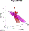
Figure 1:
Figure 1:
Angle extraction between preoperative (red) and intraoperative (magenta) carotid stem axes. For this example, the preoperative (red) carotid axis is represented by unit vector u = [0.291, −0.390, 0.873] and the intraoperative (magenta) carotid axis is represented by unit vector v = [0.096, −0.801, 0.591] in cartesian coordinates. Note that for visualization purposes, we scaled these unit vectors by a factor of 30.
Figure 2:
Figure 2:
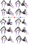
Relevant tissues in preoperative (left) vs. intraoperative (right) states for one of the patients. Osseous structures shown in gray, tongue in pink, tumor in blue, vasculature in red, and polymer laryngoscope in orange. The origin is defined by the FMV coordinate (intersection of Frankfurt, mid-sagittal, and vertical porion planes). Note that the tongue is swept to the patient’s left and that the tumor’s proximity to the tongue and vasculature changes between the preoperative and intraoperative states. Top row: Coronal views. Bottom row: Sagittal views with transparency.
Figure 3:
Figure 3:
Sagittal and axial CT images showing tumor segmentation from two Radiologists (R1 and R2). The final tumor mask used in analysis was the intersection of both the R1 and R2 image masks. The radiologists’ Intersection-over-Union (IoU) metric was computed for all six patients for both preoperative and intraoperative scans.
Figure 4:
Figure 4:
Tumor Deformation in Skull-Registered Reference Frame for Patients 1–6. Top Row: Axial/Operative Views. Bottom Row: Sagittal Views. In this skull-registered frame, all mandible bones were shifted outward from the patient and hyoid bones shifted down. Tumors in the oropharynx experienced a variety of displacement directions.
Figure 5:
Figure 5:
Vasculature Deformation (translation in mm, rotation in deg, n = 10). Displacement of the carotid bifurcation (CB) and lingual artery origin (LAO) as well as rotation of the carotid stem axis (A) are shown based on each of the three registration landmarks (skull, mandible, and hyoid). Both left (L) and right (R) sides were analyzed. Open circles represent the mean. Paired t-tests: * (p≤0.05), ** (p≤0.01), *** (p≤0.001).
Figure 6:
Figure 6:
Key Vasculature Landmarks near the Tumor Region. Black lines depict the displacement of the landmarks from the preoperative to the intraoperative state and are labeled via arrows with keys: 1. CB-L, 2. LAO-L, 3. CB-R, and 4. LAO-R. Note that the right lingual artery was not visible in the intraoperative state for this patient.
Figure 7:
Figure 7:
Vessel Deformation in Skull-registered Frame for Patients 1–11 Excluding Patient 5. Note that with the exception of Patient 2, all of the right lingual arteries were not able to be visualized on intraoperative imaging due to the right-sided compression of the tongue by the laryngoscope.
Figure 8:
Figure 8:
Tumor and Vasculature Deformations in Three Registered Frames (S: skull, M: mandible, and H: hyoid).

References

    1. Barrett WA, and Mortensen EN Interactive live-wire boundary extraction. Medical Image Analysis 1:, 1997. - PubMed
    1. Besl PJ, and McKay ND A Method for Registration of 3D-Shapes. IEEE Transactions on Pattern Analysis and Machine Inteligence 14:239–256, 1992.
    1. Bessen SY, Wu X, Sramek MT, Shi Y, Pastel D, Halter R, and Paydarfar JA Image-guided surgery in otolaryngology: A review of current applications and future directions in head and neck surgery. Head and Neck 43:, 2021. - PubMed
    1. Dice LR Measures of the Amount of Ecologic Association Between Species. Ecology 26:297–302, 1945.
    1. Gombos EC, Jayender J, Richman DM, Caragacianu DL, Mallory MA, Jolesz FA, and Golshan M Intraoperative supine breast MR imaging to quantify tumor deformation and detection of residual breast cancer: Preliminary results. Radiology 281:720–729, 2016. - PMC - PubMed